Back to Journals » Clinical Ophthalmology » Volume 18
Capsular Waves: A Warning Indicator for Potentially Malpositioned Intraocular Lenses
Authors Micheletti JM , Wang KM, Ton K, Bonem KN
Received 18 May 2024
Accepted for publication 21 August 2024
Published 2 September 2024 Volume 2024:18 Pages 2461—2466
DOI https://doi.org/10.2147/OPTH.S474591
Checked for plagiarism Yes
Review by Single anonymous peer review
Peer reviewer comments 2
Editor who approved publication: Dr Scott Fraser
J Morgan Micheletti,1 Kendrick M Wang,1,2 Khanh Ton,1 Karlie N Bonem1
1Berkeley Eye Center, Houston, TX, USA; 2Yale University, New Haven, CT, USA
Correspondence: J Morgan Micheletti, Berkeley Eye Center, 1435 Hwy 6, Suite #202, Sugar Land, Houston, Texas, 77478, USA, Tel/Fax +1 281-478-9868, Email [email protected]
Purpose: To share examination findings of the lens capsule which may act as an indicator for malpositioned intraocular lenses (IOL).
Setting: Single large multi-specialty private practice, Houston, Texas, USA.
Design: Focused, observational case series.
Methods: A review of pre-operative images of malpositioned single-piece IOLs with at least one haptic in the ciliary sulcus was conducted. The review included five cases who were referred to a single large multi-specialty private practice from June 2023 to December 2024 for an evaluation of posterior capsular opacification (PCO) and potential Nd:YAG capsulotomy.
Findings: A total of five eyes which previously had undergone cataract surgery and were referred for Nd:YAG capsulotomy for PCO were identified on slit lamp examination to have capsular waves, defined as a centripetal and circumferential striated pattern of PCO that results from a fused anterior and posterior capsule with at least part of the IOL anterior to the capsule. While one eye exhibited transillumination defects and pigment dispersion, the remainder of eyes did not. In some cases, the capsular wave was the only clue to IOL malpositioning due to a small pupil. These eyes had single-piece IOLs with at least one haptic in the sulcus and required subsequent IOL repositioning or exchange.
Conclusion: If capsular waves are seen on slit lamp exam, a thorough inspection of IOL placement should be conducted, especially before treatment with Nd:YAG capsulotomy. Capsular waves result from anterior and posterior capsule contact with an anteriorly situated IOL. This finding is a potential indicator of at least part of an IOL positioned anterior to the anterior capsule.
Keywords: IOL, sulcus, malpositioned IOL, uveitis, glaucoma, hyphema, iris atrophy, transepithelial defect
Introduction
The vast majority of cataract surgery performed involves performing an anterior capsulorhexis, removing the native lens, and inserting an intraocular lens (IOL) in the lens capsule. The lens capsule is a modified basement membrane that encapsulates the ocular lens and is mostly composed of interacting networks of laminin and type IV collagen.1 There can be instances where IOLs are inadvertently placed with either one or both haptics of a single-piece IOL in the ciliary sulcus space. Although three-piece IOLs are commonly placed in the sulcus in scenarios such as posterior capsule violation, placement of a single-piece IOL in the sulcus is typically avoided due to increased rates of post-operative complications.2 IOLs in the sulcus space compared to IOLs in the capsular bag are associated with increased rates of posterior capsular opacification (PCO), increased inflammation, and increased posterior capsular striae.3 Additionally rates of iris atrophy and transillumination defects, pigmentary dispersion, uveitis-glaucoma-hyphema syndrome are greater with single-piece IOLs in the sulcus.4–8 Common scenarios resulting in sulcus placed single-piece IOLs include inadvertent misplacement during IOL insertion, poor dilation impeding visualization of the capsulorhexis, and lack of access to three-piece IOLs for sulcus placement.
Malpositioned IOLs can precipitate various adverse consequences, encompassing image distortion, refractive errors, capsular opacification, chronic inflammation, iris damage, glaucoma, and other complications arising from iris-IOL haptic interaction. These complications may overlap with common post-operative issues following routine cataract surgery, such as the development of PCO. Discernment between the routine PCO of an in-the-bag IOL compared to capsular waves is crucial due to its impact on treatment outcomes. While both conditions may manifest as capsule opacification, visually significant PCO with in-the-bag IOLs is typically managed with Nd:YAG laser capsulotomy, whereas malpositioned IOLs typically necessitate surgical intervention for repositioning or exchange. Administering Nd:YAG laser capsulotomy in an eye later requiring IOL repositioning or exchange can significantly complicate subsequent surgery by rupturing the anterior hyaloid face and necessitating anterior vitrectomy with placement of a 3-piece IOL. Hence, accurate differentiation between PCO with an in-the-bag IOL and capsular opacification resulting from a malpositioned IOL is imperative, especially prior to Nd:YAG capsulotomy.
This manuscript aims to highlight the clinical implications of capsular waves, draw attention to their potential in signaling IOL malposition, and discuss appropriate surgical management strategies. Capsular waves could be the only subtle sign of a haptic or IOL in the sulcus, and if noted, a misplaced IOL should be ruled out prior to Nd:YAG capsulotomy. PCO that occurs with IOLs implanted in the capsular bag can be visualized as pearl-type or fibrosis-type.9 In vitro studies have demonstrated the process of cell-mediated fusion of the anterior and posterior capsule around areas where the anterior and posterior capsule make contact after cataract extraction. In these areas, F-actin organized to resemble lens fiber cells in areas of capsule fusion which were morphologically distinct from cell proliferation in areas of IOL-capsule contact.10 Menapace et al described Soemmering’s ring formation when the capsulorhexis edge meets the posterior capsule in the setting of planned sulcus fixation with PMMA or hydrogel IOLs. Posterior capsular wrinkling was described but was not found to invade centrally into the area behind the capsulorhexis, as that area remained ‘completely clear’.11 Our findings of centripetal migration and circumferential striations (capsular waves) that spread centrally and can be visually significant, have not yet been formally described in the literature as a potential warning indicator for a single-piece IOL in the sulcus.
Material and Methods
This is a retrospective chart review of a series of patients seen at a single large multi-specialty private practice in the United States from June 2023 to December 2023. The study was deemed to be category 4 exempt from institutional review board approval, and consent to review medical records was not required. Confidentiality was maintained for all patient data. All research was performed in accordance with the Declaration of Helsinki.12
Patients with PCO which subsequently were found to have at least one haptic of the implanted IOL in the ciliary sulcus were identified. The review included five cases that were referred for an evaluation of PCO and potential Nd:YAG capsulotomy. A review of slit-lamp photographs of the malpositioned IOLs and PCO pattern was conducted.
Results
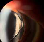
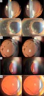
Capsular waves are defined as centripetal and circumferential striated pattern of capsular opacification. A total of 5 eyes identified during the review had findings of capsular waves (Figure 1). In these eyes, each contained a single-piece IOL where at least one haptic was located in the ciliary sulcus space (Figure 2). All five patients had been referred for evaluation and management of PCO, and all five patients had various models of hydrophobic acrylic IOLs, including SN60WF, MX60, and ZXT200. Capsular waves occur when the anterior capsule and posterior capsule come in contact and fibrosis occurs; in addition to misplaced IOLs, this can occur as a result of a larger capsulorhexis or reverse optic capture (Figure 3). Best corrected visual acuity (BCVA) of these eyes ranged from 20/40 to 20/60 prior to intervention.
 |
Figure 2 Oblique lighting of a ZXT IOL from Figure 1C showing the nasal haptic in the sulcus. |
The most common complications after initial cataract surgery were capsule opacification (100%, 5/5), glaucomatous changes in the optic nerve (60%, 3/5), decreased visual acuity (60%, 3/5), iris transillumination defects (40%, 2/5), dysphotopsias (60%, 3/5), pigment dispersion (40%, 2/5), and uveitis (20%, 1/5). All patients elected for IOL repositioning or exchange. No patients were treated with Nd:YAG capsulotomy prior to IOL revision surgery.
All patients subsequently underwent successful IOL revision surgery, resulting in either IOL exchange to a sulcus-appropriate 3-piece IOL or placement of the original IOL fully into the capsular bag. Post-operatively, all patients required subsequent Nd:YAG capsulotomy. All patients had an improvement in visual acuity from baseline after IOL repositioning and Nd:YAG capsulotomy with final BCVA ranging from 20/25 to 20/40.
Discussion
Capsular waves are a clinically significant examination finding that may show sensitivity towards malpositioned single-piece IOLs in the ciliary sulcus. The presence of capsular waves, indicated by a circumferential striated pattern of capsular opacification, should indicate to the clinician that a misplaced IOL must be ruled out. The presence of capsular waves can be exceedingly helpful in patients with poor pupillary dilation, as a misplaced IOL could be easily overlooked. Furthermore, the classic findings of transilluminating defects, uveitis, and pigment dispersion of a single-piece IOL in the ciliary sulcus are not always present. While capsular waves are possible if any part of the IOL is anterior to the anterior capsule, such as in the setting of a large capsulorhexis or reverse optic capture, malpositioned IOLs must be excluded prior to Nd:YAG capsulotomy.
The importance of accurate IOL placement is highlighted by increased adverse post-operative outcomes with sulcus based single-piece IOLs. Chang et al showed single-piece acrylic IOLs are not suited for sulcus fixation since the square-edged optic design, thick haptics, and unpolished side walls cause friction at the edge of the lens. This leads to increase adverse outcomes including iris chafing, pigment dispersion syndrome, uveitis-glaucoma-hyphema syndrome, iridocyclitis, and increased intraocular pressure.6 Thus, identifying a sulcus based single-piece acrylic IOL inciting these adverse outcomes is of clinical importance.
The treatment of PCO associated with in-the-bag single-piece IOLs compared to malpositioned IOLs are drastically different. Treatment of routine PCO is with capsulotomy, typically performed with a Nd:YAG laser. However, capsulotomy does not resolve IOL mispositioning. Subsequently performing surgery to reposition or exchange a malpositioned IOL in an eye that has a posterior capsulotomy can be more challenging with a higher risk profile than an IOL exchange with an intact posterior capsule. Furthermore, the presence of a posterior capsulotomy complicates the placement of an in-the-bag IOL, which is the ideal physiologic position.13–15 Thus, avoiding performing a capsulotomy prior to revision of malpositioned IOLs is of critical importance. This is of particular importance as more non-surgical eye care providers, such as optometrists, have started to perform Nd:YAG laser capsulotomy. Although optometrists do not perform intraocular surgical procedures, it is still important to correctly ascertain when a patient requires IOL revision instead of Nd:YAG laser capsulotomy. The identification of capsular waves as a guiding sign can help both ophthalmologists and optometrists differentiate between different patients with capsular opacification.
Repositioning a sulcus IOL into the capsule demonstrates that capsular waves resolve, indicating that this phenomenon is associated with contact between the anterior and posterior capsule. As other studies have shown, capsular opacification between an IOL and posterior capsule is morphologically distinct from capsular opacification between the anterior and posterior capsule.10 The fusion footprint in areas of anterior and posterior capsule contact form refractive structures resembling lens fiber cells and Elschnig’s pearls which likely account for the appearance of capsular waves.10 However, further investigation into the pathophysiologic formation of capsular waves is warranted.
Capsular waves, which are visualized as capsular opacification in a circumferential striated pattern, are a strong indicator of potential IOL malposition of a single-piece IOL in the ciliary sulcus. It is important to avoid performing Nd:YAG laser capsulotomy if a malpositioned IOL is identified, and ophthalmologists should instead be proceeding with IOL repositioning or exchange. These findings advocate for a thorough examination of patients presenting for PCO to confirm IOL position when capsular waves are present to help guide appropriate management and avoid complications.
Conclusion
Capsular waves, visualized as a circumferential striated pattern of capsular opacification, are a suggestive indicator to the clinicians that a malpositioned IOL must be ruled out. Although not pathognomonic of an IOL placed in the sulcus space, capsular waves can serve as a warning indicator for clinicians to investigate further. Avoiding performing a posterior capsulotomy prior to revision of malpositioned IOLs is of critical importance as it may lead to iatrogenic challenges during subsequent IOL repositioning or exchange procedures. Careful evaluation of PCO patterns to identify capsular waves can aid clinicians in performing the appropriate treatment for patients.
Funding
No financial support was supplied for the work in this manuscript.
Disclosure
JMM reports grants from ACE Vision - Consultant, Equity; Alcon – Consultant, Research Grant, Lecturer, Allergan (AbbVie) – Consultant, Research Grant, Avellino – Consultant, Bausch & Lomb – Consultant, BVI – Consultant, Centricity Vision – Consultant, Diamatrix – Consultant, Patent, Elios – Consultant, Glaukos Corp. – Consultant, Lecturer, Johnson and Johnson Vision – Consultant, Research Grant, Lenstec – Consultant, Research Grant, Lecturer, New World Medical – Consultuant, Lecturer, Nova Eye - Consultant, RxSight – Consultant, Samsara – Consultant, STAAR – Consultant, Research Grant, Lecturer, Synopic - Equity, Consultant, Tarsus – Consultant Visus Therapeutics – Consultant, Zeiss -– Consultant, Lecturer. The authors report no other conflicts of interest in this work.
References
1. Danysh BP, Duncan MK. The lens capsule. Exp Eye Res. 2009;88(2):151–164. doi:10.1016/j.exer.2008.08.002
2. Mehta R, Aref AA. Intraocular lens implantation in the ciliary sulcus: challenges and risks. Clin Ophthalmol. 2019;13:2317–2323. doi:10.2147/OPTH.S205148
3. Martin RG, Sanders DR, Souchek J, Raanan MG, DeLuca M. Effect of posterior chamber intraocular lens design and surgical placement on postoperative outcome. J Cataract Refract Surg. 1992;18(4):333–341. doi:10.1016/s0886-3350(13)80067-3
4. LeBoyer RM, Werner L, Snyder ME, Mamalis N, Riemann CD, Augsberger JJ. Acute haptic-induced ciliary sulcus irritation associated with single-piece AcrySof intraocular lenses. J Cataract Refract Surg. 2005;31(7):1421–1427. doi:10.1016/j.jcrs.2004.12.056
5. Chang SH, Lim G. Secondary pigmentary glaucoma associated with piggyback intraocular lens implantation. J Cataract Refract Surg. 2004;30(10):2219–2222. doi:10.1016/j.jcrs.2004.03.034
6. Chang DF, Masket S, Miller KM, et al. Complications of sulcus placement of single-piece acrylic intraocular lenses: recommendations for backup IOL implantation following posterior capsule rupture. J Cataract Refract Surg. 2009;35(8):1445–1458. doi:10.1016/j.jcrs.2009.04.027
7. Ollerton A, Werner L, Strenk S, et al. Pathologic comparison of asymmetric or sulcus fixation of 3-piece intraocular lenses with square versus round anterior optic edges. Ophthalmology. 2013;120(8):1580–1587. doi:10.1016/j.ophtha.2013.01.029
8. Razeghinejad MR, Havens SJ. Large Capsulorhexis Related Uveitis-Glaucoma-Hyphema Syndrome Managed by Intraocular Lens Implant Exchange and Gonioscopy Assisted Transluminal Trabeculotomy. J Ophthalmic Vis Res. 2019;14(2):215–218. doi:10.4103/jovr.jovr_122_17
9. Moreno-Montañés J, Alvarez A, Maldonado MJ. Objective quantification of posterior capsule opacification after cataract surgery, with optical coherence tomography. Invest Ophthalmol Vis Sci. 2005;46(11):3999–4006. doi:10.1167/iovs.04-1531
10. Wormstone IM, Damm NB, Kelp M, Eldred JA. Assessment of intraocular lens/capsular bag biomechanical interactions following cataract surgery in a human in vitro graded culture capsular bag model. Exp Eye Res. 2021;205:108487. doi:10.1016/j.exer.2021.108487
11. Menapace R. Posterior capsule opacification and capsulotomy rates with taco-style hydrogel intraocular lenses. J Cataract Refract Surg. 1996;22 Suppl 2:1318–1330. doi:10.1016/s0886-3350(96)80092-7
12. World Medical Association. World Medical Association Declaration of Helsinki: ethical principles for medical research involving human subjects. JAMA. 2013;310(20):2191–2194. doi:10.1001/jama.2013.281053
13. Stark WJ, Goodman G, Goodman D, Gottsch J. Posterior chamber intraocular lens implantation in the absence of posterior capsular support. Ophthalmic Surg. 1988;19(4):240–243.
14. Zhao YE, Gong XH, Zhu XN, et al. Long-term outcomes of ciliary sulcus versus capsular bag fixation of intraocular lenses in children: an ultrasound biomicroscopy study. PLoS One. 2017;12(3):e0172979. doi:10.1371/journal.pone.0172979
15. Liu Z, Lin H, Jin G, et al. In-the-Bag Versus Ciliary Sulcus Secondary Intraocular Lens Implantation for Pediatric Aphakia: a Prospective Comparative Study. Am J Ophthalmol. 2022;236:183–192. doi:10.1016/j.ajo.2021.10.006
 © 2024 The Author(s). This work is published and licensed by Dove Medical Press Limited. The
full terms of this license are available at https://www.dovepress.com/terms.php
and incorporate the Creative Commons Attribution
- Non Commercial (unported, 3.0) License.
By accessing the work you hereby accept the Terms. Non-commercial uses of the work are permitted
without any further permission from Dove Medical Press Limited, provided the work is properly
attributed. For permission for commercial use of this work, please see paragraphs 4.2 and 5 of our Terms.


