Back to Journals » Journal of Inflammation Research » Volume 17
Deciphering EIF3D’s Role in Immune Regulation and Malignant Progression: A Pan-Cancer Analysis with a Focus on Colon Adenocarcinoma
Authors Zhou Y, Chai R, Wang Y, Yu X
Received 22 May 2024
Accepted for publication 19 August 2024
Published 30 September 2024 Volume 2024:17 Pages 6847—6862
DOI https://doi.org/10.2147/JIR.S469948
Checked for plagiarism Yes
Review by Single anonymous peer review
Peer reviewer comments 2
Editor who approved publication: Professor Ning Quan
Yiming Zhou,1 Rui Chai,2 Yongxiang Wang,3 Xiaojun Yu3
1Department of Hepatopancreatobiliary Surgery, Zhejiang Cancer Hospital, Hangzhou Institute of Medicine (HIM), Chinese Academy of Sciences, Hangzhou, Zhejiang, 310022, People’s Republic of China; 2General Surgery, Cancer Center, Department of Colorectal Surgery, Zhejiang Provincial People’s Hospital (Affiliated People’s Hospital, Hangzhou Medical College), Hangzhou, Zhejiang, 310014, People’s Republic of China; 3Department of Gastric Surgery, Zhejiang Cancer Hospital, Hangzhou Institute of Medicine (HIM), Chinese Academy of Sciences, Hangzhou, Zhejiang, 310022, People’s Republic of China
Correspondence: Xiaojun Yu, Email [email protected]
Background: EIF3D, a key component of the eukaryotic translation initiation factor 3 (EIF3) complex, is critical in selectively translating mRNAs with atypical cap structures. Its relationship with colon adenocarcinoma (COAD) development and immune infiltration, however, remains under-explored. This study delves into EIF3D’s role in COAD using bioinformatics and in vitro experimentation.
Materials and Methods: We analyzed EIF3D expression levels utilizing TCGA, GTEx, CPTAC, and TISIDB databases. The TISCH database and ssGSEA method helped in assessing EIF3D’s link with the tumor immune microenvironment. EIF3D expression in CRC cells was gauged via real-time PCR. Cell proliferation was assessed using CCK8 and colony formation assays, while migration capabilities were tested through Transwell assays. Flow cytometry facilitated cell cycle distribution and apoptosis analysis. ChIP-qPCR identified transcription factors regulating EIF3D, and bulk sequencing explored EIF3D’s pathways in promoting COAD.
Results: EIF3D upregulation is a common feature in various tumors, especially in COAD, correlating with poor prognosis in many cancer types. It showed significant associations with immune cell and cancer-associated fibroblast (CAF) infiltration across multiple tumors. Additionally, it is closely associated with molecular and immune subtypes of multiple tumors, including COAD. Single-cell analyses depicted EIF3D’s distribution and proportion in CRC immune cells. In vitro findings indicated EIF3D knockdown curtailed proliferation and migration, inducing G0/G1 arrest in COAD cells. Moreover, bulk sequencing revealed EIF3D knockdown interferes with multiple cancer-related pathways, likely by curtailing cell cycle and DNA replication activities to regulate cell proliferation.
Conclusion: EIF3D emerges as a potential prognostic biomarker for tumor progression and immune infiltration, particularly in COAD, potentially predicting immunotherapy efficacy. Additionally, EIF3D represents a multifaceted target implicated in COAD’s malignant progression.
Keywords: EIF3D, pan-cancer, tumor immune microenvironment, biomarker, therapeutic target
Introduction
Cancer, recognized as a formidable global health threat, not only contributes to significant incidence and mortality rates but also stands as a major barrier to life extension.1–3 In the evolving landscape of cancer treatment, we are currently witnessing the emergence of a pan-cancer era. A plethora of cancer treatment modalities have surfaced in recent years, with immunotherapy notably thriving and attracting extensive attention in both academic and clinical realms.4,5 Immunotherapy functions by intricately orchestrating the human immune system to actively engage in the battle against tumors.6,7 The tumor immune microenvironment (TIM) is instrumental in the efficacy of immunotherapy. Research underscores that the activity status and distribution of immune and stromal cells within the TIM are critical determinants of patients’ prognosis and treatment outcomes.8,9 Specifically, T cells, especially the CD8+ subset, are frequently associated with a positive prognosis in cancer.10,11 Conversely, M2-type macrophages and T regulatory cells are widely acknowledged as adverse prognostic factors in cancer.12–14 Hence, gaining a deeper understanding of and intervening in the TIM is vital for enhancing the effectiveness of treatments. The integration of immunotherapy with conventional modalities such as radiotherapy and chemotherapy, as well as its combination with targeted therapy, has demonstrated synergistic and complementary anti-tumor effects, offering promising prospects for clinical applications.15,16 Immunotherapeutic strategies, such as immune checkpoint inhibitors (ICIs), have shown significant clinical benefits for a diverse range of cancer patients.17 However, only a minority of patients experience extended benefits, which necessitates ongoing research and development efforts to identify biomarkers that can predict treatment outcomes and facilitate more precise selection of immunotherapeutic interventions.
EIF3D plays a crucial role in the selective translation of mRNAs with non-standard cap structures.18–20 It exerts a significant influence on various cellular processes, including stress response and disease progression.21 Research has underscored the implication of EIF3D in cancer predominantly through its regulatory impact on translation.22–25 Additionally, the eIF3 subunits are associated with autophagy, cellular migration, and apoptosis, a type of programmed cell death.26,27 The interplay between apoptosis and immunotherapy is garnering increasing attention, placing EIF3D as a potential pivotal element in the TIM. Furthermore, EIF3D is instrumental in the differentiation and immune-suppressive functions of human regulatory T cells (Treg cells) through an alternative mRNA translation mechanism involving DAP5.28 Investigating EIF3D within the TIM may unveil novel perspectives for cancer research and therapy.
In this investigation, we performed an exhaustive analysis of the influence of EIF3D on prognosis and its predictive significance for immunotherapy across diverse cancers. Our findings indicated that EIF3D holds potential as a promising and novel prognostic biomarker, as well as a therapeutic target for individuals with pan-cancer.
Materials and Methods
EIF3D Expression and Pan-Cancer Analysis
We extracted RNA-sequencing data and clinical information (T stage, N stage, and pathological stage) of pan-cancer patients from PANCAN (TCGA, TARGET, GTEx) via the UCSC Xena browser (https://xenabrowser.net/) and analyzed using Sangerbox 3.0 (http://vip.sangerbox.com). EIF3D protein level expression was confirmed using the CPTAC database through the UALCAN portal (http://ualcan.path.uab.edu/analysis-prot.html).
Survival and Diagnostic Analysis
The survival and survminer in R estimated EIF3D’s correlation with overall survival (OS) rates. We generated forest maps with Sangerbox 3.029 and established diagnostic ROC curves using the PROC and timeROC packages.
Immunological Correlation
Drawing on prior research, tumor immune cell abundance was inferred from gene expression profiles using the Xcell method30 and ssGSEA, and immune/stromal scores were computed with ESTIMATE.31–33 The correlations between EIF3D, immune subtypes, and molecular subtypes were examined using TISIDB (http://cis.hku.hk/TISIDB/). The immune subtype groups are C1 (wound healing), C2 (IFN-gamma dominant), C3 (inflammatory), C4 (lymphocyte depleted), C5 (immunologically quiet), and C6 (TGF-b dominant). For single-cell immune cell data, we utilized TISCH (http://tisch.comp-genomics.org/home/). Additionally, we included immune phenotype scores (IPS) from the TCIA database to assess immunotherapy responses.
Prediction and Analysis of Proteins Interacting with EIF3D
The prediction of proteins potentially interacting with EIF3D is derived from the STRING database (https://string-db.org). We identified the top 20 interacting genes’ function pathways using the Metascape database (https://metascape.org/gp/index.html) and conducted GSEA analysis on EIF3D using Sangerbox 3.0, focusing on HALLMARK pathways.
Cell Culture and RNA Interference Techniques
HCT116 cells were purchased from Chinese Academy of Sciences (Shanghai, China) and cultured in DMEM medium containing 10% FBS and maintained at 37°C. EIF3D knockdown was achieved by transfecting EIF3D shRNA (hippobio, Zhejiang, China; sequence: 5ʹ-GCCGUUCAGCAAAGGAGAUdTdT-3ʹ) using Lipofectamine 3000 (Invitrogen, Massachusetts, USA). Knockdown efficiency was assessed via RT-qPCR 48 hours post-transfection.
Assessing Cell Viability and Colony Formation
Cell viability was measured using the CCK-8 assay (Beyotime, C0043), with readings taken every 24 hours. For colony formation assays, transfected cells were plated in six-well dishes and incubated in DMEM with 10% FBS for about two weeks. The developed colonies were treated with formaldehyde for fixation and subjected to crystal violet staining (Solarbio, China) to facilitate observation.
Flow Cytometry for Apoptosis and Cell Cycle Analysis
48 hours post-transfection, cell apoptosis and cycle were analyzed using an Annexin V-FITC kit (Invitrogen, 88–8007) and a cell cycle staining kit (YEASEN, 40301ES60), respectively. Flow cytometry analysis was conducted utilizing CytoFLEX equipment from the USA, and the acquired data were analyzed through CytExpert software (Beckman Coulter, Fullerton, CA, USA) and FlowJo software (BD).
Cell Invasion Assessment
The CHEMICON Cell Invasion Assay Kit (Sigma-Aldrich, ECM550) was employed for invasion assays. Post shRNA transduction, cells were placed in the upper chamber for 48–72 hours. Invading cells were visualized post-staining.
Chromatin Immunoprecipitation (ChIP)-qPCR
We sourced the EIF3D promoter sequence from UCSC (http://www.genome.ucsc.edu/) and identified potential transcription factor binding sites using JASPAR. The SimpleChIP Enzymatic Chromatin IP Kit (CST, 9002, USA) was employed to perform a ChIP assay, with MAZ antibody employed for DNA fragment precipitation. The qRT-PCR amplified DNA was detected using specific EIF3D promoter primers (EIF3D-Chip-325-F1 GCAGGTCACTTGGCACTTCT, EIF3D-Chip-325-R1 GAGACGCGAGAGGTGTGATT; EIF3D-Chip-239-F2 GTGGCGCATGCCTGTATT, EIF3D-Chip-239-R2 GGTTTCCTTAGCCGCCTAGT; EIF3D-Chip-200-F3 AATCACCTGAACCTGGGATG, EIF3D-Chip-200-R3 CCCGGGCTGGTCTTTAACT).
Transcriptome Sequencing and Functional Enrichment Analysis
Total RNA from HCT116 was isolated using TRIzol and quantified via NanoDrop (Thermo Fisher Scientific Inc, USA). A UID-mRNA-seq library was prepared and sequenced on a DNBSEQ-T7 (MGI Tech Co., Ltd., China). Gene expression was aligned to the GRCh38 reference genome using STAR (version 2.5.3a) and quantified by RPKM. Differential gene expression between EIF3D knockdown and control groups was calculated using the DESeq package. GO and KEGG analyses were performed using ClusterProfiler.
Statistical Analysis
Data analyses were carried out utilizing R software (version 4.1.0). Group differences were assessed using either the Wilcoxon Test or the Kruskal–Wallis Test. Survival analyses involved Kaplan-Meier curves and Log rank tests. A p-value <0.05 was considered statistically significant.
Results
EIF3D Expression and Its Prognostic and Diagnostic Significance Across Various Cancers
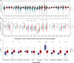
We examined the mRNA expression levels of EIF3D in human cancers using data from the TCGA and GTEx databases. As illustrated in Figure 1A and B, EIF3D was overexpressed in 19 cancers, primarily including CHOL, COAD and ESCA. However, lower expression was observed in ACC, BRCA, OV, and UCEC. Protein expression levels from CPTAC corroborated these findings (Figure 1C).
We investigated the correlation between EIF3D expression and pathological features. Elevated EIF3D expression was linked to advanced stages in cancers like HNSC, LIHC, and LUAD, while in BLCA and ESCA, lower expression levels were noted in advanced stages (Figure 2A–C). Additionally, the expression of EIF3D exhibited notable variation among distinct molecular subtypes in several tumor types, including BRCA, ESCA, and STAD (Figure 2D).
Subsequently, we evaluated the prognostic relevance of EIF3D across various cancers using Cox analysis for overall survival (OS), as illustrated in Figure 3A. Figure 3B highlighted that elevated EIF3D expression was linked to inferior OS in ACC, LIHC, KICH, and LUAD, while it was linked to a more favorable prognosis in PCPG and LGG. The diagnostic value was evaluated using ROC curves (Figure 3C), suggesting EIF3D’s potential as a diagnostic marker in several cancers, including STAD (AUC =0.760), BRCA (AUC = 0.705), COAD (AUC = 0.880), ESCA (AUC = 0.767), GBM (AUC = 0.954), HNSC (AUC = 0.790), KIRC (AUC = 0.748), LGG (AUC = 0.941), LIHC (AUC = 0.929), LUSC (AUC = 0.890), PRAD (AUC = 0.707), and READ (AUC = 0.875).
Immune-Related Characteristics of EIF3D in Pan-Cancer
The analysis also encompassed examining the relationship between EIF3D expression and immune cell infiltration in the TIM (Figure 4A). In most tumors, we noticed a pronounced positive correlation between the expression of EIF3D and the extent of infiltration by B cells, T cells, macrophages, and cancer-associated fibroblasts (CAFs). Additionally, EIF3D expression was associated with immune scores in several cancers, including CESC, COAD, and ESCA (Figure 4B; |cor| > 0.2 and P < 0.05 for all). In various other cancer types, including COAD, PRAD, LUSC, THYM, SKCM, TGCT, PCPG, and DLBC (Figure 4C; |cor| > 0.2 and P < 0.05 for all), EIF3D consistently exhibited a significant correlation with stromal scores.
Pan-cancer analysis to ascertain EIF3D’s immunological role indicated positive correlations with most immunomodulators in READ (Figure 5A). EIF3D expression was also linked to immune checkpoint-related genes across various cancers (Figure 5B). Additionally, its association with different immune subtypes in multiple cancers was established, showing significant variation between subtypes (Figure 5C).
Evaluating the Immunomodulatory Function of EIF3D in Colorectal Cancer (CRC)
In COAD, the expression of EIF3D exhibited a notable negative correlation with the infiltration of immune cells, possibly linked to immune resistance in COAD. EIF3D demonstrated a negative correlation with both immune and stromal scores (Figure 6A and B). Single-cell analysis from multiple CRC datasets indicated elevated EIF3D levels in T cells (conventional CD4 T cells, exhausted CD8 T cells, Treg cells, and proliferating T cells), suggesting its involvement in immune regulation and potential impact on the response to immunotherapy (Figure 6C and D). Moreover, comparable patterns of elevated EIF3D expression in NK cells, DC cells, and mast cells were also noted in CRC_GSE146771_Smartseq2 (Figure 6C–E).
Functional Analysis of EIF3D in COAD
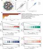
EIF3D, crucial in translation initiation, may impact cell biology and cancer progression. Low EIF3D levels correlated with worse OS and RFS in COAD patients (Figure S1A and B). PPI network analysis using STRING tool revealed a significant correlation between EIF3D and EIF3L at mRNA (r = 0.67, P < 0.001) and protein levels (r = 0.81, P < 0.001) (Figure 7A–C). Simultaneously, we observed that decreased levels of EIF3L were associated with poorer OS and RFS in patients with COAD (Figure S1C and D). Functional enrichment analysis indicated EIF3D’s involvement in translational initiation and regulation (Figure 7D), and GSEA indicated a potential involvement oxidative phosphorylation, cell cycle regulation, and genome integrity maintenance (Figure 7E).
EIF3D’s Role in Cell Proliferation and Metastasis in COAD
Initially, we confirmed EIF3D knockdown in HCT116 cells (Figure 8A). Subsequent assessments using CCK-8 and colony formation assays revealed a notable decrease in cell proliferation following the reduction of EIF3D expression (Figure 8B and C). This suppression of EIF3D induced a significant arrest in the G0/G1 phase, leading to an increased accumulation of cells in the G2/M phase, as illustrated in Figure 8D. Interestingly, Figure 8E suggests a trend towards reduced apoptosis upon EIF3D inhibition. The matrigel migration assays further substantiated that EIF3D suppression hampers the migratory capabilities of COAD cells (Figure 8F). Additionally, ChIP-qPCR experiments identified MAZ as a key transcription factor regulating EIF3D (Figure 8G).
EIF3D’s Pathway Involvement in COAD
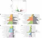
Advancing our investigation, post EIF3D knockdown, we undertook sequencing and conducted a differential gene expression analysis (Figure 9A). The analysis revealed that genes upregulated upon EIF3D suppression predominantly participate in pathways associated with environmental information processing, including the MAPK signaling pathway, PI3K-Akt signaling pathway, and the Focal adhesion pathway (Figure 9B–D). Conversely, genes downregulated post-suppression were significantly enriched in pathways pertinent to metabolism and the cell cycle (Figure 9C–E). These insights are pivotal in delineating the potential pathways through which EIF3D contributes to the progression of COAD.
Discussion
In our investigation, we performed an extensive pan-cancer analysis of EIF3D expression, integrating data from various databases. We identified notable variations in EIF3D expression across different types of cancer. Using Cox regression analysis, we established a correlation between elevated EIF3D expression and unfavorable OS in specific cancer types, while in others, it correlates with better prognosis. Furthermore, using ROC curve analysis, we discovered that EIF3D could potentially be an important diagnostic marker for various cancers, demonstrating strong predictive power. We also observed that EIF3D exhibits abnormal expression in immune cells and stromal cells and is closely associated with the expression of immune checkpoint genes, suggesting that EIF3D may play different biological roles in different types of cancer.
Earlier studies have shown that EIF3D plays a role in tumorigenesis by regulating the translation of oncogenes,22–25,34 suggesting EIF3D emerges as a promising novel biomarker and target for cancer treatment. Previous research has suggested an upregulation of EIF3D expression in various cancers, including breast cancer,35 ovarian cancer,36 gastric cancer,37 bladder cancer,38 colorectal cancer,39 lung,40 and renal cell carcinoma.41 These findings implied a potential association between EIF3D and tumor development. Our study also indicated diverse expression patterns of EIF3D across multiple cancer types. EIF3D may play diverse roles in the prognosis of patients with different types of cancer.23,37,40,42 We observed a significant association between elevated EIF3D expression and poor prognosis in ACC, LIHC, KICH, and LUAD. Conversely, high EIF3D expression was linked to a significantly better prognosis in PCPG and LGG. While the reason for the variation in these studies remains unclear and may be attributed to different cancer types, the association of EIF3D with prognosis, whether indicating improvement or deterioration, implies its potential role in therapeutic response. Additionally, we noted that EIF3D demonstrates good diagnostic efficacy in various cancers, suggesting its potential as a diagnostic marker.
The growing body of evidence indicated that the infiltration of lymphocytes in tumors is associated with cancer progression.43,44 Our results demonstrated a significant association between EIF3D and various immune cells in most cancer types. Previous studies have indicated that the DAP5/EIF3D complex is important in determining T cell lineage and function.28 Data analysis from CRC_GSE146771 revealed that EIF3D is significantly enriched in T cells.45,46 Our research findings indicated a close correlation between the expression of EIF3D and the expression of immune checkpoint genes, as well as immune cell infiltration in various tumors.
Crucially, our study further confirms that EIF3D knockdown suppresses malignant biological behaviors in colorectal cancer cells. Our findings indicated that suppressing EIF3D expression inhibits the proliferation of CRC cells, consistent with previous research.23,35,39,47 The discovery that EIF3D promotes the proliferation of CRC cells while predicting a better prognosis is intriguing yet not entirely unexpected. Previous studies in prostate cancer have found that EIF3D, while being a favorable prognostic factor, also promotes prostate cancer cell proliferation.48 Similarly, research indicates that eIF3a promotes tumorigenesis and cancer cell proliferation, but it contributes to a better prognosis.49,50 Additionally, ChIP-qPCR analysis has revealed a physical interaction between EIF3D and the MAZ transcription factor, suggesting that MAZ may mediate the function of EIF3D in tumorigenesis. Sequencing results show that following EIF3D knockdown, downregulated differential genes are mainly enriched in processes and pathways related to DNA replication and cell cycle, which is consistent with the results from public data analysis. Whereas upregulated differential genes primarily cluster in pathways such as “pathway in cancer” and “focal adhesion”. Although these signaling pathways may be regulated by EIF3D, it remains unclear whether they play a role in tumor development. Future systematic studies are needed to explore the contribution of these pathways to the function of EIF3D in cancer development.
Despite our comprehensive analysis and thorough validation of EIF3D’s role, our study acknowledges certain limitations. First, the reproducibility of our findings across different laboratories remains a subject of debate due to potential systematic biases. Second, validating the impact of EIF3D on cellular proliferation and metastasis necessitates further investigation through animal model experiments. Third, further experiments are needed to verify the signaling pathways affected by the EIF3D gene and explore the potential carcinogenic mechanisms related to EIF3D. Finally, to robustly establish the connection between EIF3D expression and the immune microenvironment, more advanced techniques such as single-cell sequencing, flow cytometry, and well-designed clinical trials are imperative. These aspects will form the cornerstone of our future research endeavors.
Conclusions
Our study underscores the notable role of EIF3D in expression, prognosis, diagnostics, and immune modulation across various cancers, particularly in COAD. This insight paves the way for novel directions in oncology research and suggests EIF3D as a promising target for immunotherapeutic interventions. Nonetheless, a deeper exploration into EIF3D’s mechanistic roles across diverse cancer types and its prospective utility in immunotherapy is crucial for advancing our understanding and therapeutic application.
Data Sharing Statement
The original manuscript contained in the research report is included in the article and Supplementary Material. Further inquiries can be made directly to the corresponding author.
Ethics Approval and Informed Consent
Not applicable, as GEO and TCGA are publicly available and have already received ethical approval. Since our study is based entirely on open-source data, the Zhejiang Cancer Hospital does not require additional ethical review.
Consent for Publication
The work has not been published previously, and it is not under consideration for publication elsewhere.
Funding
This work was supported by grants from Zhejiang Provincial Natural Science Foundation of China (LY18H30007).
Disclosure
The authors have no relevant financial or non-financial interests to disclose in this work.
References
1. Wu S, Powers S, Zhu W, Hannun YA. Substantial contribution of extrinsic risk factors to cancer development. Nature. 2016;529(7584):43–47. doi:10.1038/nature16166
2. Cortes J, Perez-García JM, Llombart-Cussac A, et al. Enhancing global access to cancer medicines. Ca a Cancer J Clinicians. 2020;70(2):105–124. doi:10.3322/caac.21597
3. Siegel RL, Miller KD, Wagle NS, Jemal A. Cancer statistics, 2023. Ca a Cancer J Clinicians. 2023;73(1):17–48. doi:10.3322/caac.21763
4. Riley RS, June CH, Langer R, Mitchell MJ. Delivery technologies for cancer immunotherapy. Nat Rev Drug Discov. 2019;18(3):175–196. doi:10.1038/s41573-018-0006-z
5. Yasunaga M. Antibody therapeutics and immunoregulation in cancer and autoimmune disease. Semi Cancer Biol. 2020;64:1–12. doi:10.1016/j.semcancer.2019.06.001
6. Hiam-Galvez KJ, Allen BM, Spitzer MH. Systemic immunity in cancer. Nat Rev Cancer. 2021;21(6):345–359. doi:10.1038/s41568-021-00347-z
7. Zhang Y, Zhang Z. The history and advances in cancer immunotherapy: understanding the characteristics of tumor-infiltrating immune cells and their therapeutic implications. Cell Mol Immunol 2020;17(8):807–821. doi:10.1038/s41423-020-0488-6
8. Pitt JM, Marabelle A, Eggermont A, Soria JC, Kroemer G, Zitvogel L. Targeting the tumor microenvironment: removing obstruction to anticancer immune responses and immunotherapy. Ann Oncolo. 2016;27(8):1482–1492. doi:10.1093/annonc/mdw168
9. Zeng D, Wu J, Luo H, et al. Tumor microenvironment evaluation promotes precise checkpoint immunotherapy of advanced gastric cancer. Journal for Immunotherapy of Cancer. 2021;9(8):e002467. doi:10.1136/jitc-2021-002467
10. Fridman WH, Pagès F, Sautès-Fridman C, Galon J. The immune contexture in human tumours: impact on clinical outcome. Nat Rev Cancer. 2012;12(4):298–306. doi:10.1038/nrc3245
11. Fridman WH, Zitvogel L, Sautès-Fridman C, Kroemer G. The immune contexture in cancer prognosis and treatment. Nat Rev Clin Oncol. 2017;14(12):717–734. doi:10.1038/nrclinonc.2017.101
12. Mendoza-Reinoso V, Baek DY, Kurutz A, et al. Unique pro-inflammatory response of macrophages during apoptotic cancer cell clearance. Cells. 2020;9(2):429. doi:10.3390/cells9020429
13. Noy R, Pollard JW. Tumor-associated macrophages: from mechanisms to therapy. Immunity. 2014;41(1):49–61. doi:10.1016/j.immuni.2014.06.010
14. Han S, Chung DC, St Paul M, et al. Overproduction of IL-2 by Cbl-b deficient CD4(+) T cells provides resistance against regulatory T cells. Oncoimmunology. 2020;9(1):1737368. doi:10.1080/2162402X.2020.1737368
15. Albini A, Sporn MB. The tumour microenvironment as a target for chemoprevention. Nat Rev Cancer. 2007;7(2):139–147. doi:10.1038/nrc2067
16. Shi J, Kantoff PW, Wooster R, Farokhzad OC. Cancer nanomedicine: progress, challenges and opportunities. Nat Rev Cancer. 2017;17(1):20–37. doi:10.1038/nrc.2016.108
17. Gong J, Chehrazi-Raffle A, Reddi S, Salgia R. Development of PD-1 and PD-L1 inhibitors as a form of cancer immunotherapy: a comprehensive review of registration trials and future considerations. Journal for Immunotherapy of Cancer. 2018;6(1):8. doi:10.1186/s40425-018-0316-z
18. Sadato D, Ono T, Gotoh-Saito S, et al. Eukaryotic translation initiation factor 3 (eIF3) subunit e is essential for embryonic development and cell proliferation. FEBS Open Bio. 2018;8(8):1188–1201. doi:10.1002/2211-5463.12482
19. Wolf DA, Lin Y, Duan H, Cheng Y. eIF-three to tango: emerging functions of translation initiation factor eIF3 in protein synthesis and disease. J mole cell biolo. 2020;12(6):403–409. doi:10.1093/jmcb/mjaa018
20. Jackson RJ, Hellen CU, Pestova TV. The mechanism of eukaryotic translation initiation and principles of its regulation. Nat Rev Mol Cell Biol. 2010;11(2):113–127. doi:10.1038/nrm2838
21. Dong Z, Zhang JT. Initiation factor eIF3 and regulation of mRNA translation, cell growth, and cancer. Crit Rev Oncol/Hematol. 2006;59(3):169–180. doi:10.1016/j.critrevonc.2006.03.005
22. Lee AS, Kranzusch PJ, Doudna JA, Cate JH. eIF3d is an mRNA cap-binding protein that is required for specialized translation initiation. Nature. 2016;536(7614):96–99. doi:10.1038/nature18954
23. Zhang F, Xiang S, Cao Y, et al. EIF3D promotes gallbladder cancer development by stabilizing GRK2 kinase and activating PI3K-AKT signaling pathway. Cell Death Dis. 2017;8(6):e2868. doi:10.1038/cddis.2017.263
24. Cieśla M, Ngoc PCT, Cordero E, et al. Oncogenic translation directs spliceosome dynamics revealing an integral role for SF3A3 in breast cancer. Molecular Cell. 2021;81(7):1453–1468.e1412. doi:10.1016/j.molcel.2021.01.034
25. de la Parra C, Ernlund A, Alard A, Ruggles K, Ueberheide B, Schneider RJ. A widespread alternate form of cap-dependent mRNA translation initiation. Nat Commun. 2018;9(1):3068. doi:10.1038/s41467-018-05539-0
26. Pakos-Zebrucka K, Koryga I, Mnich K, Ljujic M, Samali A, Gorman AM. The integrated stress response. EMBO Rep. 2016;17(10):1374–1395. doi:10.15252/embr.201642195
27. Zhong Y, Lan J. Overexpression of eukaryotic translation initiation factor 3D induces stem cell-like properties and metastasis in cervix cancer by activating FAK through inhibiting degradation of GRP78. Bioengineered. 2022;13(1):1952–1961. doi:10.1080/21655979.2021.2024336
28. Volta V, Pérez-Baos S, de la Parra C, et al. A DAP5/eIF3d alternate mRNA translation mechanism promotes differentiation and immune suppression by human regulatory T cells. Nat Commun. 2021;12(1):6979. doi:10.1038/s41467-021-27087-w
29. Shen W, Song Z, Zhong X, et al. Sangerbox: a comprehensive, interaction-friendly clinical bioinformatics analysis platform. iMeta. 2022;1(3):e36. doi:10.1002/imt2.36
30. Zeng D, Li M, Zhou R, et al. Tumor microenvironment characterization in gastric cancer identifies prognostic and immunotherapeutically relevant gene signatures. Cancer Immunol Res. 2019;7(5):737–750. doi:10.1158/2326-6066.CIR-18-0436
31. Yoshihara K, Shahmoradgoli M, Martínez E, et al. Inferring tumour purity and stromal and immune cell admixture from expression data. Nat Commun. 2013;4(1):2612. doi:10.1038/ncomms3612
32. Bu F, Li X, Zhao Y, Bai L, Zhang S, Min L. Pan-cancer patterns of cuproptosis markers reveal biologically and clinically relevant cancer subtypes. Biomarker Res. 2023;11(1):13. doi:10.1186/s40364-022-00446-5
33. Bu F, Zhao Y, Zhao Y, et al. Distinct tumor microenvironment landscapes of rectal cancer for prognosis and prediction of immunotherapy response. Cellul Oncol. 2022;45(6):1363–1381. doi:10.1007/s13402-022-00725-1
34. Zhang W, Sun Y, Bai L, et al. RBMS1 regulates lung cancer ferroptosis through translational control of SLC7A11. J Clin Invest. 2021;131(22). doi:10.1172/JCI152067
35. Fan Y, Guo Y. Knockdown of eIF3D inhibits breast cancer cell proliferation and invasion through suppressing the Wnt/β-catenin signaling pathway. Int J Clin Exp Pathol. 2015;8(9):10420–10427.
36. Lin Y, Zhang R, Zhang P. Eukaryotic translation initiation factor 3 subunit D overexpression is associated with the occurrence and development of ovarian cancer. FEBS Open Bio. 2016;6(12):1201–1210. doi:10.1002/2211-5463.12137
37. He J, Wang X, Cai J, Wang W, Qin X. High expression of eIF3d is associated with poor prognosis in patients with gastric cancer. Cancer Manage Res. 2017;9:539–544. doi:10.2147/CMAR.S142324
38. Latosinska A, Mokou M, Makridakis M, et al. Proteomics analysis of bladder cancer invasion: targeting EIF3D for therapeutic intervention. Oncotarget. 2017;8(41):69435–69455. doi:10.18632/oncotarget.17279
39. Du W, Cheng H, Peng L, Yang D, Yang C. hmiR-34c-3p upregulation inhibits the proliferation of colon cancer cells by targeting EIF3D. Anti-Cancer Drugs. 2018;29(10):975–982. doi:10.1097/CAD.0000000000000674
40. Wang D, Jia Y, Zheng W, Li C, Cui W. Overexpression of eIF3D in lung adenocarcinoma is a new independent prognostic marker of poor survival. Dis Markers. 2019;2019:6019637. doi:10.1155/2019/6019637
41. Pan XW, Chen L, Hong Y, et al. EIF3D silencing suppresses renal cell carcinoma tumorigenesis via inducing G2/M arrest through downregulation of Cyclin B1/CDK1 signaling. Int j Oncol. 2016;48(6):2580–2590. doi:10.3892/ijo.2016.3459
42. Maekawa M, Hiyoshi H, Nakayama J, et al. Cullin-3/KCTD10 complex is essential for K27-polyubiquitination of EIF3D in human hepatocellular carcinoma HepG2 cells. Biochem Biophys Res Commun. 2019;516(4):1116–1122. doi:10.1016/j.bbrc.2019.07.010
43. Denkert C, von Minckwitz G, Darb-Esfahani S, et al. Tumour-infiltrating lymphocytes and prognosis in different subtypes of breast cancer: a pooled analysis of 3771 patients treated with neoadjuvant therapy. Lancet Oncol. 2018;19(1):40–50. doi:10.1016/S1470-2045(17)30904-X
44. Luen SJ, Salgado R, Dieci MV, et al. Prognostic implications of residual disease tumor-infiltrating lymphocytes and residual cancer burden in triple-negative breast cancer patients after neoadjuvant chemotherapy. Ann Oncolo. 2019;30(2):236–242. doi:10.1093/annonc/mdy547
45. Shen S, Chen L, Liu J, et al. Current state and future of co-inhibitory immune checkpoints for the treatment of glioblastoma. Cancer Biol Medi. 2020;17(3):555–568. doi:10.20892/j.issn.2095-3941.2020.0027
46. Fan A, Wang B, Wang X, et al. Immunotherapy in colorectal cancer: current achievements and future perspective. Int J Bio Sci. 2021;17(14):3837–3849. doi:10.7150/ijbs.64077
47. Ren M, Zhou C, Liang H, Wang X, Xu L. RNAi-mediated silencing of eif3d alleviates proliferation and migration of glioma U251 and U87MG cells. Chem Biol Drug Des. 2015;86(4):715–722. doi:10.1111/cbdd.12542
48. Jiang M, Lu Y, Duan D, et al. Systematic Investigation of mRNA N (6)-methyladenosine machinery in primary prostate cancer. Dis Markers. 2020;2020:8833438. doi:10.1155/2020/8833438
49. Yin JY, Shen J, Dong ZZ, et al. Effect of eIF3a on response of lung cancer patients to platinum-based chemotherapy by regulating DNA repair. Clini Cancer Resear. 2011;17(13):4600–4609. doi:10.1158/1078-0432.CCR-10-2591
50. Ma S, Dong Z, Huang Y, Liu JY, Zhang JT. eIF3a regulation of mTOR signaling and translational control via HuR in cellular response to DNA damage. Oncogene. 2022;41(17):2431–2443. doi:10.1038/s41388-022-02262-5
 © 2024 The Author(s). This work is published and licensed by Dove Medical Press Limited. The
full terms of this license are available at https://www.dovepress.com/terms.php
and incorporate the Creative Commons Attribution
- Non Commercial (unported, 3.0) License.
By accessing the work you hereby accept the Terms. Non-commercial uses of the work are permitted
without any further permission from Dove Medical Press Limited, provided the work is properly
attributed. For permission for commercial use of this work, please see paragraphs 4.2 and 5 of our Terms.


