")
Back to Journals » Journal of Pain Research » Volume 17
Evidence-Based Clinical Practice Guidelines on Regenerative Medicine Treatment for Chronic Pain: A Consensus Report from a Multispecialty Working Group
Authors D'Souza RS , Her YF , Hussain N, Karri J, Schatman ME , Calodney AK , Lam C , Buchheit T, Boettcher BJ , Chang Chien GC, Pritzlaff SG , Centeno C, Shapiro SA, Klasova J, Grider JS, Hubbard R, Ege E , Johnson S, Epstein MH, Kubrova E , Ramadan ME, Moreira AM , Vardhan S , Eshraghi Y, Javed S, Abdullah NM, Christo PJ, Diwan S , Hassett LC, Sayed D , Deer TR
Received 30 May 2024
Accepted for publication 21 August 2024
Published 11 September 2024 Volume 2024:17 Pages 2951—3001
DOI https://doi.org/10.2147/JPR.S480559
Checked for plagiarism Yes
Review by Single anonymous peer review
Peer reviewer comments 2
Editor who approved publication: Dr Vwaire Orhurhu
Ryan S D’Souza,1 Yeng F Her,1 Nasir Hussain,2 Jay Karri,3 Michael E Schatman,4 Aaron K Calodney,5 Christopher Lam,6 Thomas Buchheit,7 Brennan J Boettcher,8 George C Chang Chien,9 Scott G Pritzlaff,10 Christopher Centeno,11 Shane A Shapiro,12 Johana Klasova,1 Jay S Grider,13 Ryan Hubbard,14 Eliana Ege,15 Shelby Johnson,8 Max H Epstein,16 Eva Kubrova,8 Mohamed Ehab Ramadan,17 Alexandra Michelle Moreira,18 Swarnima Vardhan,19 Yashar Eshraghi,20 Saba Javed,21 Newaj M Abdullah,22 Paul J Christo,17 Sudhir Diwan,23 Leslie C Hassett,24 Dawood Sayed,6 Timothy R Deer25
1Department of Anesthesiology and Perioperative Medicine, Mayo Clinic, Rochester, MN, USA; 2Department of Anesthesiology, The Ohio State Wexner Medical Center, Columbus, OH, USA; 3Departments of Orthopedic Surgery and Anesthesiology, University of Maryland School of Medicine, Baltimore, MD, USA; 4Department of Anesthesiology, Perioperative Care, & Pain Medicine, NYU Grossman School of Medicine, New York, NY, USA; 5Precision Spine Care, Tyler, TX, USA; 6Department of Anesthesiology and Pain Medicine, The University of Kansas Medical Center, Kansas City, KS, USA; 7Department of Anesthesiology, Duke University, Durham, NC, USA; 8Department of Physical Medicine and Rehabilitation, Mayo Clinic, Rochester, MN, USA; 9Department of Regenerative Medicine, GCC Institute, Torrance, CA, USA; 10Department of Anesthesiology and Pain Medicine, University of California, Davis, Sacramento, CA, USA; 11Centeno-Schultz Clinic, Broomfield, CO, USA; 12Department of Orthopedic Surgery, Mayo Clinic, Jacksonville, FL, USA; 13Department of Anesthesiology, University of Kentucky College of Medicine, Lexington, KY, USA; 14Department of Sports Medicine, Anderson Orthopedic Clinic, Arlington, VA, USA; 15Department of Physical Medicine & Rehabilitation, Baylor College of Medicine, Houston, TX, USA; 16Department of Physical Medicine & Rehabilitation, Harvard Medical School, Boston, MA, USA; 17Department of Anesthesiology and Critical Care Medicine, Johns Hopkins Hospital, Baltimore, MD, USA; 18Department of Physical Medicine & Rehabilitation, University of Miami/Jackson Memorial Hospital, Miami, FL, USA; 19Department of Internal Medicine, Yale New Haven Health – Bridgeport Hospital, Bridgeport, CT, USA; 20Department of Anesthesiology & Critical Care Medicine, Ochsner Health System, New Orleans, LA, USA; 21Department of Pain Medicine, University of Texas MD Anderson Cancer Center, Houston, TX, USA; 22Department of Anesthesiology, University of Utah, Salt Lake City, UT, USA; 23Department of Pain Medicine, Advanced Spine on Park Avenue, New York City, NY, USA; 24Mayo Clinic Libraries, Mayo Clinic, Rochester, MN, USA; 25Department of Anesthesiology and Pain Medicine, West Virginia University School of Medicine, Charleston, WV, USA
Correspondence: Ryan S D’Souza, Department of Anesthesiology and Perioperative Medicine, Mayo Clinic, 200 First St. SW, Rochester, MN, 55905, USA, Tel +1-(507)-284-9696, Fax +1-(507)-266-7732, Email [email protected]
Purpose: Injectable biologics have not only been described and developed to treat dermal wounds, cardiovascular disease, and cancer, but have also been reported to treat chronic pain conditions. Despite emerging evidence supporting regenerative medicine therapy for pain, many aspects remain controversial.
Methods: The American Society of Pain and Neuroscience (ASPN) identified the educational need for an evidence-based guideline on regenerative medicine therapy for chronic pain. The executive board nominated experts spanning multiple specialties including anesthesiology, physical medicine and rehabilitation, and sports medicine based on expertise, publications, research, and clinical practice. A steering committee selected preliminary questions, which were reviewed and refined. Evidence was appraised using the United States Preventive Services Task Force (USPSTF) criteria for evidence level and degree of recommendation. Using a modified Delphi approach, consensus points were distributed to all collaborators and each collaborator voted on each point. If collaborators provided a decision of “disagree” or “abstain”, they were invited to provide a rationale in a non-blinded fashion to the committee chair, who incorporated the respective comments and distributed revised versions to the committee until consensus was achieved.
Results: Sixteen questions were selected for guideline development. Questions that were addressed included type of injectable biologics and mechanism, evidence in treating chronic pain indications (eg, tendinopathy, muscular pathology, osteoarthritis, intervertebral disc disease, neuropathic pain), role in surgical augmentation, dosing, comparative efficacy between injectable biologics, peri-procedural practices to optimize therapeutic response and quality of injectate, federal regulations, and complications with mitigating strategies.
Conclusion: In well-selected individuals with certain chronic pain indications, use of injectable biologics may provide superior analgesia, functionality, and/or quality of life compared to conventional medical management or placebo. Future high-quality randomized clinical trials are warranted with implementation of minimum reporting standards, standardization of preparation protocols, investigation of dose–response associations, and comparative analysis between different injectable biologics.
Keywords: regenerative medicine, injectable biologics, platelet-rich plasma, mesenchymal stem cell, bone marrow aspirate concentrate, pain medicine
Introduction
Regenerative medicine is an emerging field that harnesses the body’s own healing capacity to enhance tissue recovery, decrease pain, and improve functionality. Since the first use of the human body’s own cells for hematopoietic stem cell transplantation in 1957,1 several historical advancements have been achieved in the field of regenerative medicine including: allogeneic skin tissues in 1998,2 gene therapy to treat severe combined immunodeficiency (SCID) in 1990,3 and development of cellularized scaffolds to replace tissues in 2016.4 Similarly, the integration of regenerative medicine into the field of pain has also surged over the past decade, supported by preclinical and clinical studies.
With the goal of treating painful conditions, non-cellular regenerative medicine modalities have also been developed, including prolotherapy in the 1930s, followed by platelet-rich plasma (PRP) in the 1990s for orofacial wound management,5,6 both of which have rapidly gained traction, mainly in sports medicine and orthopedic surgery. Furthermore, the identification of mesenchymal stem cells (MSCs) has introduced another promising biologic and novel avenue for clinical use.
Regenerative medicine comprises approaches such as cell-based therapy, gene therapy, and tissue engineering that influence cell proliferation, interaction, and extracellular matrix restoration. Within pain management, regenerative medicine is employed as a treatment for a spectrum of conditions ranging from musculoskeletal conditions to neuropathic pain to spinal cord injury. Proposed mechanisms of action involve mobilizing inflammatory cells and secretion of cytokines and growth factors that play pivotal roles in tissue regeneration and maturation, and acting as natural anti-inflammatory agents.7 The pillars of regenerative medicine treatments in pain management include non-culture-expanded autologous MSCs from products such as bone marrow aspirate concentrate (BMAC) or fat aspirate, PRP, and their derivates (Figure 1). Per federal guidelines, these products should meet the requirement of minimally manipulated products. Currently, only 36 more than “minimally manipulated” cell and gene therapies are approved by the Food and Drug Administration (FDA), primarily targeting cancer, inherited genetic diseases, and allogeneic umbilical cord cell transplantation.8
 |
Figure 1 Diagram displaying adult and perinatal sources of mesenchymal stem cells. Adapted with permission from Hoang DM et al. Stem cell-based therapy for human diseases. Signal Transduct Target Ther. 2022 Aug 6;7(1):272.9 |
Utilization of regenerative medicine therapy for pain in the absence of widely accepted consensus guidelines leads to inconsistency in practice, reliance on physician experience, case-by-case decision-making, and variable interpretation from outcomes of small, heterogeneous trials with mixed results. This inconsistency impedes the broad acceptance of regenerative medicine by physicians, government regulatory agencies, and health insurance providers, potentially hindering patient access to beneficial treatment.
Therefore, the American Society of Pain and Neuroscience (ASPN) commissioned a group of experts to create formal guidelines summarizing the existing evidence for regenerative medicine treatment for chronic pain indications. Notably, the American Society of Interventional Pain Physicians (ASIPP) previously issued guidelines on biologics for low back pain10 and the American Academy of Orthopedic Surgeons (AAOS) issued an overview on the use of PRP for knee osteoarthritis (OA),11 although neither addressed the full spectrum of regenerative medicine therapy in chronic pain management. Our objective was to develop pragmatic guidelines that can guide clinical care and establish a basis for future clinical research and regulatory agencies’ considerations.
Materials and Methods
Development Process
We utilized a systematic approach to appraise the evidence on regenerative medicine interventions (eg, injectable biologics) for treatment of chronic pain, including the type of injectable biologics, evidence for various chronic pain indications, best peri-procedural practices to optimize response to injectable biologics as well as yield and quality of injectable biologics, safety profile and adverse effects, and ethical considerations. We employed a similar methodology that was utilized in prior guideline articles produced by ASPN.12–14 This current consensus guideline for regenerative medicine interventions adds to the current family of consensus practice guidelines and incorporates a systematic review process.
To create this consensus guideline, the ASPN society commissioned an international multidisciplinary panel consisting of anesthesiologists, pain medicine physicians, physical medicine and rehabilitation physicians, and sports medicine physicians who are experts in the field of regenerative medicine. To qualify for inclusion on this multidisciplinary panel, each author was required to have experience with offering and performing regenerative medicine therapy, or publications or ongoing research pertaining to regenerative medicine. Further, each panel member’s impact on the field of regenerative medicine was considered.
A steering committee was tasked with selecting preliminary questions, which were reviewed and refined to 16 questions by the entire committee. Questions were assigned to 3 to 4 person modules, who collectively worked with the Committee Chairs (RSD, TRD) on preliminary versions. After completion of all sections for each question, this was subsequently distributed to the full committee.
Regenerative medicine continues to be a relatively novel and emerging field, with evolving evidence. While systematic review methodology15–18 is standard for forming consensus guidelines on established therapy, the development of new modalities, disease applications, and other innovations may also benefit from this approach and advance the field faster than randomized controlled trials (RCTs). Therefore, in instances where RCT-level data were unavailable, the authors reviewed the available evidence with lower levels of quality coupled with clinical expertise from the multidisciplinary panel to fill the gap. In this section, we outline the methodology including the search strategy, systematic review process, and consensus guideline creation and grading.
Literature Search Methods
A protocol was developed a priori outlining steps to be undertaken in this consensus guideline. A comprehensive search of several databases was performed on November 14, 2023, identifying relevant studies on regenerative medicine therapy for chronic pain. No date or language limits for the search were applied. Databases searched (and the content coverage dates) were Ovid MEDLINE(R) (1946+ including epub ahead of print, in-process, and other nonindexed citations), Ovid Embase (1974+), Ovid Cochrane Central Register of Controlled Trials (1991+), and Scopus via Elsevier (1970+). The search strategies were designed and conducted by a medical librarian (LH) with input from the study investigators. Controlled vocabulary supplemented with keywords was used. The actual strategies listing all search terms used and how they were combined are available in the Supplemental Material. We also hand-searched reference lists of identified publications to identify relevant publications not captured in the librarian-assisted search strategy.
Broad MeSH terms and Boolean operators were selected for each database search, including terms and synonyms for regenerative medicine, stem cells, injectable biologics, mesenchymal stem cells, platelet-rich plasma, bone marrow aspirate concentrate, autologous conditioned serum, stromal vascular fraction, adipose-tissue derived biologics, analgesia, chronic pain, neuropathy, and other specific chronic pain conditions.
Inclusion criteria encompassed studies that described regenerative medicine (eg, injectable biologics) for treatment of various pain conditions (eg tendinopathy, OA, muscle pathology, conditions manifesting with neuropathic pain, and other selected conditions), physiological mechanism of action of injectable biologics, technique of obtaining injectable biologics, adverse events, ethical considerations, and other aspects. Study designs that were identified and included in our search strategy included RCTs, prospective observational studies, and retrospective observational studies. If RCTs or observational studies were unavailable for certain questions addressed in this guideline, we utilized case series, case reports, or pre-clinical studies. Given the emerging body of the literature pertaining to regenerative medicine, the panel felt that these sources of evidence also held value to help inform consensus guidelines. Exclusion criteria comprised non-peer reviewed publications and certain study designs (eg, review articles). Two authors (YFH and MER) independently selected abstracts as well as full-text articles from the above listed databases using the aforementioned search strategies. A third author (RSD) adjudicated any discrepancies.
Systematic Evaluation of Evidence and Consensus Best Practices Development
A total of 8256 studies were identified with the initial search. Abstracts for each study were reviewed independently by two authors (YFH and MER) to identify studies for full-text review. In total, 970 articles were identified for full-text review and, of these, 740 articles were included for final analysis.
Given the infancy of regenerative medicine and its emerging literature, the primary goal was to fill in gaps of knowledge through consensus from our panel. Based on the reviewed evidence, a list of consensus points was created by the panel. Evidence for each consensus point was appraised using the United States Preventive Services Task Force (USPSTF) criteria for evidence level (Table 1) and degree of recommendation (Table 2). A degree of recommendation indicates the highest degree of recommendation, while D indicates the lowest degree of recommendation. A list of all consensus points reported in this guideline is included in Table 3.
 |
Table 1 Hierarchy of Studies by the Type of Design (US Preventive Services Task Force) |
 |
Table 2 Meaning of Recommendation Degrees (I.S. Preventive Services Task Force) |
 |
Table 3 List of Consensus Points |
Modified Delphi Approach and Oversight of Bias and Conflict of Interest
All authors disclosed financial conflicts of interest and were asked to recuse themselves on any issue with which they have a relationship as well as competing interests. Conflicts of interest were disclosed and managed before initiation of the guideline development process. Per ASPN protocol,20 one of the primary authors was non-conflicted (RSD) and served as the editor of the paper for bias.
Using a modified Delphi approach, the interim draft and consensus points were distributed to all collaborators en bloc and each collaborator was requested to vote on each point.21 The evaluation criteria were simplified with three potential options: “agree” or “disagree” or “abstain”. If collaborators provided a decision of “disagree” or “abstain” for any point, they were invited to provide a rationale for their decision in a non-blinded fashion to the committee chair (RSD), who incorporated the respective comments and distributed revised versions to the full committee until consensus was achieved.
Two rounds of voting were implemented. Of those allowed to vote on consensus points, an eighty percent agreement rate was required to accept the consensus point. However, recommendations with <100% agreement were revised based on feedback from those who disagreed or abstained from voting. During the second round of voting, consensus points with a pre-defined >80% agreement were accepted. In addition, the list of final consensus points and manuscript were shared with all collaborators for final approval.
Question 1: What Injectable Biologic Agents are Available for Treatment of Chronic Pain Conditions?
In this section, we provide details on specific injectable biologics including PRP, autologous-conditioned serum (ACS), bone marrow aspirate concentrate (BMAC), adipose tissue-derived biologics, mesenchymal stem cells, amniotic-based products, hollow osseous fillers, extracellular matrix substances, and extracellular vesicles. Details on preparation and processing of all injectable biologics are provided in Supplemental Table 1. A list of 501(k) cleared preparation systems and FDA approval status are provided in Supplemental Table 2. In addition, a list of companies that offer regenerative medicine therapy are displayed in Supplemental Table 3.
Platelet Rich Plasma
PRP is a blood product derived from the centrifugation of a patient’s whole blood to produce a supraphysiologic concentration of platelets suspended in blood plasma. The PRP product is then injected into the site of injury with the goal of supplementing the body’s natural healing mechanisms. Initial uses for PRP were found in dentistry and oral maxillofacial surgery as well as wound healing in dermatology, although its uses have substantially expanded to musculoskeletal disorders.22 The mechanism of PRP involves potentiation of the healing process that is mediated by the release of growth factors including epidermal growth factor (EDF), insulin-like growth factor one (IGF-1), basic fibroblast growth factor one (bFGF-1), platelet-derived growth factor alpha/beta (PDGF-alpha/beta), vascular endothelial growth factor (VEGF), and transforming growth factor beta1 (TGF-β1), amongst others. Additionally, the release of other bioactive molecules such as metabolites, chemokines, cytokines, and dense granules of platelets supplement the growth factor response.23,24 Of all injectable biologics, PRP is most commonly studied and evidence generally supports its use to treat pain from OA and ligament injuries. A list of injectable biologics, their key characteristics, and respective secreted biochemical factors are displayed in Supplemental Table 4.
Autologous Conditioned Serum (ACS)
ACS is a product that was first introduced in the 1990s and used primarily to treat musculoskeletal injuries. It is derived from incubating venous blood under controlled conditions (historically within specialized glass spheres), which promotes monocyte activation and leads to high levels of several anti-inflammatory cytokines in the product including interleukin (IL) 1Ra, IL-10, and TGF-β1.25,26 There is recent evidence that the analgesic and functional improvements with ACS are due to exosome expansion during the incubation process.27 Studies have highlighted the efficacy of ACS in treatment of musculoskeletal disorders, with OA being the most frequent indication, followed by soft tissue pathology such as tendinopathies and radiculopathies.25,28–31 The preparation of ACS involves more than minimal manipulation and therefore this product is not approved for use in the United States.
Bone Marrow Aspirate Concentrate (BMAC)
There are two types of bone marrow: red and yellow. Red bone marrow contains hematopoietic stem cells (HSCs) that give rise to red blood cells, white blood cells, and platelets. Yellow bone marrow is made mostly of adipocytes and contains stem cells that can become cartilage, fat, or bone cells. The bone marrow can produce 200 billion red blood cells, 10 billion white blood cells, and 400 billion platelets on a daily basis.32 BMAC contains a variety of progenitor cells, including HSCs, endothelial progenitor cells (EPCs) and mesenchymal stromal cells (MSCs, also known as “mesenchymal stem cells” or “medicinal signaling cells”). The frequency of these progenitor cells in whole blood is very low, unless a trauma has occurred. The use of bone marrow-derived cells in the treatment of orthopedic conditions has been shown to be secondary to both MSC and HSC effects. Furthermore, MSCs are believed to be pericytes, or, perivascular cells, with different populations and properties based on different vascular niches. These cells localize adjacent to marrow vessels and can be identified by different markers besides the typical mesenchymal markers (cluster of differentiation [CD]105, CD90, CD73).33 The process of extracting and processing BMAC is described in Supplemental Table 1 and is displayed in Figures 2–4.
 |
Figure 2 Diagram displaying bone marrow aspiration technique. Two approaches to bone marrow aspiration are displayed in this figure. We advise the approach from the posterior superior iliac spine to the anterior inferior iliac spine (novel approach) due to its safety margin away from the greater sciatic notch. Adapted with permission from D’Souza et al. A three-dimensional computed tomography study to determine the ideal method for fluoroscopically guided bone marrow aspiration from the iliac crest. Bosn J Basic Med Sci. 2021 Jun 1;21(3):370–377.34 |
 |
Figure 3 Diagram displaying bone marrow aspiration. Fluoroscopic imaging of the posterior iliac crest with the Jamshidi needle. A hammer is used to gently penetrate the cortex of the iliac crest prior to aspiration. |
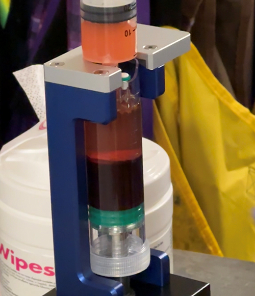 |
Figure 4 Processing of bone marrow aspirate to create bone marrow aspirate concentrate. Note the three fractions of the bone marrow aspirate after centrifugation. The fraction of interest is in the middle buffy coat directly above the dark red blood cells. |
Adipose Tissue-Derived Biologics
Adipose tissue is a readily available source of adipose-derived cells (ADCs) which contain a heterogeneous population of cells including MSCs, EPCs, regulatory T cells, macrophages, smooth muscle cells, pericytes and preadipocytes. Extraction of adipose tissue is performed by liposuction. Approximately 400,000 liposuction surgeries are performed in the United States each year. Liposuction can yield between 100 mL to >3 L of lipoaspirate tissue, which is routinely discarded.35 In general, two categories of adipose tissue are derived to concentrate MSCs: stromal vascular fraction (SVF), micro-fragmented adipose tissue (MFAT), and more recently nanofat.
SVF is a heterogeneous collection of cells contained within adipose tissue that requires the use of enzymes such as collagenase, dispase, or trypsin to break down the connective tissue. With the removal of connective tissue, there is a greater concentration of SVF in the aspirate made up of MSCs, EPCs, regulatory T cells, macrophages, smooth muscle cells, pericytes and preadipocytes. The following terminology has been used to identify the same adipose tissue cell population that is derived after enzymatic processing: Adipose-derived Stem/Stromal Cells (ASCs); Adipose Derived Adult Stem (ADAS) Cells, Adipose Derived Adult Stromal Cells, Adipose Derived Stromal Cells (ADSC), Adipose Stromal Cells (ASC), Adipose Mesenchymal Stem Cells (AdMSC), Lipoblast, Pericyte, Pre-Adipocyte, Processed Lipoaspirate (PLA) cells. The use of this diverse nomenclature has led to significant confusion in the literature.36
MFAT is a mechanical fragmentation of adipose tissue that avoids the use of enzymes. Utilizing metal beads or progressively smaller sieves, the lipoaspirate is refined to clusters of 0.2–0.8 mm. Repeated washing with saline is performed to remove lipids, blood, and tumescent solution. Techniques that have compared purely mechanical breakdown of adipose tissue, compared to the addition of collagenase, have demonstrated up to a 5-fold increase in MSCs favoring the addition of collagenase.37–41
Nanofat is an emerging ADC that is derived from emulsification and filtration of lipoaspirate.42,43 Components of nanofat approximate 400 to 600 μm. In nanofat, the product is devoid of mature adipocytes and contains microvascular fragments of arterioles, venules, and capillaries in addition to a variety of growth factors, biological peptides, and cytokines.
A disadvantage of ADCs is the processing of SVF, which is currently not allowed by the FDA. Processing of adipose tissue to create SVF is considered more than a minimally manipulated product, and thus constitutes a drug.44 Risks of liposuction include soft tissue and vascular damage, therefore requiring further training not common to the skillset of an interventional pain physician or sports medicine physician. Further, there may be a need for a liposuction system, although the quantity of lipoaspirate needed for adipose processing can be easily performed with a vacuum-locked syringe system.
Mesenchymal Stem Cells
Arnold Caplan first coined the term MSCs for their ability to differentiate into cells of the mesodermal lineage.45 The International Society for Cellular Therapy proposed a minimum criteria requirement for cells to be classified as human MSC. The cells must be able to differentiate in vitro into chondrogenic, osteogenic, or adipogenic tissue. They must express CD105, CD73, and CD90 but not express Human Leukocyte Antigen (HLA)-DR molecules, CD14, CD34, CD11b, CD45, CD79a, or CD19. Lastly, these cells must be able to adhere to plastic when preserved in standard culture conditions.46 MSCs are further classified as bone marrow-derived MSCs (BM-MSCs), adipose-derived MSCs (A-MSC), although other sources are reported as well.47 BM-MSCs are typically aspirated and harvested from bone marrow in the iliac crest or hip while A-MSCs are harvested from subcutaneous adipose tissue, often from the abdomen. Umbilical and amniotic fluid-derived MSCs have been proposed but are not as well-studied.48 Adult and perinatal sources of MSCs are displayed in Figure 1.
The therapeutic activity of MSCs is mediated by paracrine effects. It has been proposed that these cells secrete a variety of anti-inflammatory cytokines.49,50 Whereas the common misconception is that the stem cells will identify injured tissues and differentiate directly into those tissues by engraftment, it has been demonstrated that less than 1% of stem cells injected remain after as little as 7 days.51,52 MSCs respond to injured tissues and can coordinate a healing response via secretion of bioactive molecules, such as angiopoietin (Ang)-1, Ang-2, bone morphogenetic protein (BMP), brain-derived neurotrophic factor (BDNF), IL-6, and VEGF.53 MSCs have immune-privileged status, multilineage potential and the ability to support angiogenesis in a paracrine fashion.
Amniotic-Based and Other Perinatal Products
Initially utilized for treatment of burns and wound healing, amniotic products including amniotic membranes and fluid have been suggested for use as injectable biologics.54 Amniotic membrane consists of amniotic epithelial cells and amniotic MSCs.55 The latter consists of fibroblasts as well as protein complexes that collectively facilitate cellular transport. Amniotic membranes are thought to regulate amniotic fluid, form a protective layer for the fetus, and secrete bioactive molecules.55 These include fibroblast growth factors, epidermal growth factor, hyaluronic acid (HA), IL-1, IL-10, beta-defensins, TGF-β1, elafin, HLA-G, matrix metalloproteinases (MMPs), tissue inhibitors of metalloproteinases, and PDGF.55,56 Their anti-inflammatory and metabolically active properties allow for catabolism and subsequent tissue regeneration. Other perinatal products in use include umbilical cord blood and Wharton’s jelly.
Extracellular Matrix (ECM) Substances
Similar to biologic osseous fillers, extracellular matrix, also known as demineralized bone matrix (DBM), is allograft bone tissue with osteoconductive and osteoinductive properties. Harvested from donors, DBM contains mostly type 1 collagen, some type IV and type X collagen, as well as non-collagenous proteins and various osteoinductive growth factors (BMP, insulin-like growth factors, and PDGF).57 Injected or placed along bone sites of injury requiring growth and repair, the DBM is broken down by the host cells resulting in a release of growth factors promoting bone growth.58 Once obtained, DBM can be further augmented to provide a combinatorial effect, such as therapeutic drugs or bioactive materials, to enhance or augment the intrinsic activity of DBM.58
Recombinant Bone Morphologic Proteins
Bone morphogenetic protein-2 (BMP-2) is one of two recombinant BMPs utilized for bone induction in patients. Recombinant BMPs are used because endogenous human BMP from cadaveric bones have insufficiently small yields needed for clinical applications.59 BMPs are a group of proteins in the TGF-β family that bind to cell surface serine-threonine kinase receptors activating gene expression for bone creation.60 Cell culture cell lines are utilized to create recombinant BMP-2. Though effective, BMP-2 use has been controversial as it has been associated with possible neurologic impairment, heterotrophic ossification, wound complications, and carcinogenicity.61,62
Extracellular Vesicles
Extracellular vesicles (EVs) are promising therapeutic agents that are derived from platelets and progenitor cells and have been reported to support the paracrine function associated with progenitor cells.63 EVs are small particles (50 nm to 1000 nm) released by cells in the body, are involved in cell-to-cell communication in vivo and are found in all fluids and tissues in the body.64 While the mechanisms of potential therapeutic benefit of EVs in vivo are under evaluation, there is developing literature for MSC exosomes and platelet exosomes in the treatment and restoration of cartilage and even nerve injury.65,66 MSC-exosomes, the smallest class of EVs and derived from MSCs, when added to cultured nucleus pulposus cells obtained from a degenerated disc resulted in reestablishing a normal nucleus pulposus cell phenotype of the degenerated disc, suggesting its regenerative potential.67
Therapeutic Mechanisms of Injectable Biologics
There are several important sources of potential therapeutic benefit associated with injectable biologics. This section focuses on the common mechanisms of PRP, BMAC, and ADCs (eg, SVF, MFAT):
- All three preparations release EVs. The released EVs bind to target tissue-resident cells and are known to mimic the paracrine function of progenitor cells and platelets.63 EVs also amplify the therapeutic benefit of the cells and platelets present in injectable biologics.
- PRP and BMAC contain plasma, which is known to contain beneficial cytokines and functional biomolecules like IL-1Ra and α-2-Macroglobulin (a2M). IL-1Ra blocks the binding of IL-1β, which reduces pain associated with the pro-inflammatory supporting IL-1β molecule.68 a2M is an inhibitor of proteolytic enzymes like trypsin, but more importantly inhibits the collagen-degrading enzymes known as MMPs, including Collagen Type II-degrading MMP-13.69
- Platelets release growth factors and cytokines (the paracrine effect) following activation, including Epidermal Growth Factor (EGF), VEGF, Insulin-like Growth Factor (IGF), TGF-β1 and TGF-β2, adenosine, serotonin, and PDGF-BB, among others.70,71 These factors are able to promote proliferation of resident cells, enhance stem/stromal cell homing to the site of pathology, and reduce inflammation in the injected site.72
- Progenitor cells are able to respond to their environment and release a wide variety of bioactive molecules, including molecules that have important activities for enhanced tissue cell proliferation, immunomodulation, neo-angiogenesis, wound repair, rescue of dying cells (anti-apoptotic) and anti-microbial activity.73,74
- Mononuclear cells obtained from whole blood (eg, peripheral blood-derived mononuclear cells) that contain monocytes might provide a therapeutic benefit, since monocytes are known to transdifferentiate into anti-inflammatory macrophages (Type 2, M2).75 Monocytes are enriched in PRP and BMAC.
Although both the platelets in PRP and the progenitor cells in BMAC and SVF participate in the paracrine mechanism and release EVs and cytokines, once the platelets in PRP degranulate, there is no further therapeutic benefit. In contrast, progenitor cells are able to respond to their microenvironment74 and continue to release growth factors, cytokines and EVs after being delivered to the pathologic site.63 Thus, the therapeutic benefit when treating with cell-containing injectable biologics might theoretically be more robust and dynamic compared to that achieved by platelets in PRP. The therapeutic components present in injectable biologics primarily are focused on reducing the pro-inflammatory milieu within the injected site, to rebalance the catabolic and anabolic pathways. Restoring homeostatic balance may allow for the resident cells to return to their pre-degenerative status, improve the flux of nutrients, and stabilize the mechanical loading properties.76
Consensus Point 1. Patients should be advised that the mechanisms of action of injectable biologics in the treatment of chronic pain conditions are multifaceted and related to the specific injected biologic agent. Most mechanisms are centered on modulation of the injected tissue to promote an anti-inflammatory microenvironment. Common mechanisms include (1) release of anti-inflammatory cytokines, (2) release of growth factors, (3) differentiation of mononuclear cells into anti-inflammatory macrophages, and (4) release of extracellular vesicles that bind to target tissue resident cells and perform a paracrine function similar to progenitor cells (Level II-2, Grade C).
Question 2: What is the Evidence Basis for Variants of Standard Platelet Biologics?
Due to the variability of the different PRP products and production protocols (eg, preparation, dosing, delivery), as well as lack of clear description of PRP components in studies, there is no consensus regarding the most appropriate PRP product to administer for each specific pathology. Research is warranted to determine best practices regarding the number of injections needed per pathology, the interval of multiple injections, dose-effect with platelet concentration, best practices for LR-PRP versus LP-PRP products, the need for exogenous platelet activation, and the use of local anesthetics which may reduce the pain associated with the procedure but also may have a negative effect on platelet function.
Autologous Conditioned Plasma (ACP)
ACP is a specific commercially available preparation of PRP, consisting of a single-spin LP-PRP injectate (ACP, Arthrex, Naples, Florida, USA). As this is an LP-PRP, it would be expected to have similar beneficial use as other LP-PRP preparations. There has been minimal evidence to support that ACP has specific use or benefit over other PRP preparations. In a study of ACP in knee OA, intra-articular injections of ACP had mixed results, suggesting possible benefit over HA.77 However, real-world data has not demonstrated clinically beneficial effects from ACP in pain or functional outcome scores.78,79 Overall, there is insufficient evidence supporting the use of ACP over other PRP preparations, or for its specific use for musculoskeletal conditions.
Leukocyte-Rich PRP
There is debate on the utility of including leukocytes in PRP preparations due to potential for secreted cytokines to cause inflammation. However, these preparations may also have potential in preventing infection and improving healing. The evidence to support the optimal leukocyte concentration in PRP formulations is controversial and has not been well-established in vivo. In tendon pathology, a laboratory study suggests that LR-PRP may be superior to LP-PRP on tenocyte proliferation.80 This is contrary to in vitro studies that suggest the opposite effect.81 A three-arm comparison of LR-PRP versus LP-PRP versus saline showed no difference in all three treatment groups in patellar tendinopathy, with all three cohorts demonstrating similar benefit at 1 year.82 This is consistent with a study of lateral epicondylitis, in which LR-PRP, LP-PRP, and saline cohorts had similar favorable outcomes at 8 weeks.83
Leukocyte-Poor PRP
Some in vitro studies have reported that a high leukocyte concentration within PRP could increase the expression of catabolic pro-inflammatory molecules, which could be detrimental to the joint, suggesting that LP-PRP may be better suited for OA.84,85 Some clinical data suggest that LR-PRP has a more positive effect on ligament and tendon pathologies, while LP-PRP has a more positive effect on joint OA pathologies.23,31,86–91
Overall, it is most likely that both LP-PRP and LR-PRP have favorable outcomes for OA. Authors in a meta-analysis suggest that exclusion of leukocytes in LP-PRP may be favorable for less local adverse reaction and pain following injection compared to LR-PRP.92 However, outcomes comparing LR-PRP and LP-PRP for intraarticular injection are similar, with both LR-PRP and LP-PRP showing similar clinically meaningful improvements in pain and function.92 In a direct comparison of LR-PRP versus LP-PRP in knee OA, an RCT of 192 patients who received three weekly injections of LR-PRP or LP-PRP showed similar outcomes of pain intensity and functionality at 12-month follow-up, suggesting that the presence of leukocytes did not significantly impact efficacy.93
To our knowledge, there have been no studies directly comparing LR-PRP to LP-PRP for intradiscal, epidural, or facet use. There is some belief that the LR-PRP may be the preferred injectate in intradiscal injections for possible anti-bacterial properties, however this has not been supported or tested in the literature.
In summary, there is some evidence supporting the role of leukocytes in growth factor release, and several studies showing that leukocyte-reduced, and often platelet-reduced, products may not be effective. The heterogeneity or non-classification of PRP in studies makes it challenging to assess the differential efficacy of platelet derivatives.
Platelet-Rich Fibrin
PRF is a PRP derivative where autologous platelets and leukocytes are present in a complex fibrin matrix. The fibrin matrix is thought to act as a scaffold to enable cell migration to a site of repair. This has led to its use particularly in tendon pathology, as the fibrin matrix may have beneficial effects in a tendon defect. This has largely been studied as an adjunct to arthroscopic surgical tendon repairs with limited evidence of support. An RCT studying rotator cuff tears compared arthroscopic surgery versus arthroscopic surgery with PRF adjunct and reported no significant between-group differences in pain intensity or structural integrity.94 This is concordant with a meta-analysis of 219 patients showed similar re-tear rates in arthroscopic repair with use of PRF for rotator cuff injury.95 To our knowledge, there have not been prospective studies of percutaneous injection of PRF for tendon pathology outside of a surgical setting.
Platelet Lysate
PL is a cell-free platelet derivative, in which the intracellular components of the platelet have been released and collected as an injectate. It has been studied as an injectate in intra-articular joints96 and tendons,97 consistent with other platelet-derived biologics with favorable benefit. PL carries a unique safety profile compared to other platelet derivatives as it is acellular and should theoretically not carry the potential risk for platelet aggregation or vascular occlusion, although this warrants further investigation. In a study of 470 patients treated for lumbar radiculopathy, PL injection mixed with local anesthetic and corticosteroid decreased pain and increased functionality up to 2 years post-injection.98
Consensus Point 2.There is insufficient evidence regarding the most appropriate PRP product (ACP, LP-PRP, LR-PRP, PRF, PL) to administer for each specific pathology. There is some evidence supporting the role of leukocytes in growth factor release, and several studies showing that leukocyte-reduced, and often platelet-reduced, products may not be effective. Further comparative research among these products is warranted, including details on dose-effect with platelet concentration and need for exogenous platelet activation (Level III, Grade I).
Question 3: What Injectable Solutions are used in Prolotherapy for the Treatment of Chronic Pain Conditions?
Prolotherapy is a form of regenerative therapy where non-biologic agents are used to stimulate inflammation and growth. Some non-biologic agents utilized in prolotherapy include hypertonic dextrose, phenol-glycerin-glucose (P2G), sodium morrhuate, and pumice.99–101 Hypertonic dextrose is a commonly used solution for prolotherapy. Hypertonic dextrose may be used in a concentration ranging between 10% and 25% and is often diluted in local anesthetics in a 1:1, 1:2, 1:3, 1:4, or 2:5 ratio before injection.100,101 Sodium morrhuate is less commonly used and may be considered as an alternative if hypertonic dextrose is ineffective. Sodium morrhuate is found in a 5% solution composed of 2% benzyl alcohol in sodium salts of cod liver oil. Sodium morrhuate is diluted to a 0.5% to 1% solution before injection.100 Pumice is another non-biologic agent, derived from lava rock and grounded in fine powder. Pumice permanently remains in the body after injection, and therefore, is used as a last resort when all other proliferative injections fail. Finally, although phenol-glycerin-glucose is no longer used in clinical settings,99 it is used in research settings. This agent is constituted of 25% dextrose, 25% glycerin, and 2.5% phenol and is often diluted in local anesthetic prior to injection.100
The underlying mechanism for all proliferative agents used in prolotherapy involves the stimulation of inflammation and tissue growth.99,101 Glucose transporter (GLUT) 1–4 are found on the surface of many cells and are responsible for the transport of glucose.101 The entry of high concentrations of glucose or irritation from other proliferative agents such as P2G and sodium morrhuate leads to cellular lysis and release of intracellular contents. This generates temporary low-grade inflammation and the production of cytokines.101 Subsequently, these cytokines act in a paracrine fashion on the surrounding cells to promote growth and healing.102 Fibroblasts and nerve cells are all activated upon exposure to proliferative agents.101 In addition to the inflammation caused by proliferative agents, the direct trauma from the needle disrupts the cellular membrane and blood supply. This leads to the release of inflammatory factors such as calcitonin gene-related peptide (CGRP), bradykinin, and prostaglandins that are implicated in healing.103 It is noteworthy to highlight the nascent nature of research on elucidating the mechanism of various proliferative agents in prolotherapy. More work is needed to clarify the mechanism of proliferative non-biologic agents and to develop well-defined protocols for their use in pain medicine.
Consensus Point 3.Prolotherapy has primarily been utilized in research settings. The most common agent utilized in prolotherapy is hypertonic dextrose with a proposed mechanism involving generation of temporary low-grade inflammation, followed by cytokine release that promotes growth and healing (Level II-2, Grade C).
Question 4: What is the Evidence for Treatment of Tendinopathies Using Injectable Biologics?
In the current and subsequent sections of the guideline, we summarize evidence for injectable biologics for specific painful indications. Study data are also summarized in Supplemental Tables 5–21.
Lateral Epicondylitis
Lateral epicondylitis (LE), or tennis elbow, describes an overuse injury of the extensor tendons attaching to the lateral humerus. While typically self-limited, injuries failing the initial healing response can lead to chronic, degenerative tendinopathy. Consequently, the focus of treatment may shift from anti-inflammatory medication to regenerative approaches. Corticosteroid injections (CSI) have traditionally been used in LE refractory to conservative management. While they can provide short-term symptomatic relief, their use is increasingly discouraged due to questionable efficacy, high rates of recurrence, and risk of further tissue damage.104 Dextrose prolotherapy has yielded outcomes comparable or superior to CSI and placebo.104 A meta-analysis of nine trials demonstrated positive outcomes with dextrose prolotherapy, although there was a moderate risk of bias.105
The use of autologous blood products has gained interest as an alternative intervention for recalcitrant LE. RCTs comparing either leukocyte-rich PRP (LR-PRP) or leukocyte-poor PRP (LP-PRP) to placebo demonstrated significant improvements in pain and function with both types over a year of follow-up.106,107 RCTs also reported superiority of PRP injections to CSI at 3 months to 2 years.108 Some trials reported that CSI may be equivalent or more effective for pain relief in the short-term, but this effect declined over time.109 Meta-analyses have reiterated this association: CSI demonstrates efficacy for 2 to 8 weeks, while PRP remains superior in the longer term (3 months to 2 years).110 PRP injections also yielded similar benefits to surgical intervention in multiple studies.111 However, they have not demonstrated efficacy as an adjuvant to surgery or tenotomy.112 Unlike PRP, autologous conditioned plasma (ACP) injections failed to show significant improvements versus placebo over 6 months in small RCTs.113 However, they were superior to CSI in a larger RCT with 1-year follow-up.114
Administration of BMAC was associated with symptom relief from 2 to 12 weeks in one uncontrolled trial.115 Two uncontrolled pilot studies reported progressive improvements in pain, performance, and anatomical findings noted on MRI over a year with A-MSCs.116,117 The use of these biologics deserves further investigation with high-quality RCTs.
Consensus Point 4. PRP injection for lateral epicondylitis is associated with superior long-term relief (3 months to 2 years) compared to corticosteroid injection (Level I, Grade B).
Consensus Point 5. Limited evidence suggests that BMAC and adipose-derived MSC injection for lateral epicondylitis may be associated with improvement in pain, physical function, and anatomical findings (Level II-3, Grade C).
Consensus Point 6. Intra-tendinous ACP injection is likely ineffective for improving pain control or functionality in lateral epicondylitis and other tendinopathies (Level I, Grade D).
Medial Epicondylitis
Medial epicondylitis (ME), also known as golfer’s elbow or little league elbow, is an overuse injury of the flexor-pronator tendon complex at the medial elbow.118 ME manifests as a result of progressive angiofibroblastic disorganization and tendinopathy from repetitive microtrauma. Partial or complete tears of the involved tendons and ligaments, such as the ulnar collateral ligament (UCL), may accompany the degenerative process, particularly in athletes. While conservative management may lead to symptom resolution in the majority of cases, interventional approaches for recalcitrant ME include injections, tenotomy, and surgery.
CSI is the only injection type studied in an RCT specifically targeting ME. In a double-blinded RCT of 58 patients with ME, methylprednisolone injections resulted in significantly more pain relief than saline at 6 weeks, but not at 3 or 12 months.118 This short-lived effect of CSI is consistent with data from other joint and tendon disorders. As with other locations, CSI use at the medial epicondyle is associated with lipoatrophy, depigmentation, and tendon damage.119
In relation to injectable biologics for ME, some evidence is present for PRP and ACS. A systematic review of retrospective studies concluded that either LR-PRP or LP-PRP led to comparable outcomes to surgery and tenotomy for pain relief and function.120 In cases of sport-related UCL tears, two uncontrolled prospective studies reported significant improvements with PRP in functional scoring and ultrasound findings at an average follow-up of 54 to 70 weeks, with an average return to play at 12 weeks.121,122 An uncontrolled study of 14 patients reported no significant benefit from the use of ACP over 6 months, although there was a high dropout rate.123
Consensus Point 7. PRP injection for medial epicondylitis may provide long-term relief and functionality compared to surgery and tenotomy (Level II-2, Grade C).
Achilles Tendinopathy
Achilles tendinopathy (AT) involves a painful degeneration of the triceps surae tendons. Biomechanical overload and tissue-level alterations lead to a progressive loss of matrix integrity. The Victorian Institute of Sports Assessment-Achilles (VISA-A) or Achilles Tendon Rupture Score (ATRS) are often utilized to assess the severity of tendinopathy. Management of AT may include activity modification, physical therapy, orthotics, and anti-inflammatory medications. The use of CSI has fallen out of favor due to lack of efficacy and increased risk of rupture.124 Complete tendon ruptures or partial tears that fail to improve with conservative management may warrant surgical repair. In both non-surgical and surgical cases, the use of injectable biologics has been explored as an adjunct to treatment.
Multiple case series and uncontrolled trials have reported promising clinical improvements with PRP injection for AT.125 However, these results were not reproduced in several RCTs comparing PRP injection to placebo.126 Repetitive meta-analyses of these RCTs also failed to demonstrate significant long-term differences in VISA-A scores, pain, or tendon parameters on imaging.127 Some meta-analyses did find a significant short-term improvement at 6 weeks in VISA-A in the PRP group.128 In the setting of Achilles rupture, there was no difference in ATRS or rate of re-rupture with PRP versus saline injections.129 As an adjuvant to surgical repair130 and endoscopic debridement,131 the addition of PRP failed to demonstrate meaningful clinical benefit.
The use of BMAC for both chronic AT and Achilles rupture has been reported by retrospective case series.132 SVF injections compared to PRP produced similar improvements on imaging in one RCT133 and superior scoring outcomes in the short-term in another,134 but neither study was placebo-controlled and imaging did not correlate with symptoms.
Consensus Point 8. There is inconsistent and conflicting evidence on the use of PRP and BMAC for treatment of Achilles tendinopathy. Similarly, the re-rupture rate was not different with PRP injection versus saline injection (Level I, Grade C).
Plantar Fasciitis
Plantar fasciitis (PF) presents with stiffness and pain near the medial tuberosity of the calcaneus that is typically worse when initiating walking. In the majority of cases, symptoms resolve with conservative management. For recalcitrant PF, local interventions such as extracorporeal shockwave therapy (ESWT) or injections have been utilized. CSI is commonly administered to manage severe pain associated with PF. However, several trials and meta-analyses have found them to offer similar analgesia to dry needling and inferior analgesia to alternatives such as ESWT and PRP.135 CSI has also been associated with an increased risk of plantar fascia rupture.135 Meta-analyses have demonstrated low-quality evidence supporting the superiority of dextrose prolotherapy to placebo, although there was moderate-quality evidence of its inferiority to other modalities such as CSI for PF.136 Similarly, a series of five HA injections alleviated PF pain more than placebo at 5 weeks in another RCT,137 although additional studies are warranted to confirm this effect.
Most RCTs generally support that PRP injections were superior to placebo and CSI for pain relief and functional outcomes over 6 to 24 months.138 Some RCTs with shorter follow-up and lack of sham controls reported comparable outcomes to CSI in the early study phase, which may be confounded by anesthetic or placebo effect.139 Despite the heterogeneity and inconsistent blinding in these studies, multiple meta-analyses have reiterated that PRP is superior to CSI by 6 months for PF.140
Two RCTs reported significant improvements in pain and function with injection of human amniotic/chorionic membrane (HACM) versus sham injections for PF up to 3 months,141 while a pilot RCT demonstrated comparable outcomes to CSI over 12 weeks.142 A meta-analysis comparing different injections for PF found HACM likely to be superior to placebo, although this warrants further study.143
Consensus Point 9. There is low-quality evidence supporting superiority of dextrose prolotherapy compared to placebo for plantar fasciitis, although moderate-quality evidence suggests that it is inferior to corticosteroid injections. Therefore, prolotherapy is not the first recommended injection option for plantar fasciitis (Level I, Grade C).
Consensus Point 10. There is consistent evidence supporting that PRP injections for plantar fasciitis are associated with superior analgesia and physical function compared to placebo and corticosteroid injections over the long-term (6 to 24 months), although outcomes may be comparable in the short-term (Level I, Grade B).
Patellar Tendinitis
There are few, low-quality studies investigating injectable biologics for patellar tendinitis.144 A review highlighted best practices in patellar tendinopathy treatment, suggesting that PRP may not provide additional benefit beyond conventional therapy.145 Studies suggest that this may be due to lack of consistent methodology on dose, frequency, and timing coupled with variable patient selection criteria.146 Two small studies report the use of MSCs in patellar tendinopathy. Pascual-Garrido et al treated eight patients and reported high patient satisfaction with the treatment at five years.147 Conversely, Rodas et al failed to demonstrate the superiority of MSCs compared to PRP treatment, although it should be noted that both arms had improvements in reported pain and function scores.148
Consensus Point 11. There is inconsistent and conflicting evidence from low-quality studies on the use of PRP and injectable biologics for the treatment of patellar tendinopathy. Our appraisal suggests that injectable biologics are not recommended as an adjunct to conventional therapy (Level II-3, Grade D).
Rotator Cuff Tendinopathy
With regard to the use of PRP to treat partial rotator cuff tears, several RCTs showed efficacy over CSI including one study that demonstrated that intralesional injections improved rotator cuff tear size.149–153 A systematic review examining the use of PRP for partial thickness rotator cuff tears consisted of eight studies on intra-articular, subacromial or intra-tendinous injection approaches. Although the review was inconclusive of the best injection approach, PRP injection was generally associated with functional improvement in rotator cuff tendinitis.154 A significant number of studies performed PRP injection as an adjunct to surgical repair, making the assessment of PRP effect challenging. Evaluation of certainty of evidence is difficult due to the heterogeneity of techniques (eg palpation-guided vs ultrasound-guided), location of injection (intratendinous/bursa vs intra-articular), and other variables. All clinical endpoints suggested that PRP is potentially superior to CSI for rotator cuff tendinitis. The three RCTs that showed no difference or some difference in only short-term outcomes used PRP preparations that were less than two times concentrated.155–157 This is important as Marx et al have suggested that the minimum definition of PRP is 4–5 times concentrated.158
The use of BMAC injection to treat partial rotator tears versus exercise has been investigated in an RCT with a published mid-term analysis showing efficacy and reduced tear size.157 A second study of BMAC demonstrated reduced tear size and efficacy when compared to exercise.159
Consensus Point 12. The use of PRP injection for rotator cuff tendinitis is likely to provide superior analgesia compared to corticosteroid injections, including intra-articular, subacromial, and intra-tendinous approaches. Studies with non-inferior outcomes utilized less than two times the platelet concentrations, highlighting the importance of having a minimum concentration of four to five times in the PRP injectate (Level I, Grade B).
Consensus Point 13. There are limited studies suggesting analgesic utility of BMAC compared to exercise therapy for rotator cuff tendinitis, although future studies are warranted (Level II-1, Grade C).
Proximal Hamstring Tendinosis and Gluteal Tendinosis
A double-blinded RCT on proximal hamstring tendinopathy compared PRP versus whole blood injections. While patients in the cohort receiving whole blood experienced improved outcomes at 12 weeks, patients in the PRP cohort demonstrated superior analgesia and functionality at the six-month endpoint.160 Conversely, Levy et al demonstrated equivocal results at 8 weeks following PRP injection, although it is possible that the study duration of 8 weeks may have been too short for the therapeutic effect of PRP to manifest.161 A double-blinded RCT compared intratendinous PRP injection versus CSI for gluteus medius and minimus tendinopathy. The study reported superior analgesia and physical function in the PRP cohort compared to the CSI cohort at 12 and 24 weeks, which sustained improvement at two years.162
Consensus Point 14. There is inconsistent but promising evidence from a few RCTs on the use of PRP for hamstring and gluteal medius/minimus tendinopathy (Level I, Grade C).
Question 5: What is the Evidence for Treatment of Muscular Pathologies with Injectable Biologics?
Muscle Injury/Strain
Muscle strains and tears are common with the hamstring and rotator cuff muscles frequently injured. For persistent or high-grade injuries, refractory to rest, ice, and physical therapy, injectable biologics have been explored as both monotherapy or as an adjunct to surgical repair. PRP is the most extensively studied injectable biologic for muscle injuries. Multiple clinical trials and meta-analyses have demonstrated faster return to sport with PRP compared to placebo or therapy alone,163 although the quality of evidence is low due to inconsistency in findings, presence of clinical heterogeneity, and lack of blinding.
Of muscle groups, most regenerative medicine trials have focused on hamstrings. A meta-analysis of 10 RCTs reported non-significant improvements in return to play and re-injury rates in hamstring injuries treated with PRP combined with therapy instead of therapy alone or no treatment.164 For rotator cuff tears, PRP adjuncts to surgical repair significantly lowered re-tear rates and fatty infiltration on imaging, but not pain or speed of healing.165,166
One double-blind pilot RCT reported improved gluteus medius strength, volume, and histological healing response with placenta-derived MSCs versus placebo administered during and after hip arthroplasty, with no adverse events reported over a 2-year follow-up.167
Consensus Point 15. There is limited evidence to suggest that a strategy of PRP with physical therapy is superior to a strategy with just physical therapy alone for treatment of muscle injuries. Current evidence suggests there is likely minimal to no benefit (Level I, Grade C).
Question 6: What is the Evidence for Treatment of Osteoarthritis/Osteochondral Defects Using Injectable Biologics?
Despite an expanding body of literature highlighting the efficacy of regenerative medicine interventions for OA, there has been a variable degree of support within the medical community. A position paper from the American Association of Hip and Knee Surgeons (AAHKS) does not advocate using PRP in advanced hip and knee arthritis, stating that insufficient evidence supports its effectiveness.168 Conversely, the American Society of Interventional Pain Physicians (ASIPP) and American Medical Society for Sports Medicine have published position papers strongly supporting regenerative techniques, including the use of PRP and MSCs in degenerative musculoskeletal disorders.169,170 In this section, we review the current evidence for injectable biologics for each specific OA/osteochondral indications.
Knee and Hip Osteoarthritis
PRP may offer more benefit than placebo for alleviating pain and improving patient-reported outcomes related to knee OA symptoms for up to one year post-injection. However, the ability of PRP to enhance the osteoarthritic joint’s structure or function remains uncertain.
A meta-analysis171 comparing PRP to traditional injectates for knee OA examined 42 studies through 2023 and concluded that PRP had superior efficacy over HA, CSI, and placebo, particularly in reducing WOMAC and VAS pain scores, with the most significant improvement observed at the 6-month mark. The PRP cohort also sustained pain relief and functional improvement at the 12-month follow-up. Subgroup analyses indicated that multiple injections of PRP, particularly LR-PRP, yielded significantly better outcomes than single injections or LP-PRP. However, the debate regarding leukocytes’ role in PRP formulations persists, necessitating further research for clarification. Sahi et al evaluated the current state of systematic reviews concerning the use of PRP in knee OA. Their analysis involved 41 systematic reviews published between 2013 and 2023, revealing methodological deficiencies in most of these reviews. Despite the substantial number of studies included in these reviews (ranging from 5 to 43 primary studies), the overall methodological quality was deemed “critically low”.172 This underscores the need for improved methodological rigor in future research on PRP therapy for knee OA to ensure the reliability and validity of findings.
Two systematic reviews evaluated the efficacy of PRP injections for hip OA (HOA), although the data available are less robust than for knee OA. The first review, analyzing four trials with 334 participants, indicated potential intermediate-term benefits of PRP for HOA; however, its superiority over HA remains uncertain.173 Similarly, the second review, comprising five trials with 185 patients, observed improvements in patient outcomes at 6 and 12 months with PRP, but no significant difference was noted compared to HA alone.174
Meta-analyses examining MSC injections for knee OA have yielded mixed results. A meta-analysis exploring MSCs for knee OA as an alternative to joint replacement surgery reviewed 18 recent articles encompassing 1069 treated knees.175 Results indicated significant improvements in pain intensity, functional ability, walking distance, and quality of life, with earlier intervention yielding better outcomes. However, approximately 12.7% of cases experienced local complications.175 A conflicting meta-analysis,176 consisting of 13 RCTs, compared MSC injections versus placebo or HA for knee OA. The analysis showed that MSCs did not significantly improve pain, function, or stiffness scores compared to placebo, and the observed treatment effects did not exceed the minimum clinically important difference (MCID).176 These findings suggest that intra-articular MSCs may not be superior to placebo or other interventions for knee OA symptom relief, highlighting the need for further research, including direct testing and combination trials involving different types of cells, doses, and injection regimens for knee OA.
Consensus Point 16. PRP injection for knee OA is associated with superior long-term analgesia and physical functioning outcomes compared to HA, corticosteroid injection, and placebo at 6 and 12 months (Level I, Grade B).
Consensus Point 17. There is inconsistent and conflicting evidence on the use of MSCs to improve pain intensity and physical function in patients with knee OA. While some RCTs have indicated superior analgesia, functional ability, walking distance, and quality of life with MSC injection, some studies have failed to replicate this outcome (Level I, Grade C).
Consensus Point 18. Although intra-articular PRP injection may lead to improvements in analgesia and physical function in patients with hip OA, there may be no difference when compared to other interventions like HA injections (Level I, Grade C).
Shoulder Osteoarthritis
In contrast to knee OA, the evidence supporting the use of PRP in shoulder joint OA, particularly glenohumeral OA, is relatively limited with only two RCTs available.177,178 Both RCTs indicate that PRP is an effective treatment for improving pain and function in glenohumeral OA, demonstrating superiority to CSI in long-term follow-up, although there was no significant difference compared to HA.177,178 To date, no RCT has investigated the role of injectable biologics in treating acromioclavicular (AC) joint OA.
Consensus Point 19. There is consistent evidence, albeit from a few RCTs, that PRP injection for shoulder OA is associated with long-term improvement in pain and function compared to corticosteroid injection (Level I, Grade B).
Foot and Hand Osteoarthritis
A meta-analysis179 evaluating PRP for osteochondral lesions of the talus (OLTs) consisted of seven studies and found inconsistent anatomical results regarding chondral regeneration via MRI. However, pain and functionality scores consistently improved over four years with PRP treatment. Another systematic review180 included four studies and demonstrated that PRP as an adjunct to microfracture surgery significantly improved function and reduced pain compared to microfracture alone. Both reviews had limitations including inconsistency across included studies, lack of long-term data, lack of observer blinding, and lack of image guidance. Thus, despite some short-term benefits, the current evidence does not support PRP as a standard treatment for OLTs. Further, a single RCT comparing PRP versus placebo injections for ankle OA did not reveal any between-group differences in the American Orthopaedic Foot and Ankle Society (AOFAS) Ankle-hindfoot score.181
Studies have also explored BMAC injection for OLTs. A case–control study182 offered BMAC combined with bone marrow stimulation (BMS) or BMS alone. Both groups experienced significant improvements in foot and ankle outcome scores (FAOS) and physical component scores (12-item short-form survey [SF-12] PCS), with comparable clinical outcomes. However, the BMAC/BMS group exhibited notably enhanced magnetic resonance observation of cartilage repair tissue (MOCART) scores, indicative of superior cartilage repair (eg, border tissue integration, reduced fissuring). Similarly, a retrospective observational study183 compared autologous osteochondral transplantation with and without BMAC, revealing similar improvements in both cohorts in functional scores.
In terms of hand OA, a study184 explored autologous fat, PRP, a combination of both, or a saline injection for thumb OA in 95 patients. Results highlighted that a combination of autologous fat and PRP led to significant pain relief compared to saline injections. Additional benefits included improved hand function and overall quality of life.
Consensus Point 20. There is inconsistent and limited evidence regarding the use of PRP and BMAC for osteochondral lesions or OA of the foot or hand. Current evidence does not support routine use of injectable biologics for this indication compared to conventional therapy (Level I, Grade D).
Facet Osteoarthritis
Facet joints, innervated by the medial branches of dorsal rami, are commonly impacted by OA and may lead to facet-mediated pain, with a prevalence of 27% to 40% in individuals with chronic low back pain.185 The evidence supporting the application of PRP in managing facet-mediated pain is limited to small-scale retrospective observational studies and prospective clinical trials, encompassing patient cohorts ranging from 19 to 86 individuals with follow-up durations extending up to 6 months.186,187 In an RCT comparing PRP with intra-articular local anesthetic/corticosteroids, patients receiving PRP demonstrated higher satisfaction and success rates, with sustained efficacy observed over 6 months.188 Another study involving 49 patients with isolated lumbar facet syndrome revealed significant pain reduction and enhanced functionality following PRP administration after 18 months, with no reported adverse reactions, indicating long-term effectiveness in managing facet-mediated pain.189 Furthermore, in an RCT involving 144 patients over the age of 65 years with symptomatic degenerative facet joint disease, PRP demonstrated comparable efficacy to facetoplasty with HA at an average 18-month follow-up. However, the PRP group exhibited superior clinical improvement based on VAS and Oswestry Disability Index (ODI) scores and higher patient satisfaction during the late postoperative period.190 A recent systematic review of PRP for low back pain, encompassing two RCTs and three case series targeting facet-mediated pain and nine multi-target studies involving facet, disc, and the epidural space, demonstrated significant improvements in pain levels and ODI scores.72
Until recently, there were no published data on the use of MSCs in managing facet-mediated pain. A case report demonstrated the efficacy and safety of allogeneic MSC injection into lumbar facet joints and the epidural space using umbilical cord-derived MSCs, resulting in significant improvement in low back pain within 5 days.191 A prospective, open-label, non-randomized pilot study evaluated the clinical efficacy of a single injection of BM-MSC extracellular vesicle, an advanced investigational product (IP), into the lumbar facet joint space for chronic low back pain in 20 patients, showing statistically significant pain and functional improvement at the 3-month follow-up without complications.192 Additionally, a narrative review of three retrospective studies assessing intra-articular amniotic membrane and umbilical cord (AM+UC) in facet-mediated low back pain demonstrated the safety and potential efficacy of AM+UC administration.193 The evidence for MSC use in managing facet-mediated pain remains at an early stage. Further preclinical and clinical studies are imperative to elucidate the mechanisms of action of various MSC types, including amniotic membrane, umbilical cord-derived, and exosomes, in managing facet joint syndrome, to better inform therapeutic strategies in the future.
Consensus Point 21. PRP injection in the lumbar facet joints may be associated with superior analgesia, physical function, and patient satisfaction compared to local anesthetic and corticosteroid injection for lumbar facet-mediated pain (Level I, Grade B).
Consensus Point 22. There is limited evidence supporting lumbar facet injection with bone-marrow derived MSCs for lumbar facet-mediated pain, with modest improvement in pain intensity and physical function (Level II-3, Grade C).
Sacroiliac Joint Arthritis
The sacroiliac joint (SIJ) is the principal weight-bearing diarthrodial synovial axial joint. Innervation of the SIJ stems consistently from the dorsal rami of L5, alongside the lateral branches of S1 and S2. Osteoarthritic degeneration commonly underlies SIJ-mediated pain. Traditional interventional approaches for managing such pain include intra-articular or periarticular injections, as well as neurolysis of nerves innervating the SIJ.194
Few studies describe the use of PRP treatment for SIJ pain. The existing literature comprises prospective cohort studies, non-randomized trials, and RCTs with sample sizes ranging from 40 to 186 patients.195–198 Follow-up periods varied from 3 to 12 months. Saunders et al195 administered PRP into and around the dorsal interosseous ligament, whereas, in other studies, PRP was injected into the synovial portion of the joint. Across all studies, PRP treatment yielded significantly improved pain scores compared to steroid injections.195–198 A systematic review comparing the outcomes of patients treated with PRP or corticosteroids for lumbar spondylosis and SIJ arthropathy encompassed five studies (three RCTs and two non-randomized trials) comprising 242 chronic low back pain patients.199 Three studies reported a significant improvement in pain scores among the PRP cohorts. The study concluded that both PRP and corticosteroid injections are safe and effective options, however, there is some indication suggesting that PRP injections may offer superior efficacy over CSI during long-term follow-up.199 It is pertinent to note that the scope of the study was limited by the inclusion of studies analyzing diverse spine pathologies. To date, no human studies have been published to our knowledge on the use of MSCs in SIJ pain and this remains an area for future research.
Consensus Point 23. The current evidence generally supports that intra-articular sacroiliac joint injection with PRP may be associated with superior analgesia compared to intra-articular corticosteroid injection for sacroiliac joint pain (Level I, Grade B).
Question 7: What is the Evidence for Treatment of Intervertebral disc Disease and Discogenic Pain Using Injectable Biologics?
In this section, injectable biologics will be limited to autologous therapies, like PRP, BMAC, and ADCs (eg, SVF). Other therapies of a biological nature that have been used to treat intervertebral disc disease (IVDD) and/or discogenic back pain are obtained from living donors (eg, amniotic fluid), tissue culture (eg, cultured cells or exosomes released by cultured cells), or cadaveric donors (eg, disc tissue). For a variety of reasons, autologous biological treatments are more frequently used compared to allogeneic products. An important consideration is the potential for exposure to an infectious agent due to use of donor-derived materials in allogeneic products, whereas an autologous product has a lower chance of introducing an infectious agent. Although HA is an injectable biologic, without modification (eg, to create crosslinked or other forms—PEGylated), HA injected into the nucleus pulposus will be degraded.200 On the other hand, HA has been used as a carrier for injecting cultured cell preparations in several clinical studies.201
There are several anatomical structures within the disc that are associated with degeneration of the disc, including the annulus fibrosis, the nucleus pulposus, and the cartilaginous endplates, all of which can contribute to discogenic pain.202 A degenerating disc will show Modic changes,203 as well as display instability.202 Thus, a degenerating disc, while fully capable of being extremely painful on its own, may cause associated pathology in the facets, ligaments and muscles adjacent to the degenerating disc.204 Consequently, treatments of the disc and adjacent anatomical structures with injectable biologics (PRP, BMAC and SVF) will be reviewed in this section.
PRP Treatments for Discogenic Back Pain
A systematic review72 assessed the clinical evidence on the use of PRP to treat discogenic back pain and/or other pain generators in low back pain, which included the clinical studies listed in two other recent systematic reviews.205,206 Machado et al72 reviewed RCTs and prospective, single-arm trials in which PRP was injected into discs, intraspinal compartments (epidural and transforaminal), facets and muscles (either directed at a single target or multiple, simultaneously treated targets). A total of 914 study participants were treated in the 13 RCTs with an intradiscal injection and 1759 study participants were treated in the 27 prospective, single-arm trials, with approximately 29% receiving multi-target simultaneous injections. The RCTs compared the PRP treatment to a variety of control or placebo treatments, including corticosteroids, contrast agent, saline/kefazol, saline, lidocaine, HA, or contrast agent, as well as two-active treatment arms (ozone, Platelet Rich Fibrin [PRF]).72 Machado et al72 concluded that almost all studies showed positive results for the PRP treatments evaluated, with relatively few adverse events reported over the duration of the studies, which ranged from eight weeks out to two years. Their review showed that 11 RCTs demonstrated positive therapeutic benefit of the PRP treatment for both pain mitigation and improved disability, in contrast to the degree and durability of control treatments.
There are several publications that illustrate important considerations when using PRP as a therapy. In one study,207 PRP preparations with higher platelet concentrations used in treating discogenic back pain showed a positive correlation for improvement in pain (numerical rating scale [NRS] at 6-month milestone: Pearson coefficient [r] = 0.73) and disability (ODI at 6-month milestone: Pearson coefficient [r] = 0.7); both of which were assessed as statistically significant. PRP injectates, characterized as LR-PRP, with a 10-fold increase in platelet concentration were reported to be more therapeutically beneficial compared to PRP that had a platelet concentration increase of 5-fold or less.208 In contrast, Schepers et al209 used a lower volume of whole blood (32 mL) compared to the volume (60 mL) processed in Lutz et al208 and used a processing technology that resulted in a leukocyte-depleted PRP (LP-PRP), both of which might have accounted for a finding of non-superiority of the PRP treatments over the control arm.
Clearly, in view of the evaluation of a variety of types of PRP, including LR-PRP, LP-PRP, and PRF, there is no consensus as to which kind of PRP is more effective in reducing low back pain. One of the factors mentioned in favor of using LP-PRP is based on in vitro studies that found that PRP with a depleted level of leukocytes did not induce pro-inflammatory cytokines and catabolic activity in the nucleus pulposus in an animal model. In contrast, PRP with leukocytes promoted the upregulation of pro-inflammatory cytokines and enhanced catabolic activity.210,211 However, as reviewed above, clinical studies with both LP-PRP and LR-PRP injectates have demonstrated a therapeutic benefit in treating low back pain. Another challenge of the published clinical studies with PRP treatment is that most studies are limited to shorter study periods of one year or less.72 In addition, an important aspect of therapeutic benefit relates to the number of platelets in the PRP injectate. In this regard, higher doses of platelets were shown to provide a greater therapeutic benefit in a study that specifically addressed this important aspect of PRP treatments.207
Consensus Point 24. The current evidence generally suggests that intra-discal PRP injection may lead to improvement of discogenic pain (or pain from intervertebral disc disease) compared to placebo, corticosteroid injections, HA, and other injections. However, there is substantial clinical heterogeneity among studies, especially with respect to platelet concentration as well as LP-PRP versus LR-PRP formulations with non-superiority of PRP treatments in studies using low-volume leukocyte-poor injectate (Level I, Grade C).
SVF Treatments for Discogenic Back Pain
As reported in a recent review of clinical studies that use SVF as a therapeutic treatment,212 only a single clinical study213 used SVF to treat low back pain and/or discogenic low back pain. In this study, 15 study participants had a tumescent liposuction procedure performed after which adipose tissue was recovered and processed with a collagenase product. The digestate was centrifuged and washed with sterile buffered saline twice. The cell pellet was re-suspended in <3 mL of autologous PRP. The resulting SVF injectate was injected into one or more discs under fluoroscopic guidance. No serious adverse events were recorded over a 1-year study period. There were meaningful differences compared to baseline at 6 months in pain intensity and some functional domains. However, most other functional outcomes failed to reach a meaningfully significant difference compared to baseline at 2 and/or 6 months (eg, P-Flexion, L-Flexion, ODI).
Consensus Point 25. There is limited evidence that suggests that intra-discal SVF injection may lead to improvement in pain intensity from discogenic back pain, although these findings were inconsistent with other outcomes including physical function and spine range of motion failed to improve (Level II-3, Grade D).
BMAC Treatments for Discogenic Back Pain
BMAC as a treatment for discogenic back pain is not as widely studied as PRP, as shown in a recent review of BMAC to treat low back pain that listed only a few autologous BMAC-based studies, which were outnumbered by MSC cultured-cell studies.204,214 In a prospective, open-label clinical study, Pettine et al215 demonstrated that intradiscal injection with BMAC resulted in a short-term decrease in VAS and improvement in ODI, with durable therapeutic benefit for both pain mitigation and improved disability out to the 3-year endpoint of the study. Six out of the 26 study participants enrolled had a resolution of their discogenic pain at the 3-year milestone. In a prospective, open-label clinical study of the intradiscal injection of either autologous PRP or BMAC, Navani et al216 selected the biologic treatment (PRP or BMAC) based on diagnostic findings related to severity of pathology: study participants with Pfirrmann index 1 and 2 levels received PRP, while Pfirrmann index 3 and 4 levels received BMAC, either of which was injected in no more than 3 discs per participant. The authors reported that out of 20 study participants, 15 remained at the study endpoint of 18 months, with 93% of study participants reporting a >50% reduction in pain (assessed by the verbal pain score) and a >50% improvement in function (SF-36). Finally, Navani et al217 recently reported outcomes from a multi-center, prospective, RCT with PRP or BMAC injected into the painful disc(s) compared to a control group (saline trigger point injections). The study design allowed for a cross-over from the control treatment to either PRP or BMAC or from PRP/BMAC to BMAC/PRP if the participant had less than 50% improvement in NRS at the 3-month milestone. All participants in the control group crossed over to a biologic injection, while just one PRP-treated and one BMAC-treated participant crossed over to the other biologic. While there was not a meaningful difference in NRS and ODI between the PRP and BMAC cohorts at the 12-month milestone, both PRP and BMAC cohorts showed durable, statistically meaningful improvement in NRS and ODI at the 12-month milestone compared to baseline.217
Consensus Point 26. The current evidence suggests that intra-discal BMAC injection may provide long-term alleviation of pain and improvement in physical function for patients with discogenic pain, although these differences may be similar to those with intra-discal injection with PRP (Level I, Grade C).
Question 8: What is the Evidence for Treatment of Neuropathic Pain Etiologies Using Injectable Biologics?
In this section, we report the evidence for injectable biologics in treating various conditions that manifest with neuropathic pain. A systematic review218 evaluating the effectiveness of injectable biologics for neuropathic pain included 27 human studies. Indications included post-spinal cord injury (SCI) neuropathic pain, diabetic peripheral neuropathy (DPN), painful scars, pudendal neuralgia, trigeminal neuralgia, carpal tunnel syndrome, peripheral neuropathy, and radiculopathy. Overall, the review reported very low certainty of evidence in support of the efficacy of injectable biologics for treatment of neuropathic pain per The Grading of Recommendations Assessment, Development and Evaluation (GRADE) criteria.
Diabetic Peripheral Neuropathy
In one RCT219 consisting of 60 patients, PRP combined with medical treatment was compared with medical treatment only for DPN. VAS scores in the PRP cohort improved significantly from baseline and compared to the control cohort at all timepoints at 1, 3 and 6 months. Neurological symptoms and physical exam signs, as measured per the modified Toronto Clinical Neuropathy Score (mTCNS), were also superior in the PRP cohort at all timepoints.
A retrospective study reported that 8 out of 17 total patients with DPN were treated with US-guided injections of amniotic membrane/umbilical cord (AM/UC) particulate CLARIX FLO® perineurally, to common and deep peroneal nerves as well as the tibial nerve. Patients reported improvement in pain intensity by 50% at 1 month, 73% at 2 months, and 75% at 3 months after receiving an average of 2.75 injections (range 1–5).220
In summary, evidence remains limited for regenerative medicine therapy for DPN. One RCT219 supported the use of PRP in DPN with statistically significant VAS score improvement at 1–6 months, whereas one retrospective study220 reported improvement in pain intensity up to 3 months following injections with AM/UC product.
Consensus Point 27. Very limited evidence suggests that perineural injection of PRP along certain nerves (eg, median, ulnar, radial, peroneal, tibial, saphenous, and/or sural) may be associated with improvements in pain intensity and neurological symptoms (eg, numbness) in patients with diabetic peripheral neuropathy compared to conventional medical management (Level I, Grade C).
Spinal Cord Injury
Regenerative medicine interventions for SCI have included MSCs and neural stem cells. Aside from motor and sensory recovery, neuropathic pain is investigated as one of the outcomes. Neuropathic pain in SCI may be mediated by several mechanisms of increased neuronal hyperexcitability, as well as disruption of the blood–brain barrier with subsequent influx of macrophages. MSCs are proposed to modulate microglia and macrophage activity and decrease inflammatory cytokines.221 Neural stem cells may alter neuropathic pain through decrease of P2X receptors.222
All studies used culture-expanded (CE) MSCs either from umbilical cord (Wharton jelly, cord blood) or placenta, or BM-MSCs, and injection of MSCs were all performed intrathecally. One RCT223 showed no difference in chronic SCI pain up to 6 months following a single intrathecal dose of Wharton jelly MSCs, while two single-cohort studies with repeated injections of autologous CE BM-MSCs224 or CE UC-MSCs225 showed significant improvements in pain intensity from 1 to 10 months. One single-cohort study consisting of 20 patients, but only 4 with neuralgia in SCI, reported improvements in pain after 5 mL injection of 100 million autologous CE BM-MSCs at 1 month, with either intrathecal or CT-guided in-lesion injection.226
Consensus Point 28. Limited and conflicting evidence suggests that intrathecal or in-lesion injection with MSCs for spinal cord injury may decrease chronic pain intensity. Due to inconsistent and limited data, the current evidence does not support the routine use of intrathecal or in-lesion biologic injection for spinal cord injury (Level I, Grade D).
Radiculopathy
Four RCTs showed positive response to epidural injection of ACS for radicular pain, with three studies showing superior pain relief with ACS compared to CSI at 22 weeks227 to six months228 follow-up. Efficacy has been demonstrated in both lumbar and cervical epidural injections with ACS. Goni et al229 showed improved analgesia at 6 months compared to steroid, although it is unclear if statistical significance was achieved. Godek et al230 showed significant pain relief at 6 months; however, there were no between-group differences. Two single-group studies noted significant improvement in pain and other metrics at 6 months with repeat ACS injections.231,232 All studies that included functional outcome scores improved similarly with ACS injection.227,228
In terms of PRP and PRP-related products, one systematic review233 and a single-group study highlighted positive analgesic and functional outcomes with epidural injection of PRP/PRGF or platelet lysate (PL) for use in radicular pain. In the systematic review,233 use of PRP, PRGF or PL to treat radiculopathy and radicular pain was found in a total of three RCTs,234–236 and several observational studies. A total of 1092 patients received PRP or platelet-derived product. All studies noted improvement in pain scores and functional outcomes. Pain relief was sustained in both PRP and steroid groups until approximately three months; however, two of three RCTs showed long-lasting results in PRP cohorts for up to 12 months.234,235 Further, a retrospective observational study highlighted substantial analgesia lasting up to 24 months using PL, corticosteroid and local anesthetic combination injection.98 A high risk of bias was noted in two RCTs in at least one category per the Cochrane collaboration risk of bias evaluation tool, and the systematic review determined a very low certainty of evidence per GRADE analysis.
A single cohort study evaluated the effects of CE BM-MSCs for radicular pain.237 Autologous CE BM-MSCs were injected in 33 subjects at volumes 1–3 mL with PL via intradiscal access to relieve radicular pain. Numeric pain score decreased significantly from baseline (5.2) to 3 months (1.6,), 4 years (2.5), 5 (3.7), and 6 years.
Consensus Point 29. While epidural injection with ACS may improve radicular pain symptoms, some evidence suggests that it is not superior to corticosteroid injection (Level I, Grade C).
Consensus Point 30. Epidural injection with PRP or other PRP-related products (eg, PL) may alleviate radicular pain symptoms in radiculopathy, although studies had a high risk for bias (Level I, Grade C).
Consensus Point 31. There is very limited and low-quality evidence suggesting that intra-discal injection with MSCs or PL may relieve radicular pain (Level II-3, Grade C).
Trigeminal Neuralgia
Regenerative medicine injections to treat trigeminal neuralgia have been limited to case reports and series. Two case series were found to support the use of ACS or SVF injection perineurally for trigeminal neuralgia with significant decrease in pain at 3 weeks along with decrease in medication use.238 This effect was durable with autologous SVF injection with significant analgesia up to 6 months.239
Consensus Point 32. Perineural injection at the trigeminal nerve with biologics (eg, ACS, SVF) is currently not recommended for trigeminal neuralgia outside of an experimental setting due to data limited to case reports (Level III, Grade D).
Post-Herpetic Neuralgia
A single-cohort study by Sollie et al utilized autologous fat graft to treat post-herpetic neuralgia (PHN) and reported statistically significant improvements in pain intensity at 12 weeks.240 However, importantly, an RCT performed by the same group found no significant difference in pain relief between participants injected with autologous fat graft versus saline at 6 months.241 Conversely, in a comparative study administering medications (gabapentin, famciclovir, mecobalamin) with PRP versus medications alone,242 pain scores decreased significantly from baseline at each time point and were lower in the adjunct PRP cohort up to six months. Further, in a subset of patients in a single-cohort study treated with PRP for PHN (sample 23/45 participants total), a total of 88%, 100%, and 91% of participants improved in the three PHN groups at 3 months.243
Consensus Point 33. Autologous fat graft injection at the site of pain for post-herpetic neuralgia is not associated with superior analgesia compared to saline/placebo injection (Level I, Grade D).
Consensus Point 34. PRP injection at the site of pain for post-herpetic neuralgia as an adjunct to oral neuropathic analgesics may be associated with improved pain intensity compared to medications alone (Level II-2, Grade C).
Chronic Constriction Injury
Data on regenerative medicine interventions for chronic constriction injury are limited to pre-clinical models.244 MSCs alone or in conjunction with another treatment, genetically or other modified MSCs, or neural crest stem cells have been explored in animal models of post-injury neuropathic pain, displaying promising results through local, systemic, or intrathecal delivery of cells.244–248
Question 9: What is the Role of Injectable Biologics in Surgical Augmentation?
Injectable biologics are substances that may potentially enhance the healing and regeneration of tissues, such as bone, cartilage, ligament, tendon, and meniscus, in orthopedic surgery.249 Injectable biologics can be applied during or after surgery, either alone or in combination with other materials, such as scaffolds, implants, or grafts,249 which may have benefits such as improved healing rates and lower re-tear rates in various orthopedic surgeries. PRP, PRF, and cell-based therapies have been used to augment rotator cuff repair, Achilles tendon repair, anterior cruciate ligament (ACL) reconstruction, meniscal repair, and cartilage repair, with variable effects on clinical outcomes, healing rates, and re-tear rates.250,251 Growth factors have been used to enhance bone grafts, cartilage repair, and tendon-bone healing, with promising results from preclinical studies, but limited evidence from clinical trials.249
Rotator Cuff Repair
PRP, growth factors, stem cells, and exosomes may enhance the healing and regeneration of the rotator cuff tendon after injury or surgery. Preclinical studies have shown positive effects on biomechanical and histological outcomes, while clinical studies have shown variable results on clinical outcomes, healing rates, and re-tear rates. Innovative delivery techniques, such as concentrated PRP globule with fibrin matrix, may also improve the retention and efficacy of biologics at the repair site.250
These biologics are strategically applied to the junction between the tendon and bone, the critical area necessary for the healing process that is often not conducive when solely relying on conventional surgical procedures. Despite significant developments in this field, rigorous evidence suggests that the broad application of PRP fails to produce considerable impact on clinical outcomes. Innovative application strategies, for example, incorporating concentrated PRP spheres within fibrin systems, could potentially enhance biological integration at the repair site, therefore, paving the way for improved healing.252–254
In parallel, MSCs and biological grafts have demonstrated potential in the augmentation of surgical results. Studies revolving around preclinical trials have highlighted the possible benefits of MSCs in enhancing tendon repair while simultaneously reducing rates of re-tear. Such promising findings underscore the importance of conducting high-quality, RCTs for the purpose of validating consistent effectiveness across a wide spectrum of patient demographics.255–257
In addition to stem cell therapies, strategic utilization of biological grafts, including xenografts, allografts, and autografts – specifically acellular dermal matrices – have shown varied extent of success in the realm of repairing massive rotator cuff tears. The evidence garnered from these findings highlights the potential benefits of these biological agents in the improvement of surgical outcomes.258,259
While the potential benefits presented by biological agents such as PRP, MSCs, and biological grafts in aiding rotator cuff repairs are promising, the evidence is yet inconclusive.
Achilles Tendon Repair
The use of injectable biologics including PRP, growth factors, and exosomes has demonstrated potential in boosting healing and functional recovery post-injury when used to augment surgical outcomes in Achilles tendon repair. Their beneficial impacts on tissue regeneration, modulation of inflammation, and angiogenesis have been supported by preclinical research. Nonetheless, the outcomes of clinical studies have demonstrated inconsistency in terms of pain management, functional recovery, and overall healing, indicating a disconnect between experimental results and their real-world application.260,261
Emerging studies combining various injectable biologics present a hopeful synergy that could potentially redefine treatment methodologies for Achilles tendinopathy. These approaches aspire to modify the inflammatory environment and promote tissue regeneration more effectively than single-mode treatments. However, more concrete evidence gathered from larger-scale clinical trials is needed to validate these early findings and direct clinical practice. In essence, although injectable biologics like PRP present promise in enhancing recuperation in Achilles tendon repair, the inconsistent results of clinical studies highlight the urgency for continued research to develop effective treatment protocols.262
ACL Surgery
The use of biologic agents PRP and MSCs for ACL surgery signifies a promising direction to boost graft healing and integration. However, despite promising effects shown in preclinical studies on graft maturation and joint function, clinical evidence remains inconsistent. Incorporating these biologics may help reduce the surgical re-initiated inflammatory response, which is crucial to prevent post-operative complications like OA.263,264 Although some benefits, like improved graft maturation with PRP, have been suggested, there is no consistent enhancement in clinical outcomes concerning bone-graft integration and re-rupture rates.265 These reviews underscore the need for additional, high-quality research to elucidate the roles of biologics in augmenting ACL surgery, especially their optimal dosage, timing, and delivery mechanisms.
Meniscal Repair/Regeneration
Meniscus injuries are common and are associated with pain, instability, and early degenerative arthritis. Due to the role of the meniscus in overall knee health, the emphasis of surgical management is meniscus preservation. Meniscus repair, however, has a rate of failure as high as 20–24%.266 Augmentation with injectable biologics has been proposed to help stimulate meniscal healing, improve pain and functional outcomes, and reduce revision rates after meniscus surgery, with varying results.267
Several studies and systematic reviews have reported on the use of PRP augmentation in meniscus repair with conflicting results due to variability in PRP preparation and application, variability in platelet concentrations (if reported), poor methodological quality, and overall limited data.268–272 A systematic review of 8 studies, including only 3 RCTs concluded that at about 2-year follow-up, individuals with isolated meniscal repair had similar pain and functional scores as those augmented with PRP at time of surgery. There was also no difference in failure or revision rates.269 Another systematic review of 9 studies, including 2 RCTs, demonstrated a statistically significant improvement in post-operative pain and reduced rate of failure in PRP-augmented meniscal repairs compared to meniscal repairs alone.268 There were no differences in functional outcomes. In addition, Yang et al compared a cohort of 30 individuals who received a series of three intra-articular PRP injections at 2, 4, and 6 weeks after meniscal repair versus 31 individuals who only underwent meniscal repair and found similar pain, functional outcomes, and healing rates at minimum 2 year follow-up.273
Fewer studies have evaluated the use of MSCs for augmentation of meniscal repair, and results are similarly mixed. An RCT compared 55 patients who received an intra-articular injection of either 50 million allogeneic BM-MSCs suspended in HA solution, 150 million allogeneic BM-MSCs in HA solution, or HA solution alone 7–10 days following partial medial meniscectomy. At 12 months post-meniscectomy, the group injected with 50 million MSCs were found to have significantly greater meniscal gain volume on MRI (24%) compared to those in the higher dosage group (6%) and the control group (0%).274 A recent case series of 53 patients revealed sustained improvements in functional scores of individuals treated with arthroscopic matrix-based meniscal repair with BMAC augmentation at 2, 5, and 10 years with 10 year follow-up data available in 23 patients.275 Conversely, Dancy et al compared 570 patients treated with BMAC and PRP augmented meniscal repair to 2850 matched controls and found no difference in overall revision rate in the two groups.276 In addition, this study demonstrated a lower rate of revision in individuals treated with BMAC and PRP augmentation, however the number of overall revisions was low, and thus the clinical significance was thought to be negligible.276
Core Decompression and BMAC for Avascular Necrosis of the Hip
Avascular necrosis (AVN), or osteonecrosis, of the femoral head is due to inadequate blood supply, which can result in femoral head collapse and secondary OA.277 Core decompression (CD) is a hip preservation surgery for pre-collapse AVN and aims to reduce intraosseous pressure, improve vascularization, and ultimately decrease the need for total hip arthroplasty (THA).277 BM-MSCs are thought to introduce osteoprogenitor cells and therefore improve marrow osteogenesis and angiogenesis. Several studies have demonstrated positive results in regard to pain, functional outcomes, radiographic progression, and conversion to THA in patients with early-stage AVN treated with CD and BM-MSCs.278–282
A recent systematic review of 8 RCTs and 5 observational studies included 920 hips of 698 individuals with a follow-up ranging from 2 to 10 years.283 At final follow-up, statistically significant improvements in pain and hip function were demonstrated in patients treated with CD and BMAC compared to CD alone. Conversion to THA was reduced in the CD and BMAC group with an odds ratio of 2.38.283 Hernigou et al reported on 125 patients with bilateral early-stage AVN followed for an average of 25 years.282 The hip with the smaller volume of osteonecrosis on MRI was treated with CD alone, and the hip with the larger volume of osteonecrosis was treated with CD and BMAC. At final follow-up, the CD and BMAC-treated hips had a lower rate of primary THA (24%) compared to contralateral hips (76%) and demonstrated a greater volume of repair during post-procedure MRI.282
While most studies employ the use of BMAC, other regenerative medicine therapies have also been investigated as an adjunct to CD. A systematic review and Bayesian network meta-analysis of 17 studies, including 13 RCTs, evaluated disease progression and conversion to THA.284 Follow-up ranged from 1 to 6 years. In combination with CD, 245 hips were treated with BMAC, 177 hips were treated with autologous bone graft (ABG), 151 hips were treated with free-vascular autologous bone graft, 80 hips were treated with CE MSCs, 50 hips were treated with ABG and BMAC, and 25 hips were treated with PRP. CD alone was employed in 291 hips. Network meta-analysis found that radiographic progression and conversion to THA were statistically significantly reduced only in those who were treated with BMAC and MSCs when compared to CD.284 Heterogeneity of studies and small sample size were limitations, and further research is needed to determine optimal therapy and dosing.
Few studies have investigated the role of CD and injectable biologics for post-collapse AVN of the hip, which is typically managed with THA. A review of 12 studies, including 3 RCTs, compared radiographic progression and conversion to THA in a total of 270 post-collapse hips.285 This meta-analysis found no added benefit in the CD and BMAC group compared to CD alone for post-collapse hips.285
Consensus Point 35. To date, there are limited preclinical studies, small observational studies, and small clinical trials assessing the utility of surgical augmentation with injectable biologics. While some suggest improved analgesia, physical functioning, and re-tear rates, many systematic reviews also suggest no difference compared to surgery alone. Therefore, the evidence is currently inconclusive and suggests that surgical augmentation with injectable biologics should not be routinely employed in various orthopedic surgeries including rotator cuff repair, Achilles tendon repair, ACL surgery, meniscal repair, and hip surgery (Level II-1, Grade D).
Question 10: What Dose and Concentration of Injectable Biologics are Necessary for Therapeutic Efficacy?
Platelet dosing and MSC dosing have been the subject of heterogeneity in published literature.286 Calls for minimum reporting standards have been inconsistently implemented since then. Despite numerous RCTs, no systematic reviews are currently published demonstrating a dose–response relationship for platelet or MSC dose for any indications. In terms of PRP, Bansal et al287 performed a randomized study comparing PRP versus HA for knee OA, in which the authors highlighted that the correct dose of PRP (absolute count of 10 billion platelets) is critical for long-term clinical efficacy. In contrast, Bennell et al and Paget et al each performed an RCT comparing PRP to placebo for knee OA and ankle OA, respectively, and reported no statistical differences in clinical outcomes between the study arms. However, these studies also highlight the potential limitations of using low platelet dose (Bennell et al: 325×103 platelets/µL, 5 mL injectate; Paget et al: 500×103 platelets/µL, 2 mL injectate), which does not translate to clinically significant and durable outcomes.
This section focuses on RCTs for various indications, and provides an analysis for reported platelet dosing and MSC dosing of positive and negative trials. In cases where concentration ranges above whole blood platelet counts and injectate volume were reported, these numbers were used to calculate a maximum and minimum platelet dose. Various factors such as severity of OA were not included as they were felt to be beyond the scope of this guideline. In addition, this section focuses on dose and concentration of PRP or MSCs (if reported) and presents these data separately for each indication.
Shoulder Rotator Cuff Pathology - Hewavithana et al reported a platelet concentration of 654 million platelets/mL was effective in treating rotator cuff impingement, but a volume injected was not reported, and the dose is unknown.288 Kesikburun et al demonstrated that a dose of 5 billion platelets was effective.289 Cai et al demonstrated that a dose of 2–4 billion platelets was effective.290 Nejati et al demonstrated that a dose of 3.6 billion platelets was effective.291 Ilhanli et al reported that a dose range of 1.9–6.75 billion platelets was effective.292 Overall, based on the current literature, a dose of 2–6.75 billion platelets may be effective for a variety of rotator cuff pathologies.
Shoulder Osteoarthritis - Kirschner et al demonstrated that a platelet dose of 3.1–9.3 billion was effective for shoulder OA.178
Shoulder Adhesive Capsulitis - Gupta et al demonstrated a platelet dose of 1.72 billion was effective for adhesive capsulitis.293 Another study reported that a platelet dose of 7.56 billion was effective.294
Hip Osteoarthritis - Di Sante et al only tested one sample and reported a 1–1.5 fold increase in platelet count compared to baseline.295 Injected dose may range from 450 million to 2 billion platelets, and this was found to be ineffective. Nouri et al found that a platelet dose of 7–8.4 billion was effective.296 Based on limited literature, a dose of 7–8.4 billion may be effective for hip OA.
Epicondylar Tendinopathy - Despite the large number of studies investigating PRP for epicondylar tendinopathy, many do not report, or incompletely report platelet counts. In one study reporting no benefit from the PRP, the platelet dose was calculated to be 361 million platelets.297 On the other hand, six studies reporting platelet doses, or easily calculable platelet doses, noted that a dose of 978 million up to 9.3 billion platelets was found to be effective.106,107,298–301 This suggests a minimum dose of around 1 billion platelets may be effective for epicondylar tendinopathy.
Gluteal Tendinopathy - One RCT on gluteal tendinopathy demonstrated that a single injection of 5.8 to 6.7 billion platelets produces clinical relief of gluteal tendinopathy out to 2 years.162
Lumbar Spine Disorders – One RCT investigated PRP for discogenic pain, in which a dose of 1.35–6.75 billion platelets was injected in the peridiscal area, demonstrating efficacy at the 6-month point.302 For facet joint disorders, Wu et al demonstrated a dose of 300 million to 1.45 billion platelets/joint was effective at relieving symptoms at 6 months.188 For lumbar radiculopathy, Wongjanupong et al demonstrated that a 2 mL injection of 855 million to 2.56 billion platelets was effective for analgesia up to 24 weeks.303
Knee Osteoarthritis - Several reviews exist comparing MSC dose and effectiveness in knee OA. There is significant heterogeneity in cell source (allogenic or autologous) and type (BMAC, microfragmented adipose tissue, SVF, CE MSCs, etc.). Doyle et al noted that a moderate cell dose (40 x 106) was likely to achieve optimal outcomes when compared to higher doses (>100 x 106) or lower doses (<24 x 106).304 Muthu et al stratified MSC dose into four groups (<10 x 106, 10–50 x 106, 50–100×106 and >100 x 106) and found that the MSC dose with a range of 50–100×106 demonstrated the greatest effect.305 Review of MSCs for knee OA analyzed doses from 3.9×10⁶ to 150×106 from both autologous or allogenic sources. Most studies demonstrated positive effects at moderate-to-high doses (>40 x 106 of MSCs). However, not all dose escalation studies noted improved outcomes in higher dose groups, and the results are not definitively generalizable.306 In another review of human studies, doses of 2×10⁶ to 50×106 were found to be more effective when compared to higher or lower doses.307 Overall, the current literature support that MSC doses from as variety of cell sources around 50×106 may potentially produce the greatest efficacy while reducing treatment failure from lower doses and adverse effects of higher doses.308,309
Consensus Point 36. There is insufficient evidence regarding appropriate dose, concentration, and volume of injectate for injectable biologics. This is due to heterogeneity in the literature and calls for minimum reporting standards have been inconsistently implemented. We report dose, concentration, and volume ranges per indication in this guideline (Level III, Grade I).
Question 11: What is the Role of Culture Expansion of Stem Cells?
Clinical utilization of MSCs has outpaced the high-quality preclinical and translational research that would be required to allow for a comprehensive understanding of potential clinical efficacy.310 Although MSCs can be harvested from almost all human tissues, those obtained from bone marrow are the most commonly used in clinical trials.311 Culture expansion of MSCs is a complex process, with pre-market review of safety and efficacy of stem cells falling under Section 351 of the federal Public Health Service Act (PHS).44 The United States FDA considers expansion of MSCs to fall into the highest level of risk and thus is subject to the highest level of pre-market and post-market regulation.312 Cellular and tissue-based products, such as MSCs, cannot legally undergo cultural expansion, as doing so represents more than minimal manipulation.310
Considering this regulatory policy, practitioners of stem cell therapy in the United States are providing same-day treatment that involves aspiration of BM-MSCs, centrifuging the aspirate to produce BMAC, and directly injecting it into the target area. Evidence for this modality continues to develop, and it meets the FDA requirement for involving only minimal manipulation of the cells. Other practitioners are harvesting adipose tissues and enzymatically processing them, resulting in the production of SVF products containing MSCs.313 This manipulation has historically been considered beyond “minimal”, and consequently has been deemed to require pre-market review and FDA approval. However, after FDA filed to enjoin a California stem cell treatment center from practicing with SVF products,314 a legal suit was filed against the treatment center. In a bench trial, the district judge ruled against the FDA, opining that the SVF was not a “drug”, as the treatment center injected the same minimally manipulated autologous cells back into the patients from whom they came. This decision is under appeal by the FDA.315
Even if the appeal of the United States v. California Stem Cell Treatment Center by the FDA is unsuccessful, the complexity of culture expansion will limit the potential of FDA approval of culture-expanded BM-MSCs as a treatment option in the absence of additional research. Initial reactions to harvesting MSCs from adipose tissue have been quite positive, given the minimally invasive nature of adipose liposuction.316 The benefits of the relative ease and safety of adipose tissue extraction, however, perhaps pale compared to data indicating that concentrations of MSCs from adipose tissue are as high as 500-fold greater than those harvested from bone marrow.317
Consensus Point 37. Culture expansion of MSCs falls under Section 351 of the Federal Public Health Service Act. Cellular and tissue-based products, such as MSCs, cannot legally undergo cultural expansion as doing so represents more than minimal manipulation (Level N/A, Grade A).
Question 12: How do Injectable Biologics Compare with Each Other in Terms of Efficacy for Treatment of Chronic Pain?
Given the scope of this question, it will be based on literature on chronic knee OA. As alluded to in sections above, the two most common injectates for this indication include PRP and BMAC and thus the comparative efficacy of each will be explored, as well as other injectable biologics. It should be noted that few studies directly compare the efficacy from different injectable biologics.
For chronic knee OA, the data supporting the use of properly dosed PRP is strong. For example, there are dozens of RCTs on this topic with all but a handful showing efficacy while comparing PRP to HA, saline, steroid, exercise, and other treatments.171 Similarly, the use of BMAC for knee OA shows promise with several RCTs showing efficacy for both intra-articular and intraosseous techniques in moderate-to-severe OA with 2–15 year follow-ups comparing the intervention to exercise or to control knees.318–321 A recent meta-analysis of BMAC versus CSI demonstrated superiority of that treatment over CSI.322 In terms of comparative efficacy, one RCT demonstrated similar efficacy for intra-articular BMAC and PRP at one year, but the dose (colony forming units-fibroblast count) of BMAC was lower than is commonly used.323 Finally, a large RCT comparing intra-articular injection with BMAC, allogenic MSCs, SVF, and CSI showed no difference between the groups.324
In summary, the data supporting PRP are mature with the literature support for BMAC evolving but showing promise. Direct comparative studies between these two injectable biologics show no difference, but the evidence suffers from few small-sample studies and clinical heterogeneity.
Consensus Point 38. Comparative efficacy between injectable biologics, such as PRP versus MSCs, for various clinical indications (eg, knee OA) is likely of insignificant difference. Direct comparative studies suggest that PRP and MSC show no difference in analgesic and physical functioning outcomes (Level I, Grade C).
Question 13: What Peri-Procedural Best Practices are Recommended to Optimize Response to Injectable Biologics?
Non-Steroidal Anti-Inflammatory Drugs and Anti-Platelet Agents
Non-steroidal anti-inflammatory drugs (NSAIDs) are commonly used in the chronic pain patient as an initial adjunct to improve pain control. Indeed, the World Health Organization (WHO) analgesic ladder for non-cancer recommends starting these agents first in patients with chronic pain.325 With the widespread use of these agents, special care should be taken prior to proceeding with injectable biologics.
Various forms of NSAIDS exist, including those that are cyclooxygenase (COX) selective (eg, COX-1 or COX-2) and those that are non-selective. Broadly however, the critical component of NSAID interaction with injectable biologics is the potential ability to disrupt platelet aggregation, which may lead to reduced effectiveness. On a fundamental level, NSAIDs, and more specifically COX-1 specific medications, can disrupt the thromboxane A2 (TXA2) pathway which can lead to reduced platelet aggregation.326 Many of the injectable biologic agents, such as PRP, heavily rely on the platelet aggregation which stimulates the release of growth factors; however, this process may be impaired in patients who are concurrently receiving NSAID therapy.327
For instance, even after limited non-selective NSAID administration, thromboxane production and platelet aggregation have been found to be significantly reduced by greater than 95%.328,329 A similar trend has not been observed however with the administration on COX-2 selective inhibitors, such as meloxicam, whereby platelet aggregation was still maintained in healthy adults despite this medication use.330,331 This suggests that physicians wishing to utilize injectable biologics should take special caution when patients are on COX-1 selective inhibitors for pain management. In the post-operative phase, similar caution should also be used. Injectable biologics can take upwards of 8 weeks to achieve peak effect.332 Indeed, it has been found that co-administration of NSAIDs alongside PRP may lead to a reduction in release of growth factors; however, evidence is conflicting with others suggesting no significant decrease in growth factor levels after use.333,334
Based on best available evidence, a conservative strategy is to hold all NSAIDs prior to scheduling a procedure involving injectable biologics. A safe strategy is to hold all pre-procedure NSAIDs based on four to five times its respective plasma half-life to ensure that it has appropriately been excreted.335 These medications should continue to be held post-procedure given the rapid acting nature of these agents, as even one dose can impact platelet aggregation and growth factor release.328,329 The duration of holding these medications post-procedure may vary, but we recommend a hold of at least 4–8 weeks, which generally corresponds to the peak effect of injectable agents.
Consensus Point 39. It is recommended that all NSAIDs, including aspirin, are held for four to five times their respective plasma half-lives (eg, seven days for aspirin) prior to scheduling a procedure involving injectable biologics. These medications should continue to be held post-procedurally given the rapid onset of these agents, as even one dose can impact platelet aggregation and growth factor release. We recommend a hold of at least four to eight weeks, which corresponds to the peak effect of injectable agents (Level II-2, Grade B).
Nutritional Supplements and Naturopathic Medications
Many chronic pain patients may take a variety of supplements thought to have anti-oxidant and anti-inflammatory potential. These may include turmeric, fish oil and omega-3 supplements, vitamin E, and curcumin. While there is limited basic science evidence suggesting that herbal supplements may promote platelet aggregation, the broad concern regarding these supplements is the possibility for thrombocytopenia.336 While typically the procedure is contraindicated in patients who are thrombocytopenic, the theoretical concern is also a reduced post-procedure effectiveness; however, there is very limited clinical evidence to support the latter. Based on the best available evidence, we recommend that these agents be held for at least one week prior to procedure performance for this reason; however, they can be continued post-procedure due to lack of clinical evidence on the reduced effectiveness of injectable biologics in the presence of their use.
Consensus Point 40. Natural supplements and naturopathic agents should be held at least one week prior to a procedure involving injectable biologics. Due to limited evidence and variable half-lives among natural supplements and naturopathic agents, the time frame to resume these medications after the procedure is unclear and is per clinician discretion (Level II-3, Grade C).
Restriction of Disease-Modifying Anti-Inflammatory Drugs
There exists no clear evidence delineating interactions between regenerative medicine therapies and disease modifying anti-inflammatory medications (DMARDs) that are often used for autoimmune conditions.337 While complex and multifaceted in their mechanisms, most DMARDs mechanistically target B-cells, T-cells, and/or various cytokines. Consequently, DMARD-mediated platelet dysfunction or inhibition of platelet degranulation is thought to be unlikely. A case series by Badsha et al revealed promising analgesic benefits of intra-articular PRP into various small and large joints to treat inflammatory joint pain in patients with rheumatoid arthritis.338 Notably, all patients were maintained on DMARDs peri-procedurally and there were no reports of significant adverse effects.
Consensus Point 41. There is insufficient evidence to suggest holding or continuation of DMARDs during the peri-procedural period involving the use of injectable biologics (Level III, Grade I).
Post-Procedural Activity Restrictions and Return to Activity
There exist multiple practices regarding restrictions and/or return to activity following procedures with injectable biologics. Largely, these practices are influenced by the targeted pathology among other patient-specific parameters. Townsend et al recently reviewed and systematically characterized the evidence and various protocols for post-procedural restrictions and rehabilitation following treatment of tendinopathies with PRP.339 It should be noted that a vast majority of protocols (approximately 80%) provided no formal recommendations for post-procedural care and that the synthesized evidence represents clearly delineated restrictions that were reported in only a minority of protocols. Systematic characterization exploring post-procedure protocols with other injectable biologics is lacking and under-reported.
Weight Bearing and Activity Limitations
Formal weight-bearing restrictions following PRP injections were rarely reported but were described for lower extremity tendinopathies and included a range of 2–7 days in most protocols.339 Other protocols recommend simply “resting” the affected extremity, but clear weight bearing restrictions were not included. In comparison, half of studied protocols detailed activity limitations following PRP injections. More than half of the studied protocols recommend limiting activity for 2–7 days, with the next most prevalent recommendation being activity limitation for <2 days or not at all.
Consensus Point 42. Weight-bearing and activity limitations that limit stress on the injected area are recommended for two to seven days post-procedurally per clinician discretion (Level II-2, Grade C).
Orthoses or Crutches Recommendations
The use of orthoses and crutches following PRP injections was uncommonly reported, but few protocols detailed the use of orthoses mostly in the setting of lower extremity tendinopathies.339 In the upper extremity following PRP injection into the common extensor tendon, some protocols recommended the use of an upper extremity sling for an unclear duration. In the lower extremity, most protocols detailed post-PRP injection recommendations for patients with AT to use a crutch or walking boot for >1 week, while some protocols detailed walking boot use for up to 4–6 weeks. Some protocols for PF also detailed the use of a crutch or walking boot for >1 week.
Consensus Point 43. Use of orthoses and crutches is recommended per clinician discretion and may be used after injection of biologics for upper or lower extremity tendinopathy (Level II-2, Grade C).
Post-Injection Rehabilitation
As discussed earlier, weight bearing, activity limitations, and use of orthoses/crutches will typically influence any patient’s return to rehabilitation protocols.339 Most protocols discussing post-injection rehabilitation detailed two discrete phases to address stretching and range of motion (Phase 1) to be followed by strengthening (Phase 2). Most protocols with detailed recommendations specified initiation of phase 1 rehabilitation within 2–7 days and phase 2 rehabilitation 14–21 days following a PRP injection.
Consensus Point 44. Post-procedural rehabilitation may involve two discrete phases. Stretching and range of motion activities (phase 1) is recommended within two to seven days post-procedurally, and strengthening (phase 2) is recommended 14–21 days post-procedurally (Level II-2, Grade C).
Return to Play Protocols
Return to play timelines, typically in high-level athletes, can be challenging to determine as there exist various clinical and extra-clinical limitations. However, protocols that did discuss return to play detailed that most patients can be allowed to return as early as 4–6 weeks after a PRP injection, typically pending completion and progression through a physical therapy and home exercise program.
Question 14: What Peri-Procedural Best Practices are Recommended to Optimize Yield and Quality of Injectable Biologics?
Hydration Prior to PRP Blood Draw
Hydration is generally mentioned in many to most PRP protocols as a recommended practice. However, the appropriate rationale and extent for hydration varies with little to no research exploration. Certainly, PRP protocols vary significantly with preparation kits utilized, volume of blood drawn, and centrifugation parameters used are large contributors to PRP quantity and quality. One study in an equine model found that dehydration deleteriously led to increased concentrations of white blood cells, but aggressive hydration past normal was not studied as a mechanism for PRP optimization.340 Future studies should investigate hydration protocols as a measure to optimize PRP, with respect to leukocyte-rich and leukocyte-poor preparations.
Consensus Point 45. There is insufficient evidence to make reasonable conclusions regarding hydration prior to blood draw. Limited pre-clinical studies suggest that dehydration may deleteriously increase concentrations of white blood cells in the PRP product (Level II-3, Grade C).
Avoidance of Local Anesthetic Injectates Due to Toxicity
Local anesthetics are often used during regenerative medicine injections to provide topical analgesia and/or as an adjunct medication to be administered into the target tissue. However, interactions between local anesthetics and injectable biologics have yet to be clearly defined. Regardless, there is a well-regarded perception that given purported cytotoxicity, local anesthetics should not be admixed with injectable biologics. Almost all relevant studies are in vitro in nature, but well-designed in vivo studies which may better demonstrate pharmacodynamics in a physiological environment and more pragmatic considerations are lacking.
In an in vitro analysis of platelet viability, physiology, and function in donor samples exposed to various anesthetics, Dregalla et al found that bupivacaine 0.75% was thrombotoxic at 1:1 and 1:3 dilutions as it caused platelet shrinkage, calcium dysregulation, reduction of adhesion, and apoptosis.341 With regard to production of radical oxygen species, they found that while bupivacaine 0.75% was most harmful, lidocaine 1% and ropivacaine 0.5% were also detrimental to platelets relative to saline exposure. The clinical relevance of the production of radical oxygen species has not been fully studied or characterized, but the authors concluded that lidocaine 1% and ropivacaine 0.5% may be used in up to a 1:1 ratio for PRP injections.
Another similarly designed in vitro study of seven donor PRP samples by Bausett et al, however, found that ropivacaine 0.75% and lidocaine 1% led to reductions in platelet aggregation.342 These authors concluded their study recommending for avoidance of or limitation of local anesthetics in PRP procedures.
The deleterious effects of local anesthetics on MSCs were also demonstrated in various in vitro studies. Breu et al found that ropivacaine 0.75% was significantly less cytotoxic relative to bupivacaine 0.5% or mepivacaine 2%, as one hour in vitro exposure resulted in cell viability rates of 16%, 5%, and 1%, respectively.343 A recent review by Wu et al found three studies that similarly concluded that ropivacaine was the least cytotoxic and lidocaine was the most cytotoxic.344
Consensus Point 46. While topical infiltration of local anesthetic is reasonable during procedures, avoidance of local anesthetic directly at the final site of injection is recommended as this can be cytotoxic to the biologic injectate. In vitro studies suggest that ropivacaine is the least cytotoxic, although additional studies are warranted to confirm this finding (Level II-2, Grade B).
Avoidance of Tobacco, Alcohol, and Other Substances of Abuse
There exist no clear clinical studies exploring effects of alcohol, tobacco, or other substances of abuse including illicit drugs on the quality of injectable biologic therapies including platelets or stem cells. Consequently, clinical recommendations surrounding discontinuation of these substances are largely extrapolated from in vitro studies and anecdotal evidence. There exist studies detailing the toxic effects of these substances in reducing platelet aggregation and quality, and exocytosis of growth factors.345,346 Similarly, the quality and health of stem cells may also be reduced, but clear supportive literature is lacking. As a whole, despite a general consensus that substances of abuse compromise platelets and stem cells, providing evidence-based recommendations for the discontinuation and extent of cessation of these substances is significantly lacking.
Consensus Point 47. Tobacco and alcohol use have demonstrated cytotoxic effects within in vitro studies and may reduce platelet aggregation, quality, and release of growth factors. While evidence is lacking, expert consensus recommends avoidance of alcohol and tobacco during the peri-procedural period involving injectable biologics (Level III, Grade C).
Question 15: What are Ethical Considerations and Federal Regulations Surrounding the Use of Injectable Biological Products?
When injectable biologics are utilized in the clinical setting, they fall under regulatory control as outlined by Section 361 of the PHS Act and the Code of Federal Regulations (CFR) Part 271. Accordingly, only homologous use and minimal manipulation of the cells are permissible. No additions can be made to the product, and the product should not have a systemic effect. When the original characteristics of the cells are manipulated more than minimally, the recipient is at greater risk of product misfunction post-transplantation.347 Thus, adipose tissue that is harvested from one individual and undergoes enzymatic processing to become an SVF product would not meet regulatory standards, as the adipose tissue would require more than minimal manipulation. Further, if the recipient were a regenerative medicine patient, the use of adipose tissue for purposes other than providing cushioning and support would not meet the requirements of the CFR code for homologous use.348
Ethical issues associated with regenerative stem cell therapy are myriad and beyond the scope of this brief analysis. A review of the literature indicates a relative paucity of published literature on such ethical issues, with the majority of the small body of articles addressing “ethics” actually focusing far more heavily on regulatory issues. This is not to suggest that regulatory oversight is not crucial, as without it, clinicians themselves would be required to subjectively determine the degree to which cells have been manipulated.349 Irrespective, several ethical issues should be addressed.
Considerable attention has been paid to the implications of MSC therapy clinics that practice unethically, and, often, illegally. For many years, clinics providing MSC treatment operated openly, making false claims regarding the evidence bases of their treatment. Eventually, the United States Federal Trade Commission (FTC) began investigating these clinics and imposed seven-figure penalties on them for violating truth in advertising laws.350 Beyond offering scientifically unproven treatments while failing to meet FDA treatment standards for safe products, these clinics overstated the likely benefits of their treatments while understating the physical risks.351 However, these blatant violations of laws as well as medical ethics were not these clinics’ only violations, as more subtle ones should be noted, as well. In order to do so, we will discuss some of these ethical issues within the framework of principle-based bioethics, as articulated by Beauchamp and Childress.352
Beauchamp and Childress posited that clinical decisions and acts be guided by the mid-level principles of autonomy, non-maleficence, beneficence and justice. We opine that all four of these principles have been violated by illegally practicing MSC clinics. First, respect for patient autonomy requires genuine informed consent.353 By inflating purported benefits and understating risks, these clinics failed to provide patients with the accurate information necessary for them to consent to the treatments that they received with the benefit of accurate medical information. Second, the principle of nonmaleficence (“do no harm”) was violated, in that these unregulated and unproven stem cell clinics practiced unsafe medicine – resulting in documented cases of autoimmune reactions, loss of vision, pulmonary embolism354 and carcinogenicity.355 Although the owners of these clinics may have provided treatments with the potential for benefit from MSC therapy, the aforementioned harms associated from their treatment violates beneficence as well as non-maleficence. Finally, the principle of justice was violated, as MSC clinics have attempted to legitimize their unproven treatments by labeling their services as “clinical trials” – often with a sample size of one – for which patients have been expected to pay for costs. Given the high price of stem cell treatment and the for-profit orientations of rogue stem cell clinics, the “research participation fees” may be prohibitive for financially disadvantaged patients. Concerns regarding social justice implications of “one-person trials” have been expressed in the literature.356
The reports of severe physical complications and iatrogenesis discussed above suggest that regulation continues to be needed. In search of profit, hundreds of private stem cell therapy clinics operating illegally have been developed, with many of them closed by the federal government in its efforts to protect the public. In addition to failing to abide to relatively clear regulations, these clinics, as we have discussed, have also failed to adhere to accepted medical ethical principles.
Consensus Point 48. When injectable biologics are utilized in the clinical setting, they fall under regulatory control as outlined by Section 361 of the PHS Act and the Code of Federal Regulations (CFR) Part 271. Accordingly, only homologous use and minimal manipulation of the cells are permissible. No additions can be made to the product, and the product should not have a systemic effect (Level N/A, Grade A).
Question 16: What are Potential Rare Adverse Events from Injection of Biological Agents?
Summary of Adverse Reactions
As with all injections, there are common complications that may arise from using regenerative medicine therapy.357 Localized soreness or discomfort from the injection itself is a common adverse event after therapy. Similar to injections with corticosteroid, an increase in pain intensity may be observed after injections with injectable biologics, which many commonly attribute to an initial short-term pro-inflammatory cascade. There is also a risk of infection due to poor sterile technique or contamination of the blood or tissue product during transfer. Using closed systems for product manufacturing is preferred to reduce the risk of product contamination. In the case of BMAC therapy, it can be common for patients to develop harvest site pain, soreness, or bruising. These adverse effects can be treated with supportive therapy such as rest, ice, heat and oral analgesics. Skin dimpling or other cosmetic disfigurement can also occur during lipoaspiration for adipose-derived stem cells, and therefore it is important to practice good aspiration technique to prevent these complications.358
More severe adverse reactions that have been associated with stem cell-based therapy include: tumor formation, disease transmission, reactivation of latent viruses, unwanted cell growth, neoplasm formation (benign or malignant), undesired immune responses (eg, graft versus host disease), unintended physiological and anatomical consequences (arrhythmia), toxicity and lack of efficacy.358 We discuss a few of these rarer adverse events in greater detail below. The delivery method of stem cell therapy can also impact the occurrence of adverse reactions as well. Conventional delivery including intravenous injection and surgical implantation may have its disadvantages such as low delivery efficacy, inappropriate stem cell migration, hemorrhage, and systemic exposure of cells.359 Understanding the types of adverse reactions that can occur from stem cell therapy is essential for ensuring patient safety and optimizing treatment outcomes.
Tumor Formation and Unwanted Cell Types
Pluripotent stem cells (embryonic stem cells or induced pluripotent stem cells [iPSCs]) have an increased risk of teratoma formation. Deleterious mutations and inadequate DNA repair/cell cycle arrest can occur during in vitro culture. Studies suggest that with the accumulation of DNA damage through genomic instability, loss of cell cycle regulation, and deregulated epigenetic signature may arise during long-term culture under standard in vitro conditions, and eventually result in malignant transformation.360
Recent studies indicate that stem cells such as human MSCs may contribute to cancer development and progression either by acting as cancer-initiating cells or through interactions with stromal elements. If spontaneous transformation ex vivo occurs, this may jeopardize the use of MSC as therapeutic tools. Risk factors include type of stem cells used, their procurement, culturing history, level of manipulation and site of injection.358 Stem cells have the innate potency for malignant tumorigenic transformation due to its resemblance to cancer cells as they have a long life span, relative apoptosis resistance, and ability to replicate for an extended period of time. Similar growth regulators and control mechanisms are involved in both stem cell and cancer cell maintenance.358
Immune Response
The mechanisms involved in immunological recognition and rejection of transplanted donor cells are known as allorecognition. Allogeneic donor cells activate cells of the adaptive immune system, including T lymphocytes, B lymphocytes and natural killer (NK) cells. The strength of this immune system response depends on the donor and the recipient’s cell-surface antigens that serve to differentiate “self” from “non-self”. The antigens most relevant to transplantation are major histocompatibility complex (MHC) protein antigens.358
Stem cell therapy has been shown to affect the host immune system by either directly inducing an immune response or modulating the immune system. Of the stem cells used in clinical applications, MSCs and ESC-derived cells are noted to be immune-privileged with a low immunogenic profile. Allogenic administration of these cells may require no immune suppression. This is due to MSCs lacking major histocompatibility complex antigens (MHCs). MSCs have demonstrated successful avoidance of allogenic recognition in both animal and human models. However, upon differentiation these cells may become more immunogenic, such as due to upregulation of a normal set of MHC molecules.358
MSCs have been described in vitro to modulate the immune response. One study evaluated the co-transplantation of MSCs and hematopoietic stem cells and found that the MSCs helped by suppressing T-cell proliferation, inhibiting differentiation of monocyte and cord blood CD34+ cells, and inhibiting proliferation and cytotoxicity of resting NK cells and their cytokine production.361 On the other hand, as mechanisms of action from injectable biologics continue to be studied, it should be noted that some experts propose that the therapeutic effect of MSCs is entirely based on macrophage response and the immune system reaction to the injected MSCs.
Immune recognition is particularly important in non-autologous MSC transplantation and therefore careful HLA matching of donor and recipient may help reduce the risk of graft versus host disease. Graft rejection may lead to loss of function of the transplanted cells and may compromise therapeutic benefit.358 However, several experts have proposed that engraftment of cells is only a temporary effect, and therefore MSC apoptosis is expected.362,363
Transmission of Adventitious Agents
The risk of non-autologous and/or cultured MSCs harboring viruses may compromise their clinical benefit. The risk of donor-to-recipient transmission of bacterial, viral, fungal, or prior pathogens may lead to serious morbidity.
Consensus Point 49. Injectable biologics, notably PRP and MSCs, have been shown to be a safe treatment modality with minimal adverse effects related to the injection (localized soreness, bruising, infection, bleeding). Severe adverse reactions are very rare and may consist of neoplasm formation, disease transmission, reactivation of latent viruses, and graft-versus-host disease (Level I, Grade B).
Discussion
This multispecialty guideline summarizes the existing evidence for use of injectable biologics to treat various chronic pain indications, including their mechanisms of action, best peri-procedural practices to optimize injectate yield and treatment outcomes, ethical considerations, and adverse events. Further, we appraised the available evidence by providing a list of consensus points with level of evidence and degree of recommendation. Per the Delphi method, these consensus points achieved >80% consensus among the expert panel. This guideline builds on the foundation from prior guidelines on injectable biologics for low back pain10 and on the use of PRP.11
An important theme of this guideline is the mechanism of action of injectable biologics, which is specific to the type of biologic. While improvement in anatomical findings (eg OA or osteochondral defects) have been demonstrated after infiltration with injectable biologics in some studies, regeneration of new body tissue and healing of anatomical defects have not been consistently observed in every study. The improvement in pain intensity and physical functioning after treatment with injectable biologics is likely multifactorial, with a significant component attributed to promotion and secretion of cytokines, growth factors, and other cells that reduce the pro-inflammatory milieu. While this may not necessarily mean that the injected area will return to its pre-degenerative status, the injected biologic may function as an immunomodulator and may attenuate catabolic pathways, improve influx of nutrients, and stabilize the mechanical loading properties.
Another theme of this guideline was the high uncertainty of evidence for using injectable biologics for chronic pain indications. This partly relates to the lack of data on clear description of patient demographics, disease severity, and injectable biologic characteristics (eg, platelet/MSC count, leukocyte profile, preparation methods, etc). Our guideline highlights that if conservative treatments such as physical therapy have been exhausted, injectable biologics may be a reasonable approach for some indications including lateral epicondylitis, knee OA, shoulder OA, plantar fasciitis, and SIJ arthritis. However, we emphasize the need for additional high-quality, double-blinded, prospectively designed RCTs to evaluate the efficacy of injectable biologics versus other non-operative injectable treatments such as corticosteroid and HA injections. Further, due to the lack of standardization in technique, preparation, and administration of injectable biologics, we call for future trials to clearly report these elements.
In addition to clear reporting of preparation and administration, future studies should continue to elucidate the true mechanism of action that may lead to analgesia and improve functionality with injectable biologics. Studies should also investigate if biologics derived from their source need to have certain properties that are superior to their original tissue sources before injection, such as specific stimuli from the wound environment that may stimulate MSCs to secrete anti-inflammatory factors. Further, studies are warranted on creation of “universal MSCs” that may function equally regardless of origin, and outcomes based on autologous versus allogeneic biologic injectate.
Disclosure
RSD received investigator-initiated research grant funding from Nevro Corp and Saol Therapeutics paid to his institution. MES is a research consultant for Apurano, outside the submitted work. AKC is a consultant for Medtronic, Companion Spine, PainTeq, and Vertos; a speaker for Relievant; and receives research support from Medtronic, Nevro Corp, Stryker, Boston Scientific, Spine Biopharma, Biorestorative, Vivex, Vertos, DiscGenics, ReGelTec, Saol Therapeutics, PainTeq, Saluda, and Relievant. SGP is a consultant for Bioness, SPR Therapeutics, Nalu Medical, Medtronic; receives royalties from Oxford University Press and Wolters Kluwer; and receives research grant funding from Medtronic, Nevro Corp, Abbott, and Biotronik. CC is a shareholder and patent holder and chief medical officer of Regenexx LLC. SAS is on the board of directors for The Biologic Association (non-profit). YE is a research consultant for Autonomous Healthcare Inc, is on the advisory board for Saluda Medical Americas Inc and SPR Therapeutics. DS is a consultant to Abbott, Painteq, Saluda, Mainstay, Surgentec, Nevro, and holds stock options with Painteq, Neuralace, Mainstay, Vertos, and SPR Therapeutics. SD serves on the advisory board for CornerLoc. TRD is a consultant for Abbott, Vertos, SpineThera, Saluda Medical, Cornerloc, SPR Therapeutics, PainTeq, Spinal Simplicity, Aurora and Biotronik; he is an advisory board member for Abbott, Vertos, SPR Therapeutics, and Biotronik; he has a DRG Lead patent that is pending with Abbott; he has funded research with Abbott, Vertos, Saluda, Mainstay, SPR Therapeutic, Boston Scientific, and PainTeq. The authors report no other conflicts of interest in this work.
References
1. Thomas ED, Lochte HL Jr, Lu WC, Ferrebee JW. Intravenous infusion of bone marrow in patients receiving radiation and chemotherapy. N Engl J Med. 1957;257(11):491–496. doi:10.1056/NEJM195709122571102
2. Berthiaume F, Maguire TJ, Yarmush ML. Tissue engineering and regenerative medicine: history, progress, and challenges. Annu Rev Chem Biomol. 2011;2:403–430. doi:10.1146/annurev-chembioeng-061010-114257
3. Blaese RM, Culver KW, Miller AD, et al. T lymphocyte-directed gene therapy for ADA- SCID: initial trial results after 4 years. Science. 1995;270:5235):475–80. doi:10.1126/science.270.5235.475
4. Gudeman AS, Hinckel BB, Oladeji L, et al. Evaluation of commercially available knee cartilage restoration techniques stratified by FDA approval pathway. Am J Sports Med. 2022;50(13):3698–3704. doi:10.1177/03635465211037233
5. DeChellis DM, Cortazzo MH. Regenerative medicine in the field of pain medicine: prolotherapy, platelet-rich plasma therapy, and stem cell therapy—Theory and evidence. Tech Reg Anesthesia Pain Manage. 2011;15(2):74–80.
6. Mościcka P, Przylipiak A. History of autologous platelet-rich plasma: a short review. J Cosmet Dermatol. 2021;20(9):2712–2714. doi:10.1111/jocd.14326
7. Kaye AD, Edinoff AN, Rosen YE, et al. Regenerative Medicine: Pharmacological Considerations and Clinical Role in Pain Management. Curr Pain Headache Rep. 2022;26(10):751–765. doi:10.1007/s11916-022-01078-y
8. Administration USFaD. Approved Cellular and Gene Therapy Products; 2024. Available from: https://www.fda.gov/vaccines-blood-biologics/cellular-gene-therapy-products/approved-cellular-and-gene-therapy-products.
9. Hoang DM, Pham PT, Bach TQ, et al. Stem cell-based therapy for human diseases. Signal Transduct Target Ther. 2022;7(1):272. doi:10.1038/s41392-022-01134-4
10. Navani A, Manchikanti L, Albers SL, et al. Responsible, safe, and effective use of biologics in the management of low back pain: American society of interventional pain physicians (ASIPP) guidelines. Pain Physician. 2019;22(1S):S1–S74.
11. American Academy of Orthopedic Surgeons. Platelet-rich plasma (PRP) for knee osteoarthritis technology overview. Available from: https://www.aaos.org/globalassets/quality-and-practice-resources/biologics/technology-overview_prp-for-knee-oa.pdf.
12. Strand N, D’Souza RS, Hagedorn JM, et al. Evidence-based clinical guidelines from the American society of pain and neuroscience for the use of implantable peripheral nerve stimulation in the treatment of chronic pain. J Pain Res. 2022;15:2483–2504. doi:10.2147/JPR.S362204
13. Sayed D, Deer TR, Hagedorn JM, et al. A systematic guideline by the ASPN workgroup on the evidence, education, and treatment algorithm for painful diabetic neuropathy: SWEET. J Pain Res. 2024;17:1461–1501. doi:10.2147/JPR.S451006
14. Strand N, D’Souza RS, Karri J, et al. Medical Cannabis: A Review from the American Society of Pain and Neuroscience. J Pain Res. 2023;16:4217–4228. doi:10.2147/JPR.S425862
15. Barrington MJ, D’Souza RS, Mascha EJ, Narouze S, Kelley GA. Systematic reviews and meta-analyses in regional anesthesia and pain medicine (Part I): guidelines for preparing the review protocol. Reg Anesth Pain Med. 2023. doi:10.1136/rapm-2023-104801
16. Barrington MJ, D’Souza RS, Mascha EJ, Narouze S, Kelley GA. Systematic Reviews and Meta-analyses in Regional Anesthesia and Pain Medicine (Part I): Guidelines for Preparing the Review Protocol. Anesth Analg. 2024;138(2):379–394. doi:10.1213/ANE.0000000000006573
17. D’Souza RS, Barrington MJ, Sen A, Mascha EJ, Kelley GA. Systematic reviews and meta-analyses in regional anesthesia and pain medicine (Part II): guidelines for performing the systematic review. Reg Anesth Pain Med. 2023. doi:10.1136/rapm-2023-104802
18. D’Souza RS, Barrington MJ, Sen A, Mascha EJ, Kelley GA. Systematic reviews and meta-analyses in regional anesthesia and pain medicine (Part II): guidelines for performing the systematic review. Anesth Analg. 2024;138(2):395–419. doi:10.1213/ANE.0000000000006607
19. Harris RP, Helfand M, Woolf SH, et al. Methods of the US Preventive Services Task Force: a Review of the Process. Am J Prev Med. 2001;20(3 Suppl):21–35. doi:10.1016/s0749-3797(01)00261-6
20. Hunter CW, Deer TR, Jones MR, et al. Consensus Guidelines on Interventional Therapies for Knee Pain (STEP Guidelines) from the American Society of Pain and Neuroscience. J Pain Res. 2022;15:2683–2745. doi:10.2147/JPR.S370469
21. Diamond IR, Grant RC, Feldman BM, et al. Defining consensus: a systematic review recommends methodologic criteria for reporting of Delphi studies. J Clin Epidemiol. 2014;67(4):401–409. doi:10.1016/j.jclinepi.2013.12.002
22. Kubrova E, RS D, Hunt CL, Wang Q, van Wijnen AJ, Qu W. Injectable Biologics: what is the Evidence? Am J Phys Med Rehabil. 2020. doi:10.1097/PHM.0000000000001407
23. Ferreira-Dos-Santos G, Hurdle MFB, Clendenen SR, Eldrige JS, Qu W. Autologous platelet-rich plasma applications in chronic pain medicine: establishing a framework for future research - A narrative review. Pain Physician. 2022;25(1):15–27.
24. Rendu F, Brohard-Bohn B. The platelet release reaction: granules’ constituents, secretion and functions. Platelets. 2001;12(5):261–273. doi:10.1080/09537100120068170
25. Baltzer AW, Moser C, Jansen SA, Krauspe R. Autologous conditioned serum (Orthokine) is an effective treatment for knee osteoarthritis. Osteoarthritis Cartilage. 2009;17(2):152–160. doi:10.1016/j.joca.2008.06.014
26. Rutgers M, Saris DB, Dhert WJ, Creemers LB. Cytokine profile of autologous conditioned serum for treatment of osteoarthritis, in vitro effects on cartilage metabolism and intra-articular levels after injection. Arthritis Res Ther. 2010;12(3):R114. doi:10.1186/ar3050
27. Buchheit T, Huh Y, Breglio A, et al. Intrathecal administration of conditioned serum from different species resolves Chemotherapy-Induced neuropathic pain in mice via secretory exosomes. Brain Behav Immun. 2023;111:298–311. doi:10.1016/j.bbi.2023.04.013
28. Ajrawat P, Dwyer T, Chahal J. Autologous interleukin 1 receptor antagonist blood-derived products for knee osteoarthritis: a systematic review. Arthroscopy. 2019;35(7):2211–2221. doi:10.1016/j.arthro.2018.12.035
29. Kilinc BE, Y Ö. Evaluation of the autologous conditioned serum in the treatment of osteoarthritis. Arch ClinExperiment Med. 2019;4. doi:10.25000/acem.569936
30. Pishgahi A, Abolhasan R, Shakouri SK, et al. Effect of Dextrose Prolotherapy, Platelet Rich Plasma and Autologous Conditioned Serum on Knee Osteoarthritis: A Randomized Clinical Trial. Iran J Allergy Asthma Immunol. 2020;19(3):243–252. doi:10.18502/ijaai.v19i3.3452
31. Raeissadat SA, Rayegani SM, Jafarian N, Heidari M. Autologous conditioned serum applications in the treatment of musculoskeletal diseases: a narrative review. Future Sci OA. 2022;8(2):FSO776. doi:10.2144/fsoa-2021-0088
32. Cooper B. The origins of bone marrow as the seedbed of our blood: from antiquity to the time of Osler. Proc Bayl Univ Med Cent. 2011;24(2):115–118. doi:10.1080/08998280.2011.11928697
33. Mangialardi G, Madeddu P. Bone Marrow-Derived Stem Cells: a Mixed Blessing in the Multifaceted World of Diabetic Complications. Curr Diab Rep. 2016;16(5):43. doi:10.1007/s11892-016-0730-x
34. D’Souza RS, Li L, Leng S, et al. A three-dimensional computed tomography study to determine the ideal method for fluoroscopically-guided bone marrow aspiration from the iliac crest. Bosn J Basic Med Sci. 2021;21(3):370–377. doi:10.17305/bjbms.2020.4744
35. Katz AJ, Llull R, Hedrick MH, Futrell JW. Emerging approaches to the tissue engineering of fat. Clin Plast Surg. 1999;26(4):587–603.
36. Bunnell BA, Flaat M, Gagliardi C, Patel B, Ripoll C. Adipose-derived stem cells: isolation, expansion and differentiation. Methods. 2008;45(2):115–120. doi:10.1016/j.ymeth.2008.03.006
37. Ye Y, Zou J, Tan M, Hu K, Jiang J. Phenotypic and cellular characteristics of a stromal vascular fraction/extracellular matrix gel prepared using mechanical shear force on human fat. Front Bioeng Biotechnol. 2021;9:638415. doi:10.3389/fbioe.2021.638415
38. Bourin P, Bunnell BA, Casteilla L, et al. Stromal cells from the adipose tissue-derived stromal vascular fraction and culture expanded adipose tissue-derived stromal/stem cells: a joint statement of the International Federation for Adipose Therapeutics and Science (IFATS) and the International Society for Cellular Therapy (ISCT). Cytotherapy. 2013;15(6):641–648. doi:10.1016/j.jcyt.2013.02.006
39. Nguyen A, Guo J, Banyard DA, et al. Stromal vascular fraction: A regenerative reality? Part 1: Current concepts and review of the literature. J Plast Reconstr Aesthet Surg. 2016;69(2):170–179. doi:10.1016/j.bjps.2015.10.015
40. Rezai Rad M, Bohloli M, Akhavan Rahnama M, Anbarlou A, Nazeman P, Khojasteh A. Impact of tissue harvesting sites on the cellular behaviors of adipose-derived stem cells: implication for bone tissue engineering. Stem Cells Int. 2017;2017:2156478. doi:10.1155/2017/2156478
41. Condé-Green A, Rodriguez RL, Slezak S, Singh DP, Goldberg NH, McLenithan J. Comparison between stromal vascular cells’ isolation with enzymatic digestion and mechanical processing of aspirated adipose tissue. Plast Reconst Surg. 2014;134(4S–1):54. doi:10.1097/01.prs.0000455394.06800.62
42. Jeyaraman M, Muthu S, Sharma S, Ganta C, Ranjan R, Jha SK. Nanofat: A therapeutic paradigm in regenerative medicine. World J Stem Cells. 2021;13(11):1733–1746. doi:10.4252/wjsc.v13.i11.1733
43. Suh A, Pham A, Cress MJ, et al. Adipose-derived cellular and cell-derived regenerative therapies in dermatology and aesthetic rejuvenation. Ageing Res Rev. 2019;54:100933. doi:10.1016/j.arr.2019.100933
44. Yano K, Speidel AT, Yamato M. Four food and drug administration draft guidance documents and the REGROW Act: a litmus test for future changes in human cell- and tissue-based products regulatory policy in the United States? J Tissue Eng Regen Med. 2018;12(7):1579–1593. doi:10.1002/term.2683
45. Caplan AI. Mesenchymal stem cells. J Orthop Res. 1991;9(5):641–650. doi:10.1002/jor.1100090504
46. Dominici M, Le Blanc K, Mueller I, et al. Minimal criteria for defining multipotent mesenchymal stromal cells. The International Society for Cellular Therapy position statement. Cytotherapy. 2006;8(4):315–317. doi:10.1080/14653240600855905
47. Lopa S, Colombini A, Moretti M, de Girolamo L. Injective mesenchymal stem cell-based treatments for knee osteoarthritis: from mechanisms of action to current clinical evidences. Knee Surg Sports Traumatol Arthrosc. 2019;27(6):2003–2020. doi:10.1007/s00167-018-5118-9
48. Wang W, Cao W. Treatment of osteoarthritis with mesenchymal stem cells. Sci China Life Sci. 2014;57(6):586–595. doi:10.1007/s11427-014-4673-7
49. Lu L, Dai C, Zhang Z, et al. Treatment of knee osteoarthritis with intra-articular injection of autologous adipose-derived mesenchymal progenitor cells: a prospective, randomized, double-blind, active-controlled, Phase IIb clinical trial. Stem Cell Res Ther. 2019;10(1):143. doi:10.1186/s13287-019-1248-3
50. Freitag J, Bates D, Boyd R, et al. Mesenchymal stem cell therapy in the treatment of osteoarthritis: reparative pathways, safety and efficacy - A review. BMC Musculoskelet Disord. 2016;17:230. doi:10.1186/s12891-016-1085-9
51. Terrovitis JV, Smith RR, Marbán E. Assessment and optimization of cell engraftment after transplantation into the heart. Circ Res. 2010;106(3):479–494. doi:10.1161/CIRCRESAHA.109.208991
52. Yu S, Liu H, Liao N, Liu X. Enhancing mesenchymal stem cell survival and homing capability to improve cell engraftment efficacy for liver diseases. Stem Cell Res Ther. 2023;14(1):235. doi:10.1186/s13287-023-03476-4
53. Motavaf M, Pakravan K, Babashah S, Malekvandfard F, Masoumi M, Sadeghizadeh M. Therapeutic application of mesenchymal stem cell-derived exosomes: A promising cell-free therapeutic strategy in regenerative medicine. Cell Mol Biol (Noisy-le-grand). 2016;62(7):74–79.
54. DiDomenico LA, Orgill DP, Galiano RD, et al. Use of an aseptically processed, dehydrated human amnion and chorion membrane improves likelihood and rate of healing in chronic diabetic foot ulcers: a prospective, randomised, multi-centre clinical trial in 80 patients. Int Wound J. 2018;15(6):950–957. doi:10.1111/iwj.12954
55. Huddleston HP, Cohn MR, Haunschild ED, Wong SE, Farr J, Yanke AB. Amniotic Product Treatments: Clinical and Basic Science Evidence. Curr Rev Musculoskelet Med. 2020;13(2):148–154. doi:10.1007/s12178-020-09614-2
56. Hao Y, Ma DH, Hwang DG, Kim WS, Zhang F. Identification of antiangiogenic and antiinflammatory proteins in human amniotic membrane. Cornea. 2000;19(3):348–352. doi:10.1097/00003226-200005000-00018
57. Gruskin E, Doll BA, Futrell FW, Schmitz JP, Hollinger JO. Demineralized bone matrix in bone repair: history and use. Adv Drug Deliv Rev. 2012;64(12):1063–1077. doi:10.1016/j.addr.2012.06.008
58. Holt DJ, Grainger DW. Demineralized bone matrix as a vehicle for delivering endogenous and exogenous therapeutics in bone repair. Adv Drug Deliv Rev. 2012;64(12):1123–1128. doi:10.1016/j.addr.2012.04.002
59. De Long WG, Einhorn TA, Koval K, et al. Bone grafts and bone graft substitutes in orthopaedic trauma surgery. A critical analysis. J Bone Joint Surg Am. 2007;89(3):649–658. doi:10.2106/JBJS.F.00465
60. Fernandez L, Petrizzo A. The use of bone morphogenetic protein 2 (BMP-2) in spine surgery is it valuable? Bull Hosp Jt Dis. 2023;81(1):40–45.
61. Skovrlj B, Koehler SM, Anderson PA, et al. Association Between BMP-2 and Carcinogenicity. Spine. 2015;40(23):1862–1871. doi:10.1097/BRS.0000000000001126
62. Bravo D, Jazrawi L, Cardone DA, et al. Orthobiologics A comprehensive review of the current evidence and use in orthopedic subspecialties. Bull Hosp Jt Dis. 2018;76(4):223–231.
63. McLaughlin C, Datta P, Singh YP, et al. Mesenchymal stem cell-derived extracellular vesicles for therapeutic use and in bioengineering applications. Cells. 2022;11(21). doi:10.3390/cells11213366
64. Zhao X, Xu B, Duan W, et al. Insights into exosome in the intervertebral disc: Emerging role for disc homeostasis and normal function. Int J Med Sci. 2022;19(11):1695.
65. Song Y, Hu J, Ma C, Liu H, Li Z, Yang Y. Macrophage-derived exosomes as advanced therapeutics for inflammation: current progress and future perspectives. Int J Nanomed. 2024;19:1597–1627. doi:10.2147/IJN.S449388
66. Lopez-Verrilli MA, Picou F, Court FA. Schwann cell-derived exosomes enhance axonal regeneration in the peripheral nervous system. Glia. 2013;61(11):1795–1806. doi:10.1002/glia.22558
67. Zhang X, Hu Y, Hao D, et al. New strategies for the treatment of intervertebral disc degeneration: cell, exosome, gene, and tissue engineering. Am J Transl Res. 2022;14(11):8031–8048.
68. Harrell CR, Markovic BS, Fellabaum C, Arsenijevic N, Djonov V, Volarevic V. The role of Interleukin 1 receptor antagonist in mesenchymal stem cell-based tissue repair and regeneration. Biofactors. 2020;46(2):263–275. doi:10.1002/biof.1587
69. Wang S, Wei X, Zhou J, et al. Identification of α2‐macroglobulin as a master inhibitor of cartilage‐degrading factors that attenuates the progression of posttraumatic osteoarthritis. Arthritis Rheumatol. 2014;66(7):1843–1853.
70. Alsousou J, Keene DJ, Hulley PA, et al. Platelet rich Plasma in Achilles Tendon Healing 2 (PATH-2) trial: protocol for a multicentre, participant and assessor-blinded, parallel-group randomised clinical trial comparing platelet-rich plasma (PRP) injection versus placebo injection for Achilles tendon rupture. BMJ Open. 2017;7(11):e018135. doi:10.1136/bmjopen-2017-018135
71. Ostermeier B, Soriano-Sarabia N, Maggirwar SB. Platelet-Released factors: their role in viral disease and applications for extracellular vesicle (EV) therapy. Int J Mol Sci. 2022;23(4). doi:10.3390/ijms23042321
72. Machado ES, Soares FP, Vianna de Abreu E, et al. Systematic review of platelet-rich plasma for low back pain. Biomedicines. 2023;11(9). doi:10.3390/biomedicines11092404
73. Murphy MB, Moncivais K, Caplan AI. Mesenchymal stem cells: environmentally responsive therapeutics for regenerative medicine. Exp Mol Med. 2013;45(11):e54. doi:10.1038/emm.2013.94
74. Caplan AI. New MSC: mSCs as pericytes are Sentinels and gatekeepers. J Orthop Res. 2017;35(6):1151–1159. doi:10.1002/jor.23560
75. Chung YH, Hu MH, Kao SC, et al. Preclinical Animal study and pilot clinical trial of using enriched peripheral blood-derived mononuclear cells for intervertebral disc degeneration. Cell Transplant. 2024;33:9636897231219733. doi:10.1177/09636897231219733
76. McDonnell EE, Wilson N, Barcellona MN, et al. Preclinical to clinical translation for intervertebral disc repair: effects of species-specific scale, metabolism, and matrix synthesis rates on cell-based regeneration. JOR Spine. 2023;6(3):e1279. doi:10.1002/jsp2.1279
77. Cerza F, Carni S, Carcangiu A, et al. Comparison between hyaluronic acid and platelet-rich plasma, intra-articular infiltration in the treatment of gonarthrosis. Am J Sports Med. 2012;40(12):2822–2827. doi:10.1177/0363546512461902
78. Korpershoek JV, Vonk LA, De Windt TS, et al. Intra-articular injection with autologous conditioned plasma does not lead to a clinically relevant improvement of knee osteoarthritis: a prospective case series of 140 patients with 1-year follow-up. Acta Orthop. 2020;91(6):743–749. doi:10.1080/17453674.2020.1795366
79. Korpershoek JV, Vonk LA, Filardo G, et al. Effect of autologous conditioned plasma injections in patients with knee osteoarthritis. Orthop J Sports Med. 2023;11(7):23259671231184848. doi:10.1177/23259671231184848
80. Lin KY, Chen P, Chen AC, Chan YS, Lei KF, Chiu CH. Leukocyte-Rich Platelet-Rich Plasma Has Better Stimulating Effects on Tenocyte Proliferation Compared With Leukocyte-Poor Platelet-Rich Plasma. Orthop J Sports Med. 2022;10(3):23259671221084706. doi:10.1177/23259671221084706
81. Zhang L, Chen S, Chang P, et al. Harmful effects of leukocyte-rich platelet-rich plasma on rabbit tendon stem cells in vitro. Am J Sports Med. 2016;44(8):1941–1951. doi:10.1177/0363546516644718
82. Scott A, LaPrade RF, Harmon KG, et al. Platelet-Rich Plasma for Patellar Tendinopathy: A Randomized Controlled Trial of Leukocyte-Rich PRP or Leukocyte-Poor PRP Versus Saline. Am J Sports Med. 2019;47(7):1654–1661. doi:10.1177/0363546519837954
83. Yerlikaya M, Talay Calis H, Tomruk Sutbeyaz S, et al. Comparison of Effects of Leukocyte-Rich and Leukocyte-Poor Platelet-Rich Plasma on Pain and Functionality in Patients With Lateral Epicondylitis. Arch Rheumatol. 2018;33(1):73–79. doi:10.5606/ArchRheumatol.2018.6336
84. Cavallo C, Filardo G, Mariani E, et al. Comparison of platelet-rich plasma formulations for cartilage healing: an in vitro study. J Bone Joint Surg Am. 2014;96(5):423–429. doi:10.2106/JBJS.M.00726
85. McCarrel TM, Minas T, Fortier LA. Optimization of leukocyte concentration in platelet-rich plasma for the treatment of tendinopathy. J Bone Joint Surg Am. 2012;94(19):e143. doi:10.2106/JBJS.L.00019
86. Caiado A, Ferreira-Dos-Santos G, Goncalves S, Horta L, Soares Branco P. Proposal of a new standardized freeze-thawing technical protocol for leucocyte-poor platelet-rich plasma preparation and cryopreservation. Cureus. 2020;12(7):e8997. doi:10.7759/cureus.8997
87. Elder KE. PRP: A Roadmap for Success; 2020:64.
88. Navani A, Li G, Chrystal J. Platelet Rich Plasma in Musculoskeletal Pathology: A Necessary Rescue or a Lost Cause? Pain Physician. 2017;20(3):E345–E356.
89. Hammond JW, Hinton RY, Curl LA, Muriel JM, Lovering RM. Use of autologous platelet-rich plasma to treat muscle strain injuries. Am J Sports Med. 2009;37(6):1135–1142. doi:10.1177/0363546508330974
90. Nguyen RT, Borg-Stein J, McInnis K. Applications of platelet-rich plasma in musculoskeletal and sports medicine: an evidence-based approach. PM R. 2011;3(3):226–250. doi:10.1016/j.pmrj.2010.11.007
91. Raeissadat SA, Rayegani SM, Ahangar AG, Abadi PH, Mojgani P, Ahangar OG. Efficacy of intra-articular injection of a newly developed plasma rich in growth factor (PRGF) versus hyaluronic acid on pain and function of patients with knee osteoarthritis: a single-blinded randomized clinical trial. Clin Med Insights Arthritis Musculoskelet Disord. 2017;10:1179544117733452. doi:10.1177/1179544117733452
92. Kim JH, Park YB, Ha CW, Roh YJ, Park JG. Adverse reactions and clinical outcomes for leukocyte-poor versus leukocyte-rich platelet-rich plasma in knee osteoarthritis: a systematic review and meta-analysis. Orthop J Sports Med. 2021;9(6):23259671211011948. doi:10.1177/23259671211011948
93. Di Martino A, Boffa A, Andriolo L, et al. Leukocyte-rich versus leukocyte-poor platelet-rich plasma for the treatment of knee osteoarthritis: a double-blind randomized trial. Am J Sports Med. 2022;50(3):609–617. doi:10.1177/03635465211064303
94. Weber SC, Kauffman JI, Parise C, Weber SJ, Katz SD. Platelet-rich fibrin matrix in the management of arthroscopic repair of the rotator cuff: a prospective, randomized, double-blinded study. Am J Sports Med. 2013;41(2):263–270. doi:10.1177/0363546512467621
95. Mao XH, Zhan YJ. The efficacy and safety of platelet-rich fibrin for rotator cuff tears: a meta-analysis. J Orthop Surg Res. 2018;13(1):202. doi:10.1186/s13018-018-0881-3
96. Hosseini S, Soltani-Zangbar MS, Zamani M, et al. Comparative evaluation of autologous platelet-rich plasma and platelet lysate in patients with knee osteoarthritis. Growth Factors. 2023;41(3):165–177. doi:10.1080/08977194.2023.2227273
97. Tan XX, Ju HY, Yan W, et al. Autologous platelet lysate local injections for the treatment of refractory lateral epicondylitis. J Orthop Surg Res. 2016;11:17. doi:10.1186/s13018-016-0349-2
98. Centeno C, Markle J, Dodson E, et al. The use of lumbar epidural injection of platelet lysate for treatment of radicular pain. J Exp Orthop. 2017;4(1):38. doi:10.1186/s40634-017-0113-5
99. Schepker C, Habibi B, Yao KV. Prolotherapy. In: Cooper G, Herrera J, Kirkbride J, Perlman Z, editors. Regenerative Medicine for Spine and Joint Pain. Springer International Publishing; 2020:87–102.
100. Zhao AT, Caballero CJ, Nguyen LT, Vienne HC, Lee C, Kaye AD. A Comprehensive Update of Prolotherapy in the Management of Osteoarthritis of the Knee. Orthop Rev. 2022;14(4):33921. doi:10.52965/001c.33921
101. Hsu C, Vu K, Borg-Stein J. Prolotherapy: A Narrative Review of Mechanisms, Techniques, and Protocols, and Evidence for Common Musculoskeletal Conditions. Phys Med Rehabil Clin N Am. 2023;34(1):165–180. doi:10.1016/j.pmr.2022.08.011
102. Cigan AD, Nims RJ, Albro MB, et al. Insulin, ascorbate, and glucose have a much greater influence than transferrin and selenous acid on the in vitro growth of engineered cartilage in chondrogenic media. Tissue Eng Part A. 2013;19(17–18):1941–1948. doi:10.1089/ten.TEA.2012.0596
103. Jensen KT, Rabago DP, Best TM, Patterson JJ, Vanderby R. Early inflammatory response of knee ligaments to prolotherapy in a rat model. J Orthop Res. 2008;26(6):816–823. doi:10.1002/jor.20600
104. Dong W, Goost H, Lin XB, et al. Injection therapies for lateral epicondylalgia: a systematic review and Bayesian network meta-analysis. Br J Sports Med. 2016;50(15):900–908. doi:10.1136/bjsports-2014-094387
105. Arias-Vazquez PI, Castillo-Avila RG, Tovilla-Zarate CA, Quezada-Gonzalez HR, Arcila-Novelo R, Loeza-Magana P. Efficacy of prolotherapy in pain control and function improvement in individuals with lateral epicondylitis: A Systematic Review and Meta-analysis. ARP Rheumatol. 2022;1(2):152–167.
106. Behera P, Dhillon M, Aggarwal S, Marwaha N, Prakash M. Leukocyte-poor platelet-rich plasma versus bupivacaine for recalcitrant lateral epicondylar tendinopathy. J Orthop Surg. 2015;23(1):6–10. doi:10.1177/230949901502300102
107. Mishra AK, Skrepnik NV, Edwards SG, et al. Efficacy of platelet-rich plasma for chronic tennis elbow: a double-blind, prospective, multicenter, randomized controlled trial of 230 patients. Am J Sports Med. 2014;42(2):463–471. doi:10.1177/0363546513494359
108. Kamble P, Prabhu RM, Jogani A, Mohanty SS, Panchal S, Dakhode S. Is Ultrasound (US)-Guided Platelet-Rich Plasma Injection More Efficacious as a Treatment Modality for Lateral Elbow Tendinopathy Than US-Guided Steroid Injection?: A Prospective Triple-Blinded Study with Midterm Follow-up. Clin Orthop Surg. 2023;15(3):454–462. doi:10.4055/cios22128
109. Peerbooms JC, Sluimer J, Bruijn DJ, Gosens T. Positive effect of an autologous platelet concentrate in lateral epicondylitis in a double-blind randomized controlled trial: platelet-rich plasma versus corticosteroid injection with a 1-year follow-up. Am J Sports Med. 2010;38(2):255–262. doi:10.1177/0363546509355445
110. Mi B, Liu G, Zhou W, et al. Platelet rich plasma versus steroid on lateral epicondylitis: meta-analysis of randomized clinical trials. Phys Sportsmed. 2017;45(2):97–104. doi:10.1080/00913847.2017.1297670
111. Kim CH, Park YB, Lee JS, Jung HS. Platelet-rich plasma injection vs. operative treatment for lateral elbow tendinosis: a systematic review and meta-analysis. J Shoulder Elbow Surg. 2022;31(2):428–436. doi:10.1016/j.jse.2021.09.008
112. Martin TG. Peripheral neuropathy experience in patients with relapsed and/or refractory multiple myeloma treated with carfilzomib. Oncology (Williston Park). 2013;27(3):4–10.
113. Montalvan B, Le Goux P, Klouche S, Borgel D, Hardy P, Breban M. Inefficacy of ultrasound-guided local injections of autologous conditioned plasma for recent epicondylitis: results of a double-blind placebo-controlled randomized clinical trial with one-year follow-up. Rheumatology (Oxford). 2016;55(2):279–285. doi:10.1093/rheumatology/kev326
114. Lebiedzinski R, Synder M, Buchcic P, Polguj M, Grzegorzewski A, Sibinski M. A randomized study of autologous conditioned plasma and steroid injections in the treatment of lateral epicondylitis. Int Orthop. 2015;39(11):2199–2203. doi:10.1007/s00264-015-2861-0
115. Singh A, Gangwar DS, Singh S. Bone marrow injection: A novel treatment for tennis elbow. J Nat Sci Biol Med. 2014;5(2):389–391. doi:10.4103/0976-9668.136198
116. Lee SY, Kim W, Lim C, Chung SG. Treatment of lateral epicondylosis by using allogeneic adipose-derived mesenchymal stem cells: a pilot study. Stem Cells. 2015;33(10):2995–3005. doi:10.1002/stem.2110
117. Khoury M, Tabben M, Rolon AU, Levi L, Chamari K, D’Hooghe P. Promising improvement of chronic lateral elbow tendinopathy by using adipose derived mesenchymal stromal cells: a pilot study. J Exp Orthop. 2021;8(1):6. doi:10.1186/s40634-020-00320-z
118. Stahl S, Kaufman T. The efficacy of an injection of steroids for medial epicondylitis. A prospective study of sixty elbows. J Bone Joint Surg Am. 1997;79(11):1648–1652. doi:10.2106/00004623-199711000-00006
119. Stahl S, Kaufman T. Ulnar nerve injury at the elbow after steroid injection for medial epicondylitis. J Hand Surg. 1997;22(1):69–70.
120. Alzahrani WM. Platelet-rich plasma injections as an alternative to surgery in treating patients with medial epicondylitis: a systematic review. Cureus. 2022;14(8):e28378. doi:10.7759/cureus.28378
121. Podesta L, Crow SA, Volkmer D, Bert T, Yocum LA. Treatment of partial ulnar collateral ligament tears in the elbow with platelet-rich plasma. Am J Sports Med. 2013;41(7):1689–1694. doi:10.1177/0363546513487979
122. Kato Y, Yamada S, Chavez J. Can platelet-rich plasma therapy save patients with ulnar collateral ligament tears from surgery? Regen Ther. 2019;10:123–126. doi:10.1016/j.reth.2019.02.004
123. Glanzmann MC, Audige L. Efficacy of platelet-rich plasma injections for chronic medial epicondylitis. J Hand Surg Eur Vol. 2015;40(7):744–745. doi:10.1177/1753193414567012
124. Coombes BK, Bisset L, Vicenzino B. Efficacy and safety of corticosteroid injections and other injections for management of tendinopathy: a systematic review of randomised controlled trials. Lancet. 2010;376(9754):1751–1767. doi:10.1016/S0140-6736(10)61160-9
125. Filardo G, Kon E, Di Matteo B, et al. Platelet-rich plasma injections for the treatment of refractory Achilles tendinopathy: results at 4 years. Blood Transfus. 2014;12(4):533–540. doi:10.2450/2014.0289-13
126. de Jonge S, de Vos RJ, Weir A, et al. One-year follow-up of platelet-rich plasma treatment in chronic Achilles tendinopathy: a double-blind randomized placebo-controlled trial. Am J Sports Med. 2011;39(8):1623–1629. doi:10.1177/0363546511404877
127. Nauwelaers AK, Van Oost L, Peers K. Evidence for the use of PRP in chronic midsubstance Achilles tendinopathy: a systematic review with meta-analysis. Foot Ankle Surg. 2021;27(5):486–495. doi:10.1016/j.fas.2020.07.009
128. Desouza C, Dubey R, Shetty V. Platelet-rich plasma in chronic Achilles tendinopathy. Eur J Orthop Surg Traumatol. 2023;33(8):3255–3265. doi:10.1007/s00590-023-03570-6
129. Boksh K, Elbashir M, Thomas O, Divall P, Mangwani J. Platelet-rich plasma in acute achilles tendon ruptures: a systematic review and meta-analysis. Foot (Edinb). 2022;53:101923. doi:10.1016/j.foot.2022.101923
130. Filardo G, Presti ML, Kon E, Marcacci M. Nonoperative biological treatment approach for partial Achilles tendon lesion. Orthopedics. 2010;33(2):120–123. doi:10.3928/01477447-20100104-31
131. Thermann H, Fischer R, Gougoulias N, Cipollaro L, Maffulli N. Endoscopic debridement for non-insertional Achilles tendinopathy with and without platelet-rich plasma. J Sport Health Sci. 2023;12(2):275–280. doi:10.1016/j.jshs.2020.06.012
132. Stein BE, Stroh DA, Schon LC. Outcomes of acute Achilles tendon rupture repair with bone marrow aspirate concentrate augmentation. Int Orthop. 2015;39(5):901–905. doi:10.1007/s00264-015-2725-7
133. Albano D, Messina C, Usuelli FG, et al. Magnetic resonance and ultrasound in achilles tendinopathy: predictive role and response assessment to platelet-rich plasma and adipose-derived stromal vascular fraction injection. Eur J Radiol. 2017;95:130–135. doi:10.1016/j.ejrad.2017.08.006
134. Usuelli FG, Grassi M, Maccario C, et al. Intratendinous adipose-derived stromal vascular fraction (SVF) injection provides a safe, efficacious treatment for Achilles tendinopathy: results of a randomized controlled clinical trial at a 6-month follow-up. Knee Surg Sports Traumatol Arthrosc. 2018;26(7):2000–2010. doi:10.1007/s00167-017-4479-9
135. David JA, Sankarapandian V, Christopher PR, Chatterjee A, Macaden AS. Injected corticosteroids for treating plantar heel pain in adults. Cochrane Database Syst Rev. 2017;6(6):CD009348. doi:10.1002/14651858.CD009348.pub2
136. Ahadi T, Cham MB, Mirmoghtadaei M, Raissi GR, Janbazi L, Zoghi G. The effect of dextrose prolotherapy versus placebo/other non-surgical treatments on pain in chronic plantar fasciitis: a systematic review and meta-analysis of clinical trials. J Foot Ankle Res. 2023;16(1):5. doi:10.1186/s13047-023-00605-3
137. Kumai T, Samoto N, Hasegawa A, et al. Short-term efficacy and safety of hyaluronic acid injection for plantar fasciopathy. Knee Surg Sports Traumatol Arthrosc. 2018;26(3):903–911. doi:10.1007/s00167-017-4467-0
138. Monto RR. Platelet-rich plasma efficacy versus corticosteroid injection treatment for chronic severe plantar fasciitis. Foot Ankle Int. 2014;35(4):313–318. doi:10.1177/1071100713519778
139. Jain SK, Suprashant K, Kumar S, Yadav A, Kearns SR. Comparison of Plantar Fasciitis Injected With Platelet-Rich Plasma vs Corticosteroids. Foot Ankle Int. 2018;39(7):780–786. doi:10.1177/1071100718762406
140. Fei X, Lang L, Lingjiao H, Wei C, Zhou X. Platelet-rich plasma has better mid-term clinical results than traditional steroid injection for plantar fasciitis: A systematic review and meta-analysis. Orthop Traumatol Surg Res. 2021;107(6):103007. doi:10.1016/j.otsr.2021.103007
141. Cazzell S, Stewart J, Agnew PS, et al. Randomized Controlled Trial of Micronized Dehydrated Human Amnion/Chorion Membrane (dHACM) Injection Compared to Placebo for the Treatment of Plantar Fasciitis. Foot Ankle Int. 2018;39(10):1151–1161. doi:10.1177/1071100718788549
142. Hanselman AE, Tidwell JE, Santrock RD. Cryopreserved human amniotic membrane injection for plantar fasciitis: a randomized, controlled, double-blind pilot study. Foot Ankle Int. 2015;36(2):151–158. doi:10.1177/1071100714552824
143. Tsikopoulos K, Vasiliadis HS, Mavridis D. Injection therapies for plantar fasciopathy (‘plantar fasciitis’): a systematic review and network meta-analysis of 22 randomised controlled trials. Br J Sports Med. 2016;50(22):1367–1375. doi:10.1136/bjsports-2015-095437
144. Masiello F, Pati I, Veropalumbo E, Pupella S, Cruciani M, De Angelis V. Ultrasound-guided injection of platelet-rich plasma for tendinopathies: a systematic review and meta-analysis. Blood Transfus. 2023;21(2):119–136. doi:10.2450/2022.0087-22
145. Llombart R, Mariscal G, Barrios C, Llombart-Ais R. The best current research on patellar tendinopathy: a review of published meta-analyses. Sports. 2024;12(2). doi:10.3390/sports12020046
146. Moraes VY, Lenza M, Tamaoki MJ, Faloppa F, Belloti JC. Platelet-rich therapies for musculoskeletal soft tissue injuries. Cochrane Database Syst Rev. 2014;2014(4):CD010071. doi:10.1002/14651858.CD010071.pub3
147. Pascual-Garrido C, Rolón A, Makino A. Treatment of Chronic Patellar Tendinopathy with Autologous Bone Marrow Stem Cells: A 5-Year-Followup. Stem Cells International. 2012;2012:953510. doi:10.1155/2012/953510
148. Rodas G, Soler-Rich R, Rius-Tarruella J, et al. Effect of Autologous Expanded Bone Marrow Mesenchymal Stem Cells or Leukocyte-Poor Platelet-Rich Plasma in Chronic Patellar Tendinopathy (With Gap >3 mm): Preliminary Outcomes After 6 Months of a Double-Blind, Randomized, Prospective Study. Am J Sports Med. 2021;49(6):1492–1504. doi:10.1177/0363546521998725
149. Dadgostar H, Fahimipour F, Pahlevan Sabagh A, Arasteh P, Razi M. Corticosteroids or platelet-rich plasma injections for rotator cuff tendinopathy: a randomized clinical trial study. J Orthop Surg Res. 2021;16(1):333. doi:10.1186/s13018-021-02470-x
150. Rha DW, Park GY, Kim YK, Kim MT, Lee SC. Comparison of the therapeutic effects of ultrasound-guided platelet-rich plasma injection and dry needling in rotator cuff disease: a randomized controlled trial. Clin Rehabil. 2013;27(2):113–122. doi:10.1177/0269215512448388
151. Sari A, Eroglu A. Comparison of ultrasound-guided platelet-rich plasma, prolotherapy, and corticosteroid injections in rotator cuff lesions. J Back Musculoskelet Rehabil. 2020;33(3):387–396. doi:10.3233/BMR-191519
152. Tanpowpong T, Thepsoparn M, Numkarunarunrote N, Itthipanichpong T, Limskul D, Thanphraisan P. Effects of platelet-rich plasma in tear size reduction in partial-thickness tear of the supraspinatus tendon compared to corticosteroids injection. Sports Med Open. 2023;9(1):11. doi:10.1186/s40798-023-00556-w
153. Thepsoparn M, Thanphraisan P, Tanpowpong T, Itthipanichpong T. Comparison of a Platelet-Rich Plasma Injection and a Conventional Steroid Injection for Pain Relief and Functional Improvement of Partial Supraspinatus Tears. Orthop J Sports Med. 2021;9(9):23259671211024937. doi:10.1177/23259671211024937
154. Dakkak M, Saleem A, Patel D, et al. A systematic review of randomized control trials looking at functional improvement of rotator cuff partial thickness tears following platelet-rich-plasma injection: a comparison of glenohumeral joint vs subacromial bursa vs intratendinous injection locations. JSES International. 2024. doi:10.1016/j.jseint.2024.01.003
155. Kwong CA, Woodmass JM, Gusnowski EM, et al. Platelet-Rich Plasma in Patients With Partial-Thickness Rotator Cuff Tears or Tendinopathy Leads to Significantly Improved Short-Term Pain Relief and Function Compared With Corticosteroid Injection: A Double-Blind Randomized Controlled Trial. Arthroscopy. 2021;37(2):510–517. doi:10.1016/j.arthro.2020.10.037
156. Schwitzguebel AJ, Kolo FC, Tirefort J, et al. Efficacy of Platelet-rich plasma for the treatment of interstitial supraspinatus tears: a double-blinded, randomized controlled trial. Am J Sports Med. 2019;47(8):1885–1892. doi:10.1177/0363546519851097
157. Shams A, El-Sayed M, Gamal O, Ewes W. Subacromial injection of autologous platelet-rich plasma versus corticosteroid for the treatment of symptomatic partial rotator cuff tears. Eur J Orthop Surg Traumatol. 2016;26(8):837–842. doi:10.1007/s00590-016-1826-3
158. Marx RE. Platelet-rich plasma (PRP): what is PRP and what is not PRP? Implant Dent. 2001;10(4):225–228. doi:10.1097/00008505-200110000-00002
159. Kim SJ, Kim EK, Kim SJ, Song DH. Effects of bone marrow aspirate concentrate and platelet-rich plasma on patients with partial tear of the rotator cuff tendon. J Orthop Surg Res. 2018;13(1):1. doi:10.1186/s13018-017-0693-x
160. Davenport KL, Campos JS, Nguyen J, Saboeiro G, Adler RS, Moley PJ. Ultrasound-guided intratendinous injections with platelet-rich plasma or autologous whole blood for treatment of proximal hamstring tendinopathy: a double-blind randomized controlled trial. J Ultrasound Med. 2015;34(8):1455–1463. doi:10.7863/ultra.34.8.1455
161. Levy GM, Lucas P, Hope N. Efficacy of a platelet-rich plasma injection for the treatment of proximal hamstring tendinopathy: a pilot study. J Sci Med Sport. 2019;22(3):247–252. doi:10.1016/j.jsams.2018.08.001
162. Fitzpatrick J, Bulsara MK, O’Donnell J, Zheng MH. Leucocyte-rich platelet-rich plasma treatment of gluteus medius and minimus tendinopathy: a double-blind randomized controlled trial with 2-year follow-up. Am J Sports Med. 2019;47(5):1130–1137. doi:10.1177/0363546519826969
163. Grassi A, Napoli F, Romandini I, et al. Is platelet-rich plasma (prp) effective in the treatment of acute muscle injuries? A systematic review and meta-analysis. Sports Med. 2018;48(4):971–989. doi:10.1007/s40279-018-0860-1
164. Seow D, Shimozono Y, Tengku Yusof TNB, Yasui Y, Massey A, Kennedy JG. Platelet-rich plasma injection for the treatment of hamstring injuries: a systematic review and meta-analysis with best-worst case analysis. Am J Sports Med. 2021;49(2):529–537. doi:10.1177/0363546520916729
165. Jo CH, Shin JS, Shin WH, Lee SY, Yoon KS, Shin S. Platelet-rich plasma for arthroscopic repair of medium to large rotator cuff tears: a randomized controlled trial. Am J Sports Med. 2015;43(9):2102–2110. doi:10.1177/0363546515587081
166. Zhang C, Cai YZ, Wang Y. Injection of leukocyte-poor platelet-rich plasma for moderate-to-large rotator cuff tears does not improve clinical outcomes but reduces retear rates and fatty infiltration: a prospective, single-blinded randomized study. Arthroscopy. 2022;38(8):2381–2388e1. doi:10.1016/j.arthro.2022.02.007
167. Winkler T, Perka C, von Roth P, et al. Immunomodulatory placental-expanded, mesenchymal stromal cells improve muscle function following Hip arthroplasty. J Cachexia, Sarcopenia Muscle. 2018;9(5):880–897. doi:10.1002/jcsm.12316
168. Browne JA, Nho SJ, Goodman SB, Della Valle CJ. American Association of Hip and Knee Surgeons, Hip Society, and Knee Society Position Statement on Biologics for Advanced Hip and Knee Arthritis. J Arthroplasty. 2019;34(6):1051–1052. doi:10.1016/j.arth.2019.03.068
169. Finnoff JT, Awan TM, Borg-Stein J, et al. American Medical Society for Sports Medicine Position Statement: Principles for the Responsible Use of Regenerative Medicine in Sports Medicine. Clin J Sport Med. 2021;31(6):530–541. doi:10.1097/JSM.0000000000000973
170. Manchikanti L, Centeno CJ, Atluri S, et al. Bone marrow concentrate (BMC) therapy in musculoskeletal disorders: evidence-based policy position statement of American society of interventional pain physicians (ASIPP). Pain Physician. 2020;23(2):E85–E131.
171. Khalid S, Ali A, Deepak F, et al. Comparative effectiveness of intra-articular therapies in knee osteoarthritis: a meta-analysis comparing platelet-rich plasma (PRP) with other treatment modalities. Ann Med Surg Lond. 2024;86(1):361–372. doi:10.1097/MS9.0000000000001615
172. Sahi G, Du JT, Abbas A, Dhotar HS. Current state of systematic reviews for platelet-rich plasma use in knee osteoarthritis. Orthop Traumatol Surg Res. 2024;110(1):103735. doi:10.1016/j.otsr.2023.103735
173. Medina-Porqueres I, Ortega-Castillo M, Muriel-Garcia A. Effectiveness of platelet-rich plasma in the management of hip osteoarthritis: a systematic review and meta-analysis. Clin Rheumatol. 2021;40(1):53–64. doi:10.1007/s10067-020-05241-x
174. Berney M, McCarroll P, Glynn L, Lenehan B. Platelet-rich plasma injections for hip osteoarthritis: a review of the evidence. Ir J Med Sci. 2021;190(3):1021–1025. doi:10.1007/s11845-020-02388-z
175. Migliorini F, Rath B, Colarossi G, et al. Improved outcomes after mesenchymal stem cells injections for knee osteoarthritis: results at 12-months follow-up: a systematic review of the literature. Arch Orthop Trauma Surg. 2020;140(7):853–868. doi:10.1007/s00402-019-03267-8
176. Dai W, Leng X, Wang J, et al. Intra-articular mesenchymal stromal cell injections are no different from placebo in the treatment of knee osteoarthritis: a systematic review and meta-analysis of randomized controlled trials. Arthroscopy. 2021;37(1):340–358. doi:10.1016/j.arthro.2020.10.016
177. Saif DS, Serag DM, El Tabl MA. Comparative study between platelet-rich plasma injection and steroid injection in mild–moderate shoulder osteoarthritis and their relation to quality of life. Egypt Rheumatol Rehab. 2019;46(1):55–61. doi:10.4103/err.err_17_18
178. Kirschner JS, Cheng J, Creighton A, et al. Efficacy of Ultrasound-Guided Glenohumeral Joint Injections of Leukocyte-Poor Platelet-Rich Plasma Versus Hyaluronic Acid in the Treatment of Glenohumeral Osteoarthritis: A Randomized, Double-Blind Controlled Trial. Clin J Sport Med. 2022;32(6):558–566. doi:10.1097/JSM.0000000000001029
179. Elghawy AA, Sesin C, Rosselli M. Osteochondral defects of the talus with a focus on platelet-rich plasma as a potential treatment option: a review. BMJ Open Sport Exerc Med. 2018;4(1):e000318. doi:10.1136/bmjsem-2017-000318
180. Yausep OE, Madhi I, Trigkilidas D. Platelet rich plasma for treatment of osteochondral lesions of the talus: a systematic review of clinical trials. J Orthop. 2020;18:218–225. doi:10.1016/j.jor.2020.01.046
181. Paget LDA, Reurink G, de Vos RJ, et al. Effect of Platelet-Rich Plasma Injections vs Placebo on Ankle Symptoms and Function in Patients With Ankle Osteoarthritis: A Randomized Clinical Trial. JAMA. 2021;326(16):1595–1605. doi:10.1001/jama.2021.16602
182. Hannon CP, Ross KA, Murawski CD, et al. Arthroscopic bone marrow stimulation and concentrated bone marrow aspirate for osteochondral lesions of the talus: a case-control study of functional and magnetic resonance observation of cartilage repair tissue outcomes. Arthroscopy. 2016;32(2):339–347. doi:10.1016/j.arthro.2015.07.012
183. Shimozono Y, Yasui Y, Hurley ET, Paugh RA, Deyer TW, Kennedy JG. Concentrated Bone Marrow Aspirate May Decrease Postoperative Cyst Occurrence Rate in Autologous Osteochondral Transplantation for Osteochondral Lesions of the Talus. Arthroscopy. 2019;35(1):99–105. doi:10.1016/j.arthro.2018.06.047
184. Winter R, Tuca AC, Justich I, et al. Minimally invasive treatment of trapeziometacarpal osteoarthritis: results of a blinded randomized controlled trial. Plast Reconstr Surg. 2023;152(6):1277–1285. doi:10.1097/PRS.0000000000010516
185. Datta S, Lee M, Falco FJ, Bryce DA, Hayek SM. Systematic assessment of diagnostic accuracy and therapeutic utility of lumbar facet joint interventions. Pain Physician. 2009;12(2):437–460.
186. Wu J, Du Z, Lv Y, et al. A New Technique for the Treatment of Lumbar Facet Joint Syndrome Using Intra-articular Injection with Autologous Platelet Rich Plasma. Pain Physician. 2016;19(8):617–625.
187. Kirchner F, Anitua E. Intradiscal and intra-articular facet infiltrations with plasma rich in growth factors reduce pain in patients with chronic low back pain. J Craniov Junct Sp. 2016;7(4):250–256. doi:10.4103/0974-8237.193260
188. Wu J, Zhou J, Liu C, et al. A prospective study comparing platelet-rich plasma and local anesthetic (LA)/corticosteroid in intra-articular injection for the treatment of lumbar facet joint syndrome. Pain Pract. 2017;17(7):914–924. doi:10.1111/papr.12544
189. Byvaltsev VA, Kalinin AA, Okoneshnikova AK, Satardinova EE. Analysis of the clinical efficacy of platelet-rich plasma therapy in the treatment of patients with isolated facet-syndrome of the lumbar spine. Zh Nevrol Psikhiatr Im S S Korsakova. 2019;119(11):27–31. doi:10.17116/jnevro201911911127
190. Byvaltsev VA, Kalinin AA, Okoneshnikova AK. Comparative analysis of the effectiveness of PRP therapy and facetoplasty in older patients with isolated lumbar facet syndrome: long-term results of a randomized controlled trial. Adv Gerontol. 2019;32(5):804–811.
191. Sharan J, Barmada A, Prodromos C, Candido K. First human report of relief of lumbar and cervical discogenic and arthritic back pain after epidural and facet joint mesenchymal stem cell injection: a case report. Curr Stem Cell Res Ther. 2023;18(7):1013–1015. doi:10.2174/1574888X17666220628123115
192. Wilson JE, Today BA, Salazar M, et al. Safety of bone marrow derived mesenchymal stem cell extracellular vesicle injection for lumbar facet joint pain. Regener Med. 2024;19(1):19–26. doi:10.2217/rme-2023-0110
193. Gupta A, Maffulli N. Amniotic membrane and/or umbilical cord tissue for treatment of facet joint syndrome: a narrative review. J Orthop Surg Res. 2023;18(1):744. doi:10.1186/s13018-023-04241-2
194. Forst SL, Wheeler MT, Fortin JD, Vilensky JA. The sacroiliac joint: anatomy, physiology and clinical significance. Pain Physician. 2006;9(1):61–67.
195. Saunders J, Cusi M, Hackett L, Van der Wall H. A comparison of ultrasound guided PRP injection and prolotherapy for mechanical dysfunction of the sacroiliac joint. J Proloth. 2018;10:e992–9.
196. Singla V, Batra YK, Bharti N, Goni VG, Marwaha N. Steroid vs. Platelet-rich plasma in ultrasound-guided sacroiliac joint injection for chronic low back pain. Pain Pract. 2017;17(6):782–791. doi:10.1111/papr.12526
197. Mohi Eldin M, Sorour OO, Hassan ASA, Baraka M, Ahmed MF. Percutaneous injection of autologous platelet-rich fibrin versus platelet-rich plasma in sacroiliac joint dysfunction: an applied comparative study. J Back Musculoskelet Rehabil. 2019;32(3):511–518. doi:10.3233/BMR-181366
198. Wallace P, Bezjian Wallace L, Tamura S, Prochnio K, Morgan K, Hemler D. Effectiveness of ultrasound-guided platelet-rich plasma injections in relieving sacroiliac joint dysfunction. Am J Phys Med Rehabil. 2020;99(8):689–693. doi:10.1097/PHM.0000000000001389
199. Ling JF, Wininger AE, Hirase T. Platelet-rich plasma versus corticosteroid injection for lumbar spondylosis and sacroiliac arthropathy: a systematic review of comparative studies. Cureus. 2021;13(3):e14062. doi:10.7759/cureus.14062
200. Mohd Isa IL, Abbah SA, Kilcoyne M, et al. Implantation of hyaluronic acid hydrogel prevents the pain phenotype in a rat model of intervertebral disc injury. Sci Adv. 2018;4(4):eaaq0597. doi:10.1126/sciadv.aaq0597
201. Miranda L, Quaranta M, Oliva F, Maffulli N. Stem cells and discogenic back pain. Br Med Bull. 2023;146(1):73–87. doi:10.1093/bmb/ldad008
202. Creighton D, Fausone D, Swanson B, et al. Myofascial and discogenic origins of lumbar pain: a critical review. J Man Manip Ther. 2023;31(6):435–448. doi:10.1080/10669817.2023.2237739
203. Fujii K, Yamazaki M, Kang JD, et al. Discogenic back pain: literature review of definition, diagnosis, and treatment. JBMR Plus. 2019;3(5):e10180. doi:10.1002/jbm4.10180
204. Her YF, Kubrova E, Martinez Alvarez GA, D’Souza RS. The analgesic efficacy of intradiscal injection of bone marrow aspirate concentrate and culture-expanded bone marrow mesenchymal stromal cells in discogenic pain: a systematic review. J Pain Res. 2022;15:3299–3318. doi:10.2147/JPR.S373345
205. Peng B, Xu B, Wu W, Du L, Zhang T, Zhang J. Efficacy of intradiscal injection of platelet-rich plasma in the treatment of discogenic low back pain: a single-arm meta-analysis. Medicine. 2023;102(10):e33112. doi:10.1097/MD.0000000000033112
206. Singjie LC, Kusuma SA, Saleh I, Kholinne E. The Potency of Platelet-Rich Plasma for Chronic Low Back Pain: A Systematic Review and Metaanalysis of Randomized Controlled Trial. Asian Spine J. 2023;17(4):782–789. doi:10.31616/asj.2022.0209
207. Jain D, Goyal T, Verma N, Paswan AK, Dubey RK. Intradiscal platelet-rich plasma injection for discogenic low back pain and correlation with platelet concentration: a prospective clinical trial. Pain Med. 2020;21(11):2719–2725. doi:10.1093/pm/pnaa254
208. Lutz C, Cheng J, Prysak M, Zukofsky T, Rothman R, Lutz G. Clinical outcomes following intradiscal injections of higher-concentration platelet-rich plasma in patients with chronic lumbar discogenic pain. Int Orthop. 2022;46(6):1381–1385. doi:10.1007/s00264-022-05389-y
209. Schepers M, Groot D, Kleinjan E, Pol M, Mylenbusch H, Klopper-Kes A. Effectiveness of intradiscal platelet rich plasma for discogenic low back pain without Modic changes: a randomized controlled trial. Interventional Pain Medicine. 2022;1(1):100011.
210. Jia J, Wang SZ, Ma LY, Yu JB, Guo YD, Wang C. The differential effects of leukocyte-containing and pure platelet-rich plasma on nucleus pulposus-derived mesenchymal stem cells: implications for the clinical treatment of intervertebral disc degeneration. Stem Cells Int. 2018;2018:7162084. doi:10.1155/2018/7162084
211. Wang SZ, Fan WM, Jia J, Ma LY, Yu JB, Wang C. Is exclusion of leukocytes from platelet-rich plasma (PRP) a better choice for early intervertebral disc regeneration? Stem Cell Res Ther. 2018;9(1):199. doi:10.1186/s13287-018-0937-7
212. Ferreira MY, Carvalho Junior JDC, Ferreira LM. Evaluating the quality of studies reporting on clinical applications of stromal vascular fraction: a systematic review and proposed reporting guidelines (CLINIC-STRA-SVF). Regen Ther. 2023;24:332–342. doi:10.1016/j.reth.2023.08.003
213. Comella K, Silbert R, Parlo M. Effects of the intradiscal implantation of stromal vascular fraction plus platelet rich plasma in patients with degenerative disc disease. J Transl Med. 2017;15(1):12. doi:10.1186/s12967-016-1109-0
214. Soufi KH, Castillo JA, Rogdriguez FY, DeMesa CJ, Ebinu JO. Potential Role for Stem Cell Regenerative Therapy as a Treatment for Degenerative Disc Disease and Low Back Pain: A Systematic Review. Int J Mol Sci. 2023;24(10). doi:10.3390/ijms24108893
215. Pettine KA, Suzuki RK, Sand TT, Murphy MB. Autologous bone marrow concentrate intradiscal injection for the treatment of degenerative disc disease with three-year follow-up. Int Orthop. 2017;41(10):2097–2103. doi:10.1007/s00264-017-3560-9
216. Navani A, Ambach M, Navani R, Wei J. Biologics for lumbar discogenic pain: 18 month follow-up for safety and efficacy. Int Pain Manag Rep. 2018;2:111–118.
217. Navani A, Ambach M, Calodney A, et al. The safety and effectiveness of orthobiologic injections for discogenic chronic low back pain: a multicenter prospective, crossover, randomized controlled trial with 12 months follow-up. Pain Physician. 2024;27(1):E65–E77.
218. Bies M, Ashmore Z, Qu W, Hunt C. Injectable biologics for neuropathic pain: a systematic review. Pain Med. 2022;23(10):1733–1749. doi:10.1093/pm/pnac066
219. Hassanien M, Elawamy A, Kamel EZ, et al. Perineural platelet-rich plasma for diabetic neuropathic pain, could it make a difference? Pain Med. 2020;21(4):757–765. doi:10.1093/pm/pnz140
220. Buksh AB. Ultrasound-guided injections of amniotic membrane/umbilical cord particulate for painful neuropathy of the lower extremity. Cogent Med. 2020;7(1):1724067.
221. Watanabe S, Uchida K, Nakajima H, et al. Early transplantation of mesenchymal stem cells after spinal cord injury relieves pain hypersensitivity through suppression of pain-related signaling cascades and reduced inflammatory cell recruitment. Stem Cells. 2015;33(6):1902–1914. doi:10.1002/stem.2006
222. Du XJ, Chen YX, Zheng ZC, Wang N, Wang XY, Kong FE. Neural stem cell transplantation inhibits glial cell proliferation and P2X receptor-mediated neuropathic pain in spinal cord injury rats. Neural Regen Res. 2019;14(5):876–885. doi:10.4103/1673-5374.249236
223. Albu S, Kumru H, Coll R, et al. Clinical effects of intrathecal administration of expanded Wharton jelly mesenchymal stromal cells in patients with chronic complete spinal cord injury: a randomized controlled study. Cytotherapy. 2021;23(2):146–156. doi:10.1016/j.jcyt.2020.08.008
224. Vaquero J, Zurita M, Rico MA, et al. Intrathecal administration of autologous bone marrow stromal cells improves neuropathic pain in patients with spinal cord injury. Neurosci Lett. 2018;670:14–18. doi:10.1016/j.neulet.2018.01.035
225. Liu J, Han D, Wang Z, et al. Clinical analysis of the treatment of spinal cord injury with umbilical cord mesenchymal stem cells. Cytotherapy. 2013;15(2):185–191. doi:10.1016/j.jcyt.2012.09.005
226. Jiang PC, Xiong WP, Wang G, et al. A clinical trial report of autologous bone marrow-derived mesenchymal stem cell transplantation in patients with spinal cord injury. Exp Ther Med. 2013;6(1):140–146. doi:10.3892/etm.2013.1083
227. Becker C, Heidersdorf S, Drewlo S, de Rodriguez SZ, Krämer J, Willburger RE. Efficacy of epidural perineural injections with autologous conditioned serum for lumbar radicular compression: an investigator-initiated, prospective, double-blind, reference-controlled study. Spine. 2007;32(17):1803–1808. doi:10.1097/BRS.0b013e3181076514
228. Hashemi SM, Moemenzadeh S, Taheri M, Dadkhah P, Hojjati S, Vojdani S. Transforaminal Epidural Autologous Conditioned Serum Injection in the Treatment of Unilateral Lumbar Radicular Pain: A Randomized, Controlled, Double-Blind Clinical Trial GMJ. Galen Med J. 2022;11. doi:10.31661/gmj.v11i.2324
229. Goni VG, Singh Jhala S, Gopinathan NR, et al. Efficacy of Epidural Perineural Injection of Autologous Conditioned Serum in Unilateral Cervical Radiculopathy: a Pilot Study. Spine. 2015;40(16):E915–21. doi:10.1097/brs.0000000000000924
230. Godek P, Szczepanowska-Wolowiec B, Golicki D. Comparison of analgesic efficacy between epidural and perineural administration of autologous conditioned serum in the conservative treatment of low back pain due to lumbar degenerative disc disease: a randomized, open-label, controlled clinical trial. Brain Sci. 2023;13(5). doi:10.3390/brainsci13050749
231. Kumar R, HS GVG, KB Y. Autologous conditioned serum as a novel alternative option in the treatment of unilateral lumbar radiculopathy: a prospective study. Asian Spine J. 2015;9(6):916–922. doi:10.4184/asj.2015.9.6.916
232. Godek P. Use of autologous serum in treatment of lumbar radiculopathy pain. pilot study. Ortop Traumatol Rehabil. 2016;18(1):11–20. doi:10.5604/15093492.1198829
233. Kubrova E, Martinez Alvarez GA, Her YF, Pagan-Rosado R, Qu W, D’Souza RS. Platelet Rich Plasma and Platelet-Related Products in the Treatment of Radiculopathy-A Systematic Review of the Literature. Biomedicines. 2022;10(11). doi:10.3390/biomedicines10112813
234. Núñez PP, Cedeño JL, Oliver TG, et al.Efficacy of parasargital translaminar epidural application of growth factors derived from Platelet Rich Plasma as a treatment for unilateral root pain caused by multisegmental disc disease. Invest Medicoquir. 2021;13(1).
235. Ruiz-Lopez R, Tsai YC. A randomized double-blind controlled pilot study comparing leucocyte-rich platelet-rich plasma and corticosteroid in caudal epidural injection for complex chronic degenerative spinal pain. Pain Pract. 2020;20(6):639–646. doi:10.1111/papr.12893
236. Xu Z, Wu S, Li X, Liu C, Fan S, Ma C. Ultrasound-guided transforaminal injections of platelet-rich plasma compared with steroid in lumbar disc herniation: a prospective, randomized, controlled study. Neural Plast. 2021;2021:5558138. doi:10.1155/2021/5558138
237. Centeno C, Markle J, Dodson E, et al. Treatment of lumbar degenerative disc disease-associated radicular pain with culture-expanded autologous mesenchymal stem cells: a pilot study on safety and efficacy. J Transl Med. 2017;15(1):197. doi:10.1186/s12967-017-1300-y
238. Aghamohammadi D, Sharifi S, Shakouri SK, Eslampour Y, Dolatkhah N. Autologous conditioned serum (Orthokine) injection for treatment of classical trigeminal neuralgia: results of a single-center case series. J Med Case Rep. 2022;16(1):183. doi:10.1186/s13256-022-03393-9
239. Vickers ER, Karsten E, Flood J, Lilischkis R. A preliminary report on stem cell therapy for neuropathic pain in humans. J Pain Res. 2014;7:255–263. doi:10.2147/jpr.S63361
240. Sollie M, Thomsen JB, Sørensen JA. Autologous fat grafting seems to alleviate postherpetic neuralgia – a feasibility study investigating patient-reported levels of pain. J Plast Reconstruct Aesthetic Surg. 2021;74(2):350–356. doi:doi:10.1016/j.bjps.2020.08.038
241. Sollie M, Thomsen JB, Sørensen JA. Autologous fat grafting is not superior to placebo as treatment of postherpetic neuralgia: a double-blind randomized clinical trial. Plast Reconstr Surg. 2023;152(6):1053e–1062e. doi:10.1097/prs.0000000000010462
242. Zhou Z, Hu X, Yan F, et al. Observation on the effect of platelet-rich plasma combined with drugs in the treatment of herpes zoster neuralgia. Int J Neurosci. 2022:1–7. doi:10.1080/00207454.2022.2138381
243. Correa J, Cortés H, Ceballos E, Pizarro L. Platelet-rich plasma in treating peripheral neuropathic pain. Preliminary report. Revista de la Sociedad Española del Dolor. 2017;25. doi:10.20986/resed.2017.3625/2017
244. Genc D, Zibandeh N, Yildiz Y, Aslan S, Demirtas F. Effects of mesenchymal stromal cells on the neuropathic pain induced by chronic constriction injury in rats. J Pain Relief. 2017;6(302):2167–0846.1000302.
245. Chen KH, Lin HS, Li YC, et al. Synergic Effect of Early Administration of Probiotics and Adipose-Derived Mesenchymal Stem Cells on Alleviating Inflammation-Induced Chronic Neuropathic Pain in Rodents. Int J Mol Sci. 2022;23(19). doi:10.3390/ijms231911974
246. Chiang CY, Liu SA, Sheu ML, et al. Feasibility of human amniotic fluid derived stem cells in alleviation of neuropathic pain in chronic constrictive injury nerve model. PLoS One. 2016;11(7):e0159482. doi:10.1371/journal.pone.0159482
247. Schäfer S, Berger JV, Deumens R, Goursaud S, Hanisch UK, Hermans E. Influence of intrathecal delivery of bone marrow-derived mesenchymal stem cells on spinal inflammation and pain hypersensitivity in a rat model of peripheral nerve injury. J Neuroinflammation. 2014;11:157. doi:10.1186/s12974-014-0157-8
248. Xu Q, Zhang M, Liu J, Li W. Intrathecal transplantation of neural stem cells appears to alleviate neuropathic pain in rats through release of GDNF. Ann Clin Lab Sci. 2013;43(2):154–162.
249. Chen G, Deng C, Li YP. TGF-beta and BMP signaling in osteoblast differentiation and bone formation. Int J Biol Sci. 2012;8(2):272–288. doi:10.7150/ijbs.2929
250. Castricini R, Longo UG, De Benedetto M, et al. Platelet-rich plasma augmentation for arthroscopic rotator cuff repair: a randomized controlled trial. Am J Sports Med. 2011;39(2):258–265. doi:10.1177/0363546510390780
251. de Vos RJ, Windt J, Weir A. Strong evidence against platelet-rich plasma injections for chronic lateral epicondylar tendinopathy: a systematic review. Br J Sports Med. 2014;48(12):952–956. doi:10.1136/bjsports-2013-093281
252. Smith KM, Le ADK, Costouros JG, Dragoo JL. Biologics for rotator cuff repair: a critical analysis review. JBJS Rev. 2018;6(10):e8. doi:10.2106/JBJS.RVW.17.00185
253. Brooks CH, Revell WJ, Heatley FW. A quantitative histological study of the vascularity of the rotator cuff tendon. J Bone Joint Surg Br. 1992;74(1):151–153. doi:10.1302/0301-620X.74B1.1732247
254. Galatz LM, Sandell LJ, Rothermich SY, et al. Characteristics of the rat supraspinatus tendon during tendon-to-bone healing after acute injury. J Orthop Res. 2006;24(3):541–550. doi:10.1002/jor.20067
255. Ruiz-Moneo P, Molano-Muñoz J, Prieto E, Algorta J. Plasma rich in growth factors in arthroscopic rotator cuff repair: a randomized, double-blind, controlled clinical trial. Arthroscopy. 2013;29(1):2–9. doi:10.1016/j.arthro.2012.08.014
256. Hernigou P, Flouzat Lachaniette CH, Delambre J, et al. Biologic augmentation of rotator cuff repair with mesenchymal stem cells during arthroscopy improves healing and prevents further tears: a case-controlled study. Int Orthop. 2014;38(9):1811–1818. doi:10.1007/s00264-014-2391-1
257. Barber FA, Burns JP, Deutsch A, Labbé MR, Litchfield RB. A prospective, randomized evaluation of acellular human dermal matrix augmentation for arthroscopic rotator cuff repair. Arthroscopy. 2012;28(1):8–15. doi:10.1016/j.arthro.2011.06.038
258. Cho NS, Yi JW, Rhee YG. Arthroscopic biceps augmentation for avoiding undue tension in repair of massive rotator cuff tears. Arthroscopy. 2009;25(2):183–191. doi:10.1016/j.arthro.2008.09.012
259. Steinhaus ME, Makhni EC, Cole BJ, Romeo AA, Verma NN. Outcomes After Patch Use in Rotator Cuff Repair. Arthroscopy. 2016;32(8):1676–1690. doi:10.1016/j.arthro.2016.02.009
260. Liu F, Meng Q, Yin H, Yan Z. Stem cells in rotator cuff injuries and reconstructions: a systematic review and meta-analysis. Curr Stem Cell Res Ther. 2019;14(8):683–697. doi:10.2174/1574888X14666190617143952
261. Kovacevic D, Suriani RJ, Levine WN, Thomopoulos S. Augmentation of rotator cuff healing with orthobiologics. J Am Acad Orthop Surg. 2022;30(5):e508–e516. doi:10.5435/JAAOS-D-20-01011
262. Ramires LC, Jeyaraman M, Muthu S, et al. Application of orthobiologics in achilles tendinopathy: a review. Life. 2022;12(3). doi:10.3390/life12030399
263. Lawson J, Sequeira S, Imbergamo C, Guyton GP, Hembree WC, Gould HP. Biomechanical Comparison of Open vs Percutaneous Techniques for Primary Mid-Substance Achilles Tendon Repair: A Systematic Review and Meta-Analysis. Foot Ankle Orthopaed. 2023;8(4):2473011423S00243. doi:10.1177/2473011423s00243
264. Di Matteo B, Loibl M, Andriolo L, et al. Biologic agents for anterior cruciate ligament healing: a systematic review. World J Orthop. 2016;7(9):592–603. doi:10.5312/wjo.v7.i9.592
265. Kon E, Di Matteo B, Altomare D, et al. Biologic agents to optimize outcomes following ACL repair and reconstruction: A systematic review of clinical evidence. J Orthop Res. 2022;40(1):10–28. doi:10.1002/jor.25011
266. Nepple JJ, Dunn WR, Wright RW. Meniscal repair outcomes at greater than five years: a systematic literature review and meta-analysis. J Bone Joint Surg Am. 2012;94(24):2222–2227. doi:10.2106/JBJS.K.01584
267. Knapik DM, Evuarherhe A, Frank RM, et al. Nonoperative and operative soft-tissue and cartilage regeneration and orthopaedic biologics of the knee: an orthoregeneration network (ON) foundation review. Arthroscopy. 2021;37(8):2704–2721. doi:10.1016/j.arthro.2021.04.002
268. Li Z, Weng X. Platelet-rich plasma use in meniscus repair treatment: a systematic review and meta-analysis of clinical studies. J Orthop Surg Res. 2022;17(1):446. doi:10.1186/s13018-022-03293-0
269. Migliorini F, Cuozzo F, Cipollaro L, Oliva F, Hildebrand F, Maffulli N. Platelet-rich plasma (PRP) augmentation does not result in more favourable outcomes in arthroscopic meniscal repair: a meta-analysis. J Orthop Traumatol. 2022;23(1):8. doi:10.1186/s10195-022-00630-1
270. Xie Y, Xing Q, Wang S, Yang Z, Hu A, Wu Q. Can platelet-rich plasma enhance the effect of meniscus repair? A meta-analysis of randomized controlled trials Platelet-rich plasma and meniscus repair. J Orthop Surg. 2022;30(3):10225536221131483. doi:10.1177/10225536221131483
271. Xie YL, Jiang H, Wang S, et al. Effect of platelet-rich plasma on meniscus repair surgery: a meta-analysis of randomized controlled trials. Medicine. 2022;101(33):e30002. doi:10.1097/MD.0000000000030002
272. Kaminski R, Kulinski K, Kozar-Kaminska K, et al. A Prospective, Randomized, Double-Blind, Parallel-Group, Placebo-Controlled Study Evaluating Meniscal Healing, Clinical Outcomes, and Safety in Patients Undergoing Meniscal Repair of Unstable, Complete Vertical Meniscal Tears (Bucket Handle) Augmented with Platelet-Rich Plasma. Biomed Res Int. 2018;2018:9315815. doi:10.1155/2018/9315815
273. Yang CP, Hung KT, Weng CJ, Chen AC, Hsu KY, Chan YS. Clinical Outcomes of Meniscus Repair with or without Multiple Intra-Articular Injections of Platelet Rich Plasma after Surgery. J Clin Med. 2021;10(12). doi:10.3390/jcm10122546
274. Vangsness CT, Farr J, Boyd J, Dellaero DT, Mills CR, LeRoux-Williams M. Adult human mesenchymal stem cells delivered via intra-articular injection to the knee following partial medial meniscectomy: a randomized, double-blind, controlled study. J Bone Joint Surg Am. 2014;96(2):90–98. doi:10.2106/JBJS.M.00058
275. Bakowski P, Mieloch AA, Porzucek F, et al. Meniscus repair via collagen matrix wrapping and bone marrow injection: clinical and biomolecular study. Int Orthop. 2023;47(10):2409–2417. doi:10.1007/s00264-023-05711-2
276. Dancy ME, Marigi EM, Krych AJ, Werner BC, Camp CL. Impact of biologic augmentation on revision surgery rates after meniscus repair: a matched-cohort analysis of 3420 patients. Orthop J Sports Med. 2023;11(8):23259671231186990. doi:10.1177/23259671231186990
277. Mont MA, Cherian JJ, Sierra RJ, Jones LC, Lieberman JR. Nontraumatic Osteonecrosis of the Femoral Head: Where Do We Stand Today? A Ten-Year Update. J Bone Joint Surg Am. 2015;97(19):1604–1627. doi:10.2106/JBJS.O.00071
278. Yousif NG, Al Kilabi AEK, Hatem KK, et al. Autologous hematopoietic bone marrow and concentrated growth factor transplantation combined with core decompression in patients with avascular necrosis of the femoral head. J Med Life. 2023;16(1):76–90. doi:10.25122/jml-2022-0342
279. Lyu J, Ma T, Huang X, et al. Core decompression with beta-tri-calcium phosphate grafts in combination with platelet-rich plasma for the treatment of avascular necrosis of femoral head. BMC Musculoskelet Disord. 2023;24(1):40. doi:10.1186/s12891-022-06120-z
280. Baghdadi S, Chern I, Hanstein R, Mehraban Alvandi L, Fornari E. Femoral head core decompression and bone marrow concentrate injection in pediatric sickle-cell related avascular necrosis. J Pediatr Orthop. 2023;43(6):e433–e439. doi:10.1097/BPO.0000000000002415
281. Sadat-Ali M, Al-Omran AS, AlTabash K, Acharya S, Hegazi TM, Al Muhaish MI. The clinical and radiological effectiveness of autologous bone marrow derived osteoblasts (ABMDO) in the management of avascular necrosis of femoral head (ANFH) in sickle cell disease (SCD). J Exp Orthop. 2022;9(1):18. doi:10.1186/s40634-022-00449-z
282. Hernigou P, Dubory A, Homma Y, et al. Cell therapy versus simultaneous contralateral decompression in symptomatic corticosteroid osteonecrosis: a thirty year follow-up prospective randomized study of one hundred and twenty five adult patients. Int Orthop. 2018;42(7):1639–1649. doi:10.1007/s00264-018-3941-8
283. Wang J, Xu P, Zhou L. Comparison of current treatment strategy for osteonecrosis of the femoral head from the perspective of cell therapy. Front Cell Dev Biol. 2023;11:995816. doi:10.3389/fcell.2023.995816
284. Wang X, Hu L, Wei B, Wang J, Hou D, Deng X. Regenerative therapies for femoral head necrosis in the past two decades: a systematic review and network meta-analysis. Stem Cell Res Ther. 2024;15(1):21. doi:10.1186/s13287-024-03635-1
285. Jindal K, Aggarwal S, Kumar P, Rathod P. Core decompression with bone marrow aspirate concentrate in post collapse avascular necrosis of Hip: a systematic review and meta-analysis. J Clin Orthop Trauma. 2021;17:78–87. doi:10.1016/j.jcot.2021.02.010
286. Murray IR, Geeslin AG, Goudie EB, Petrigliano FA, LaPrade RF. Minimum information for studies evaluating biologics in orthopaedics (MIBO): platelet-rich plasma and mesenchymal stem cells. J Bone Joint Surg Am. 2017;99(10):809–819. doi:10.2106/JBJS.16.00793
287. Bansal H, Leon J, Pont JL, et al. Platelet-rich plasma (PRP) in osteoarthritis (OA) knee: Correct dose critical for long term clinical efficacy. Sci Rep. 2021;11(1):3971. doi:10.1038/s41598-021-83025-2
288. Hewavithana PB, Wettasinghe MC, Hettiarachchi G, et al. Effectiveness of single intra-bursal injection of platelet-rich plasma against corticosteroid under ultrasonography guidance for shoulder impingement syndrome: a randomized clinical trial. Skeletal Radiol. 2024;53(1):51–58. doi:10.1007/s00256-023-04373-w
289. Kesikburun S, Tan AK, Yilmaz B, Yasar E, Yazicioglu K. Platelet-rich plasma injections in the treatment of chronic rotator cuff tendinopathy: a randomized controlled trial with 1-year follow-up. Am J Sports Med. 2013;41(11):2609–2616. doi:10.1177/0363546513496542
290. Cai YU, Sun Z, Liao B, Song Z, Xiao T, Zhu P. Sodium hyaluronate and platelet-rich plasma for partial-thickness rotator cuff tears. Med Sci Sports Exerc. 2019;51(2):227–233. doi:10.1249/MSS.0000000000001781
291. Nejati P, Ghahremaninia A, Naderi F, Gharibzadeh S, Mazaherinezhad A. Treatment of Subacromial Impingement Syndrome: Platelet-Rich Plasma or Exercise Therapy? A Randomized Controlled Trial. Orthop J Sports Med. 2017;5(5):2325967117702366. doi:10.1177/2325967117702366
292. Ilhanli I, Guder N, Gul M. Platelet-rich plasma treatment with physical therapy in chronic partial supraspinatus tears. Iran Red Crescent Med J. 2015;17(9):e23732. doi:10.5812/ircmj.23732
293. Gupta GK, Shekhar S, Haque ZU, Halder S, Manjhi AK, Rai A. Comparison of the efficacy of platelet-rich plasma (PRP) and local corticosteroid injection in periarthritis shoulder: a prospective, randomized, open, blinded end-point (PROBE) study. Cureus. 2022;14(9):e29253. doi:10.7759/cureus.29253
294. Lin J. Platelet-rich plasma injection in the treatment of frozen shoulder: A randomized controlled trial with 6-month follow-up. Int J Clin Pharmacol Ther. 2018;56(8):366–371. doi:10.5414/CP203262
295. Di Sante L, Villani C, Santilli V, et al. Intra-articular hyaluronic acid vs platelet-rich plasma in the treatment of Hip osteoarthritis. Med Ultrason. 2016;18(4):463–468. doi:10.11152/mu-874
296. Nouri F, Babaee M, Peydayesh P, Esmaily H, Raeissadat SA. Comparison between the effects of ultrasound guided intra-articular injections of platelet-rich plasma (PRP), high molecular weight hyaluronic acid, and their combination in Hip osteoarthritis: a randomized clinical trial. BMC Musculoskelet Disord. 2022;23(1):856. doi:10.1186/s12891-022-05787-8
297. Linnanmaki L, Kanto K, Karjalainen T, Leppanen OV, Lehtinen J. Platelet-rich plasma or autologous blood do not reduce pain or improve function in patients with lateral epicondylitis: a randomized controlled trial. Clin Orthop Relat Res. 2020;478(8):1892–1900. doi:10.1097/CORR.0000000000001185
298. Raeissadat SA, Rayegani SM, Hassanabadi H, Rahimi R, Sedighipour L, Rostami K. Is Platelet-rich plasma superior to whole blood in the management of chronic tennis elbow: one year randomized clinical trial. BMC Sports Sci Med Rehabil. 2014;6:12. doi:10.1186/2052-1847-6-12
299. Thanasas C, Papadimitriou G, Charalambidis C, Paraskevopoulos I, Papanikolaou A. Platelet-rich plasma versus autologous whole blood for the treatment of chronic lateral elbow epicondylitis: a randomized controlled clinical trial. Am J Sports Med. 2011;39(10):2130–2134. doi:10.1177/0363546511417113
300. Creaney L, Wallace A, Curtis M, Connell D. Growth factor-based therapies provide additional benefit beyond physical therapy in resistant elbow tendinopathy: a prospective, single-blind, randomised trial of autologous blood injections versus platelet-rich plasma injections. Br J Sports Med. 2011;45(12):966–971. doi:10.1136/bjsm.2010.082503
301. Martin JI, Atilano L, Merino J, et al. Platelet-rich plasma versus lidocaine as tenotomy adjuvants in people with elbow epicondylopathy: a randomized controlled trial. J Orthop Surg Res. 2019;14(1):109. doi:10.1186/s13018-019-1153-6
302. Singh GK, Talawar P, Kumar A, Sharma RS, Purohit G, Bhandari B. Effect of autologous platelet-rich plasma (PRP) on low back pain in patients with prolapsed intervertebral disc: a randomised controlled trial. Indian J Anaesth. 2023;67(3):277–282. doi:10.4103/ija.ija_821_22
303. Wongjarupong A, Pairuchvej S, Laohapornsvan P, et al. “Platelet-Rich Plasma” epidural injection an emerging strategy in lumbar disc herniation: a Randomized Controlled Trial. BMC Musculoskelet Disord. 2023;24(1):335. doi:10.1186/s12891-023-06429-3
304. Doyle EC, Wragg NM, Wilson SL. Intraarticular injection of bone marrow-derived mesenchymal stem cells enhances regeneration in knee osteoarthritis. Knee Surg Sports Traumatol Arthrosc. 2020;28(12):3827–3842. doi:10.1007/s00167-020-05859-z
305. Muthu S, Mir AA, Kumar R, Yadav V, Jeyaraman M, Khanna M. What is the clinically significant ideal mesenchymal stromal cell count in the management of osteoarthritis of the knee? - Meta-analysis of randomized controlled trials. J Clin Orthop Trauma. 2022;25:101744. doi:10.1016/j.jcot.2021.101744
306. Copp G, Robb KP, Viswanathan S. Culture-expanded mesenchymal stromal cell therapy: does it work in knee osteoarthritis? A pathway to clinical success. Cell Mol Immunol. 2023;20(6):626–650. doi:10.1038/s41423-023-01020-1
307. Wang G, Xing D, Liu W, et al. Preclinical studies and clinical trials on mesenchymal stem cell therapy for knee osteoarthritis: A systematic review on models and cell doses. Int J Rheum Dis. 2022;25(5):532–562. doi:10.1111/1756-185X.14306
308. Khalifeh Soltani S, Forogh B, Ahmadbeigi N, et al. Safety and efficacy of allogenic placental mesenchymal stem cells for treating knee osteoarthritis: a pilot study. Cytotherapy. 2019;21(1):54–63. doi:10.1016/j.jcyt.2018.11.003
309. Matas J, Orrego M, Amenabar D, et al. Umbilical Cord-Derived Mesenchymal Stromal Cells (MSCs) for Knee Osteoarthritis: Repeated MSC Dosing Is Superior to a Single MSC Dose and to Hyaluronic Acid in a Controlled Randomized Phase I/II Trial. Stem Cells Transl Med. 2019;8(3):215–224. doi:10.1002/sctm.18-0053
310. Kingery MT, Manjunath AK, Anil U, Strauss EJ. Bone marrow mesenchymal stem cell therapy and related bone marrow-derived orthobiologic therapeutics. Curr Rev Musculoskelet Med. 2019;12(4):451–459. doi:10.1007/s12178-019-09583-1
311. Levy O, Kuai R, Siren EMJ, et al. Shattering barriers toward clinically meaningful MSC therapies. Sci Adv. 2020;6(30):eaba6884. doi:10.1126/sciadv.aba6884
312. U.S. Food and Drug Administration. Code of Federal Regulation Title 21, Part 1271 Human, tissue, and cellular and tissue‐based products. Available from: https://www.accessdata.fda.gov/scripts/cdrh/cfdocs/cfcfr/CFRSearch.cfm?CFRPart=1271.
313. Zuk PA, Zhu M, Mizuno H, et al. Multilineage cells from human adipose tissue: implications for cell-based therapies. Tissue Eng. 2001;7(2):211–228. doi:10.1089/107632701300062859
314. United States v. Cal. Stem cell treatment Ctr. No. EDCV 18-1005 JGB, 2022 U.S. Dist. LEXIS 156714 C.D. Cal. Aug. 30, 2022; 2022.
315. Walsh AK. United States v. California Stem Cell Treatment Center. Food and Drug Law Institute; 2023. Available from: https://www.fdli.org/2023/06/united-states-v-california-stem-cell-treatment-center/.
316. Jeske R, Yuan X, Fu Q, Bunnell BA, Logan TM, Li Y. In vitro culture expansion shifts the immune phenotype of human adipose-derived mesenchymal stem cells. Front Immunol. 2021;12:621744. doi:10.3389/fimmu.2021.621744
317. Fraser JK, Wulur I, Alfonso Z, Hedrick MH. Fat tissue: an underappreciated source of stem cells for biotechnology. Trends Biotechnol. 2006;24(4):150–154. doi:10.1016/j.tibtech.2006.01.010
318. Centeno C, Sheinkop M, Dodson E, et al. A specific protocol of autologous bone marrow concentrate and platelet products versus exercise therapy for symptomatic knee osteoarthritis: a randomized controlled trial with 2 year follow-up. J Transl Med. 2018;16(1):355. doi:10.1186/s12967-018-1736-8
319. Hernigou P, Bouthors C, Bastard C, Flouzat Lachaniette CH, Rouard H, Dubory A. Subchondral bone or intra-articular injection of bone marrow concentrate mesenchymal stem cells in bilateral knee osteoarthritis: what better postpone knee arthroplasty at fifteen years? A randomized study. Int Orthop. 2021;45(2):391–399. doi:10.1007/s00264-020-04687-7
320. Hernigou P, Delambre J, Quiennec S, Poignard A. Human bone marrow mesenchymal stem cell injection in subchondral lesions of knee osteoarthritis: a prospective randomized study versus contralateral arthroplasty at a mean fifteen year follow-up. Int Orthop. 2021;45(2):365–373. doi:10.1007/s00264-020-04571-4
321. Shapiro SA, Kazmerchak SE, Heckman MG, Zubair AC, O’Connor MI. A prospective, single-blind, placebo-controlled trial of bone marrow aspirate concentrate for knee osteoarthritis. Am J Sports Med. 2017;45(1):82–90. doi:10.1177/0363546516662455
322. Mautner K, Gottschalk M, Boden SD, et al. Cell-based versus corticosteroid injections for knee pain in osteoarthritis: a randomized Phase 3 trial. Nat Med. 2023;29(12):3120–3126. doi:10.1038/s41591-023-02632-w
323. Anz AW, Hubbard R, Rendos NK, Everts PA, Andrews JR, Hackel JG. Bone marrow aspirate concentrate is equivalent to platelet-rich plasma for the treatment of knee osteoarthritis at 1 year: a prospective, randomized trial. Orthopaedic journal of sports medicine. 2020;8(2):2325967119900958.
324. Jawanda H, Khan ZA, Warrier AA, et al. Platelet-rich plasma, bone marrow aspirate concentrate, and hyaluronic acid injections outperform corticosteroids in pain and function scores at a minimum of 6 months as intra-articular injections for knee osteoarthritis: a systematic review and network meta-analysis. Arthroscopy. 2024. doi:10.1016/j.arthro.2024.01.037
325. Yang J, Bauer BA, Wahner-Roedler DL, Chon TY, Xiao L. The Modified WHO Analgesic Ladder: Is It Appropriate for Chronic Non-Cancer Pain? J Pain Res. 2020;13:411–417. doi:10.2147/JPR.S244173
326. Curtis E, Fuggle N, Shaw S, et al. Safety of Cyclooxygenase-2 Inhibitors in Osteoarthritis: Outcomes of a Systematic Review and Meta-Analysis. Drugs Aging. 2019;36(Suppl 1):25–44. doi:10.1007/s40266-019-00664-x
327. Alsousou J, Thompson M, Hulley P, Noble A, Willett K. The biology of platelet-rich plasma and its application in trauma and orthopaedic surgery: a review of the literature. J Bone Joint Surg Br. 2009;91(8):987–996. doi:10.1302/0301-620X.91B8.22546
328. Munsterhjelm E, Niemi TT, Syrjala MT, Ylikorkala O, Rosenberg PH. Propacetamol augments inhibition of platelet function by diclofenac in volunteers. Br J Anaesth. 2003;91(3):357–362. doi:10.1093/bja/aeg195
329. Schippinger G, Pruller F, Divjak M, et al. Autologous Platelet-Rich Plasma Preparations: Influence of Nonsteroidal Anti-inflammatory Drugs on Platelet Function. Orthop J Sports Med. 2015;3(6):2325967115588896. doi:10.1177/2325967115588896
330. Rinder HM, Tracey JB, Souhrada M, Wang C, Gagnier RP, Wood CC. Effects of meloxicam on platelet function in healthy adults: a randomized, double-blind, placebo-controlled trial. J Clin Pharmacol. 2002;42(8):881–886. doi:10.1177/009127002401102795
331. Leese PT, Hubbard RC, Karim A, Isakson PC, Yu SS, Geis GS. Effects of celecoxib, a novel cyclooxygenase-2 inhibitor, on platelet function in healthy adults: a randomized, controlled trial. J Clin Pharmacol. 2000;40(2):124–132. doi:10.1177/00912700022008766
332. Kemmochi M. Consecutive injections of leukocyte-rich platelet-rich plasma are effective in not only mild but also severe knee degeneration. J Orthop. 2022;29:31–37. doi:10.1016/j.jor.2022.01.003
333. Frey C, Yeh PC, Jayaram P. Effects of Antiplatelet and Nonsteroidal Anti-inflammatory Medications on Platelet-Rich Plasma: A Systematic Review. Orthop J Sports Med. 2020;8(4):2325967120912841. doi:10.1177/2325967120912841
334. Magruder M, Rodeo SA. Is antiplatelet therapy contraindicated after platelet-rich plasma treatment? A narrative review. Orthop J Sports Med. 2021;9(6):23259671211010510. doi:10.1177/23259671211010510
335. Davies NM, Skjodt NM. Choosing the right nonsteroidal anti-inflammatory drug for the right patient: a pharmacokinetic approach. Clin Pharmacokinet. 2000;38(5):377–392. doi:10.2165/00003088-200038050-00001
336. Royer DJ, George JN, Terrell DR. Thrombocytopenia as an adverse effect of complementary and alternative medicines, herbal remedies, nutritional supplements, foods, and beverages. Eur J Haematol. 2010;84(5):421–429. doi:10.1111/j.1600-0609.2010.01415.x
337. Chellamuthu G, Muthu S, Khanna M, Khanna V. “Platelet-rich plasma holds promise in management of rheumatoid arthritis”-systematic review. Rheumatol Int. 2021;41(11):1895–1903. doi:10.1007/s00296-021-04849-9
338. Badsha H, Harifi G, Murrell WD. Platelet Rich Plasma for Treatment of Rheumatoid Arthritis: Case Series and Review of Literature. Case Rep Rheumatol. 2020;2020:8761485. doi:10.1155/2020/8761485
339. Townsend C, Von Rickenbach KJ, Bailowitz Z, Gellhorn AC. Post-Procedure Protocols Following Platelet-Rich Plasma Injections for Tendinopathy: a Systematic Review. PM R. 2020;12(9):904–915. doi:10.1002/pmrj.12347
340. Rinnovati R, Romagnoli N, Gentilini F, Lambertini C, Spadari A. The influence of environmental variables on platelet concentration in horse platelet-rich plasma. Acta, Vet, Scand. 2016;58(1):45. doi:10.1186/s13028-016-0226-3
341. Dregalla RC, Uribe Y, Bodor M. Effect of local anesthetics on platelet physiology and function. J Orthop Res. 2021;39(12):2744–2754. doi:10.1002/jor.25019
342. Bausset O, Magalon J, Giraudo L, et al. Impact of local anaesthetics and needle calibres used for painless PRP injections on platelet functionality. Muscles Ligaments Tendons J. 2014;4(1):18–23.
343. Breu A, Eckl S, Zink W, Kujat R, Angele P. Cytotoxicity of local anesthetics on human mesenchymal stem cells in vitro. Arthroscopy. 2013;29(10):1676–1684. doi:10.1016/j.arthro.2013.06.018
344. Wu T, Smith J, Nie H, et al. Cytotoxicity of local anesthetics in mesenchymal stem cells. Am J Phys Med Rehabil. 2018;97(1):50–55. doi:10.1097/PHM.0000000000000837
345. Rubin R, Rand ML. Alcohol and platelet function. Alcohol Clin Exp Res. 1994;18(1):105–110. doi:10.1111/j.1530-0277.1994.tb00888.x
346. Zhang QH, Das K, Siddiqui S, Myers AK. Effects of acute, moderate ethanol consumption on human platelet aggregation in platelet-rich plasma and whole blood. Alcohol Clin Exp Res. 2000;24(4):528–534.
347. U.S. Food and Drug Administration. Proposed Approach to Regulation of Cellular and Tissue-based Products; 1997. Available from: https://www.fda.gov/media/70704/download.
348. U.S. Food and Drug Administration. Regulatory Considerations for Human Cells, Tissues, and Cellular and Tissue Based Products: Minimal Manipulation and Homologous Use, July, 2020. Available from: https://www.fda.gov/media/109176/download.
349. Harris AR, Walker MJ, Gilbert F. Ethical and regulatory issues of stem cell-derived 3-dimensional organoid and tissue therapy for personalised regenerative medicine. BMC Med. 2022;20(1):499. doi:10.1186/s12916-022-02710-9
350. Federal Trade Commission. FTC stops deceptive health claims by a stem cell therapy clinic; 2018. Available from: https://www.ftc.gov/news-events/press-releases/2018/10/ftc-stops-deceptive-health-claims-stem-cell-therapy-clinic.
351. Pean CA, Kingery MT, Strauss E, Bosco JA, Halbrecht J. Direct-to-consumer advertising of stem cell clinics: ethical considerations and recommendations for the health-care community. J Bone Joint Surg Am. 2019;101(19):e103. doi:10.2106/JBJS.19.00266
352. Beauchamp TL, Childress JF. Principles of biomedical ethics.
353. Cocanour CS. Informed consent-It’s more than a signature on a piece of paper. Am J Surg. 2017;214(6):993–997. doi:10.1016/j.amjsurg.2017.09.015
354. Bauer G, Elsallab M, Abou-El-Enein M. Concise review: a comprehensive analysis of reported adverse events in patients receiving unproven stem cell-based interventions. Stem Cells Transl Med. 2018;7(9):676–685. doi:10.1002/sctm.17-0282
355. Avril P, Le Nail LR, Brennan MA, et al. Mesenchymal stem cells increase proliferation but do not change quiescent state of osteosarcoma cells: potential implications according to the tumor resection status. J Bone Oncol. 2016;5(1):5–14. doi:10.1016/j.jbo.2015.11.002
356. Yap KK. Inequality issues in stem cell medicine. Stem Cells Transl Med. 2016;5(2):265–266. doi:10.5966/sctm.2015-0232
357. Eliasberg CD, Nemirov DA, Mandelbaum BR, et al. Complications Following Biologic Therapeutic Injections: A Multicenter Case Series. Arthroscopy. 2021;37(8):2600–2605. doi:10.1016/j.arthro.2021.03.065
358. Herberts CA, Kwa MS, Hermsen HP. Risk factors in the development of stem cell therapy. J Transl Med. 2011;9:29. doi:10.1186/1479-5876-9-29
359. Noh S, Jeon S, Kim E, et al. A Biodegradable Magnetic Microrobot Based on Gelatin Methacrylate for Precise Delivery of Stem Cells with Mass Production Capability. Small. 2022;18(25):e2107888. doi:10.1002/smll.202107888
360. Tarte K, Gaillard J, Lataillade JJ, et al. Clinical-grade production of human mesenchymal stromal cells: occurrence of aneuploidy without transformation. Blood. 2010;115(8):1549–1553. doi:10.1182/blood-2009-05-219907
361. Nasef A, Ashammakhi N, Fouillard L. Immunomodulatory effect of mesenchymal stromal cells: possible mechanisms. Regener Med. 2008;3(4):531–546. doi:10.2217/17460751.3.4.531
362. Pang SHM, D’Rozario J, Mendonca S, et al. Mesenchymal stromal cell apoptosis is required for their therapeutic function. Nat Commun. 2021;12(1):6495. doi:10.1038/s41467-021-26834-3
363. Giacomini C, Granéli C, Hicks R, Dazzi F. The critical role of apoptosis in mesenchymal stromal cell therapeutics and implications in homeostasis and normal tissue repair. Cell Mol Immunol. 2023;20(6):570–582. doi:10.1038/s41423-023-01018-9
 © 2024 The Author(s). This work is published and licensed by Dove Medical Press Limited. The
full terms of this license are available at https://www.dovepress.com/terms.php
and incorporate the Creative Commons Attribution
- Non Commercial (unported, 3.0) License.
By accessing the work you hereby accept the Terms. Non-commercial uses of the work are permitted
without any further permission from Dove Medical Press Limited, provided the work is properly
attributed. For permission for commercial use of this work, please see paragraphs 4.2 and 5 of our Terms.
© 2024 The Author(s). This work is published and licensed by Dove Medical Press Limited. The
full terms of this license are available at https://www.dovepress.com/terms.php
and incorporate the Creative Commons Attribution
- Non Commercial (unported, 3.0) License.
By accessing the work you hereby accept the Terms. Non-commercial uses of the work are permitted
without any further permission from Dove Medical Press Limited, provided the work is properly
attributed. For permission for commercial use of this work, please see paragraphs 4.2 and 5 of our Terms.