")
Back to Journals » Journal of Inflammation Research » Volume 17
Higher Intraocular Levels of Inflammatory Factors are Related to Retinal Vascular and Neurodegeneration in Myopic Retinopathy
Authors Zeng L, Yang Z, Pan W , Lin D, Tang Y, Chen B, Xu H , Li X
Received 13 August 2024
Accepted for publication 15 November 2024
Published 11 December 2024 Volume 2024:17 Pages 10889—10900
DOI https://doi.org/10.2147/JIR.S484338
Checked for plagiarism Yes
Review by Single anonymous peer review
Peer reviewer comments 3
Editor who approved publication: Professor Ning Quan
Ling Zeng,1– 4 Zhikuan Yang,1– 3 Wei Pan,1,3 Ding Lin,2 Yao Tang,1– 3 Baihua Chen,4 Heping Xu,1,3,5 Xiaoning Li1– 3,6
1Aier Academy of Ophthalmology, Central South University, Changsha, People’s Republic of China; 2Changsha Aier Eye Hospital, Changsha, People’s Republic of China; 3Aier Institute of Optometry and Vision Science, Aier Eye Hospital Group, Changsha, People’s Republic of China; 4The Second Xiangya Hospital, Central South University, Changsha, People’s Republic of China; 5The Wellcome-Wolfson Institute for Experimental Medicine, School of Medicine, Dentistry and Biomedical Sciences, Queen’s University Belfast, Belfast, UK; 6Aier School of Optometry and Vision Science, Hubei University of Science and Technology, Xianning, People’s Republic of China
Correspondence: Heping Xu; Xiaoning Li, Tel +44(0)28909 76463, Email [email protected]; [email protected]
Purpose: In this study, we aimed to investigate the relationship between the intraocular levels of inflammatory factors and myopia-related retinal vascular and neuronal degeneration.
Patients and Methods: One hundred and forty-seven patients with Implantable Collamer Lens (ICL) implantation were enrolled and all participants received comprehensive ophthalmic examination. About 100~150 ul of aqueous humor was collected immediately before ICL surgery. The levels of inflammatory factors including Aggrecan, April, BAFF, CCL5, CD163, Chi3l1, gp130, IL-6Rα, IL-8, IL-10, IL-11, IL-12, IL-19, IL-27, IL-28A, IL-34, IFN-β, IFN-γ, MMP-1, MMP-2, MMP-3 and PTX3 in the aqueous humor were measured using the Luminex Multiplexing system.
Results: Results showed that aqueous humor levels of pro-inflammatory factors Chi3l1, IL-6Rα, IL-8, IL-12, IL-27, inflammation-related cytokines April, BAFF and IL-34 progressively increased from the progression of myopic retinopathy. Conversely, the aqueous levels of IL-11 and Aggrecan gradually decreased from the progression of myopic retinopathy. Correlation analysis showed that the intraocular levels of Chi3l1, IL-6Rα, IL-8, IL-27 and BAFF were negatively correlated with retinal vascular density. The intraocular level of IL-6Rα was negatively correlated with retinal neuronal thickness. Protein–Protein Interaction (PPI) analysis revealed that Chi3l1 and Aggrecan were the upstream cytokines that affect IL-10 and IL-8 in the pathological myopic eyes. KEGG pathway analysis showed that cytokine–cytokine receptor interaction, JAK-STAT signaling pathway, rheumatoid arthritis, and chagas disease were influenced by these altered inflammatory factors (adjusted p-value< 0.001).
Conclusion: The production of inflammatory factors in the eyes of individuals with high myopia and pathological myopia was altered, and the elevated levels of intraocular pro-inflammatory factors such as Chi3l1, IL-6Rα, and IL-8 were closely associated with myopia-related retinal microvascular and neurodegeneration.
Keywords: aqueous humor, inflammation, posterior staphyloma, myopic chorioretinal atrophy
A Letter to the Editor has been published for this article.
A Response to Letter by Mr Cao has been published for this article.
Introduction
Myopia is an eye disorder characterized by an excessive increase in axial length relative to the refractive power of the eye.1 The high prevalence of myopia, particularly in Asian countries, is a serious public health-related problem. High myopia increases the risk of ocular pathological changes such as posterior staphyloma, lacquer cracks, optic disc abnormalities, chorioretinal atrophy, choroidal neovascularization, and myopic maculopathy, which may eventually cause irreversible visual impairment.2–4 The pathogenesis of myopia-related retinal degeneration is poorly understood, and no effective therapies are available.
Inflammation is a protective mechanism by the higher organism to remove the insults and initiate the healing process.5 In myopia, progressive visual axial elongation is a process of continued tissue remodeling. Previous studies have unveiled the association between inflammation and myopia development.6–10 High myopia is thought to be inflammation-related disease and inflammation is closely related to myopia-mediated retinal complications such as myopic choroidal neovascularization (CNV).11,12 Inflammation is also known to be involved in the susceptibility to myopia progression.11,13 Patients with autoimmune diseases such as type 1 diabetes mellitus, systemic lupus erythematosus, uveitis, and allergic conjunctivitis have a higher prevalence of myopia.6 Moreover, animal studies have shown that myopia progression can be reduced by immunosuppressive agents but enhanced by stimulating inflammation.6,7 However, the status of intraocular inflammation in myopic eyes remains controversial and the role of inflammation in myopia-induced retinal degeneration remains poorly defined. We have previously reported that the inflammation-related pathways were activated in the guinea pig model of early-stage myopia-induced retinal degeneration and intraocular complement proteins were significantly elevated in patients with myopic retinopathy.14,15 In this study, we measured 22 cytokines in the aqueous humor from myopic patients. We further investigated the relationship between intraocular cytokines levels and myopic-related retinal neuronal and vascular degeneration.
Materials and Methods
Clinical Samples
The clinical study was performed following the principles of the Declaration of Helsinki. Informed consent was obtained from all participants and the Institutional Review Board (IRB) at the Aier Eye Hospital Group approved the study (IRB number: AIER 2019IRB03). This study recruited 147 myopic patients who were willing to participate in the study and underwent ICL surgery for the first time between November 2019 and October 2020 at Changsha Aier Eye Hospital, Hunan, China. Subjects with systemic inflammatory or autoimmune diseases, or a history of trauma were excluded from this study. The subgroups including the degree of myopia (ie, high myopia and low myopia) and the type of pathological myopia (ie, simple myopia (SM), posterior staphyloma (PS) or with myopic chorioretinal atrophy (PS+CA)) were all based on our previous study (see our previous study).14 All subjects received comprehensive ophthalmic examinations including logMAR BCVA, slit lamp examination, intraocular pressure, axial length, spherical equivalent, ocular B-scan ultrasonography and fundus examination (more details were shown in our previous study).14 It should be emphasized that the retinal neuronal thickness and vascular density in the macular zone and the optic nerve head included the below parameters: Macular zone: (1) superficial retinal vessel density (SRVD); (2) deep retinal vessel density (DRVD); (3) superficial retinal thickness (SRT); (4) deep retinal thickness (DRT); (5) foveal vessel density (FD); (6) foveal avascular zone (FAZ); (7) FAZ perimeter (PERIM); (8) average thickness of ganglion cell complex (GCC). Optic nerve head: (1) average thickness of retinal nerve fiber layers (average RNFL); (2) optic nerve head all vessel density (ONHVD-all vessels); (3) optic nerve head small vessel density (ONHVD-small vessels); (4) inside disc all vessel density (IDVD-all vessels); (5) inside disc small vessel density (IDVD-small vessels); (6) Peripapillary all vessel density (Peripapillary VD-all vessels); (7) peripapillary small vessel density (Peripapillary VD-small vessels) (see our previous study).14
Aqueous Sample Collection and Laboratory Measurement
Approximately 100~150 µL of aqueous humor was drawn from the anterior chamber through the limbus using a disposable sterile syringe before ICL implantation. Then, the sample was transferred into a sterile Eppendorf and stored at −80°C until measurement. The levels of inflammatory factors including Aggrecan, April/TNFSF13, BAFF/BlyS/TNFSF13B, CCL5/RANTES, CD163, Chitinase3-like 1 (Chi3l1), gp130, IL-6Rα, IL-8/CXCL8, IL-10, IL-11, IL-12/IL-23 p40, IL-19, IL-27, IL-28A/IFN-lambda2, IL-34, IFN-β, IFN-γ, MMP-1, MMP-2, MMP-3 and Pentraxin 3 (PTX3)/TSG-14 in the aqueous humor were assayed with the xMAP kits (No. LXSAHM-22, Lot. 139193, Rnd, USA) using the Luminex XMAP system following manufacturer’s instructions (Luminex Technology, Austin, USA). About 25 µL of aqueous humor from each sample diluted with RD6-52 (1:1 dilution) was used to measure the above inflammatory factors.
Bioinformatics Analysis
A protein–protein interaction (PPI) network of the aqueous humor differentially expressed proteins (DEPs) was performed using the STRING database with a confidence score >0.4. Cytohubba plugin calculated the highest degree of connectivity, and the protein–protein interaction network was visualized using the Cytoscape (Version 3.10.1, Oracle, Redwood City, CA, United States). Kyoto Encyclopedia of Genes and Genomes (KEGG) enrichment pathway analysis of DEPs was conducted using the online platform Omicshare (https://www.omicshare.com) and the pathways with adjusted p-value <0.05 were considered to be significant.
Animal Study
We previously developed a form deprivation-induced high myopia (FDM) in pigmented guinea pigs and the animals presented early signs of retinal neuronal degeneration.15 We used samples from this study to evaluate the expression of cytokine genes (determined by results from a patient-based study). The animal study protocols were approved by the Animal Care and Ethics Committee of the Central South University (Ref: 2021SYDW0026) and all procedures were performed according to the Association for Research in Vision and Ophthalmology (ARVO) statement for the use of animals in ophthalmic and vision research.
Quantitative Real-Time PCR Analysis of Inflammatory Factor Expression in Form Deprivation-Induced Myopic Retinal Degeneration
We used mRNA samples from our previous study,15 and conducted qRT-PCR analysis on inflammation-related genes, including Aggrecan, BAFF, CCL5, CD163, Chi3l1, IL-6r, IL-10, IL-27, MMP-2 and PTX3. Nine retinas were included in each group. 400 ng RNA from each retina sample was used to synthesize cDNA using a Reverse Transcription kit (Vazyme, Nanjing, China). We eventually diluted the cDNA to 1.25 ng/ul for the subsequent qRT-PCR and conducted qTR-PCR in 96-well plates using a lightcycle96@Real-Time PCR System (Roche, Germany). Each sample was triplicated. The primers were purchased from Tsingke Biotechnology (Beijing, China) and GADPH was used as a house-keeping gene. For details see Supplementary Table 1. The reaction was conducted by 45 cycles of 95 °C for 10s, 60 °C for 15s and 72 °C for 10s (single acquire), in which each reaction (10 µL of volume) contained 1 µL of primer (10 µM), 2 µL cDNA, 5 µL Maxima SYBR Green Master Mix (2X) (Cat. K0252, Thermo Fisher Scientific, MA, USA) and 2 µL ddH2O. The relative expression of candidate genes was obtained using the comparative threshold cycle (2-ΔΔCt) method.16
Statistics
Data analyses were performed using the Statistical Package for Social Sciences software (SPSS, V. 21.0, IBM Corp., USA). The Kolmogorov–Smirnov test was used to determine the normality of the clinical data. The continuous variables with homogeneity were compared by one-way Analysis of Variance (ANOVA), followed by the Bonferroni’ post hoc analysis. For those variables that were not homogeneous, the Kruskal–Wallis test was used. Due to the large range of variables, we rescaled the features with distribution values between 0 and 1, including axial length, spherical equivalent, corneal curvature, parameters in the optic nerve head and the macular zone and all inflammatory factors. The general equation is shown below: X norm= X-min(x)/Max(x)-Min(x). Unadjusted linear regression was employed to examine the coefficients of two dependent variables. Chi-square analysis was used to analyze categorical variables such as gender, family history of myopia, and history of photocoagulation, etc. All continuous values were expressed as mean ± SD or mean ± SEM, and the categorical values were expressed as N (%). P value< 0.05 was considered statistically significant.
Results
Patient-Based Study
Clinical Characteristics and Retinal Neuronal and Vascular Degeneration of the Participants
The basic clinical characteristics, including age, gender, BCVA, family history of myopia, spherical equivalent (SE), axial length and parameters in the optic nerve head and the macular zone of the 147 participants were detailed in our previous study.14 The subjects’ age, axial length and SE exhibited normal distributions (as shown in the Supplement Figure 1). Additionally, the age, gender distribution, BCVA, and family history of myopia did not differ between different groups of participants.
Inflammatory Factor Levels in the Aqueous Humor in Eyes with Different Degrees of Myopia
Out of the 22 inflammatory/inflammation-related cytokines, 20 were detected in all aqueous humor, including six pro-inflammatory factors (CCL5, Chi3l1, IL-6Rα, IL-8, IL-12, IL-27), eight anti-inflammatory factors (CD163, IL-10, IL-11, IL-19, IL-28A, MMP-2, MMP-3, Pentraxin 3), and six inflammation-related cytokines (Aggrecan, April, BAFF, gp130, IFN-β, IL-34). IFN-γ and MMP-1 were below the sensitivity limits (ie, 2.7 pg/mL and 0.4 pg/mL respectively).
When comparing the aqueous humor levels of pro-inflammatory factors in high myopia with those in low myopia, Chi3l1, IL-6Rα, IL-8, IL-12 and IL-27 were significantly higher in high myopic eyes, CCL5 did not differ between the two groups (Table 1). The anti-inflammatory factors IL-10, IL-11 and IL-28A were significantly lower in the high myopic eyes. The levels of CD163, IL-19, MMP-2, MMP-3 and Pentraxin3 were comparable among the high and low myopic eyes. Other inflammation-related cytokines, including April and BAFF, were statistically higher in higher myopia, but Aggrecan and IL-34 were higher in low myopic eyes (Table 1).
 |
Table 1 Inflammatory Cytokines Levels in the Aqueous Humor of Patients with High Myopia and Low Myopia (pg/mL) |
Inflammatory Factor Levels in the Aqueous Humor in Eyes with Different Degrees of Pathological Myopia
To investigate the link between intraocular inflammatory factors and myopia-related retinal pathologies, we further classified the subjects into three groups: simple myopia (SM, without pathological myopia, n = 78), posterior staphyloma (PS, n = 39) and posterior staphyloma with myopic chorioretinal atrophy (PS+CA, n = 30) (see detailed classification in our previous study).14 The aqueous levels of inflammatory factors Chi3l1, IL-6Rα, IL-8, IL-12, IL-27, April, BAFF, and IL-34 progressively increased from SM to PS, and to PS+CA group. Conversely, the aqueous levels of IL-11 and Aggrecan gradually decrease from SM to PS, with the lowest values in the PS+CA group. Pentraxin 3 showed the highest value in PS group with no significant difference between SM and PS+CA (Table 2). Our results suggest that with the progression of pathological myopia, intraocular pro-inflammatory mediators increase, and the anti-inflammatory factors decrease.
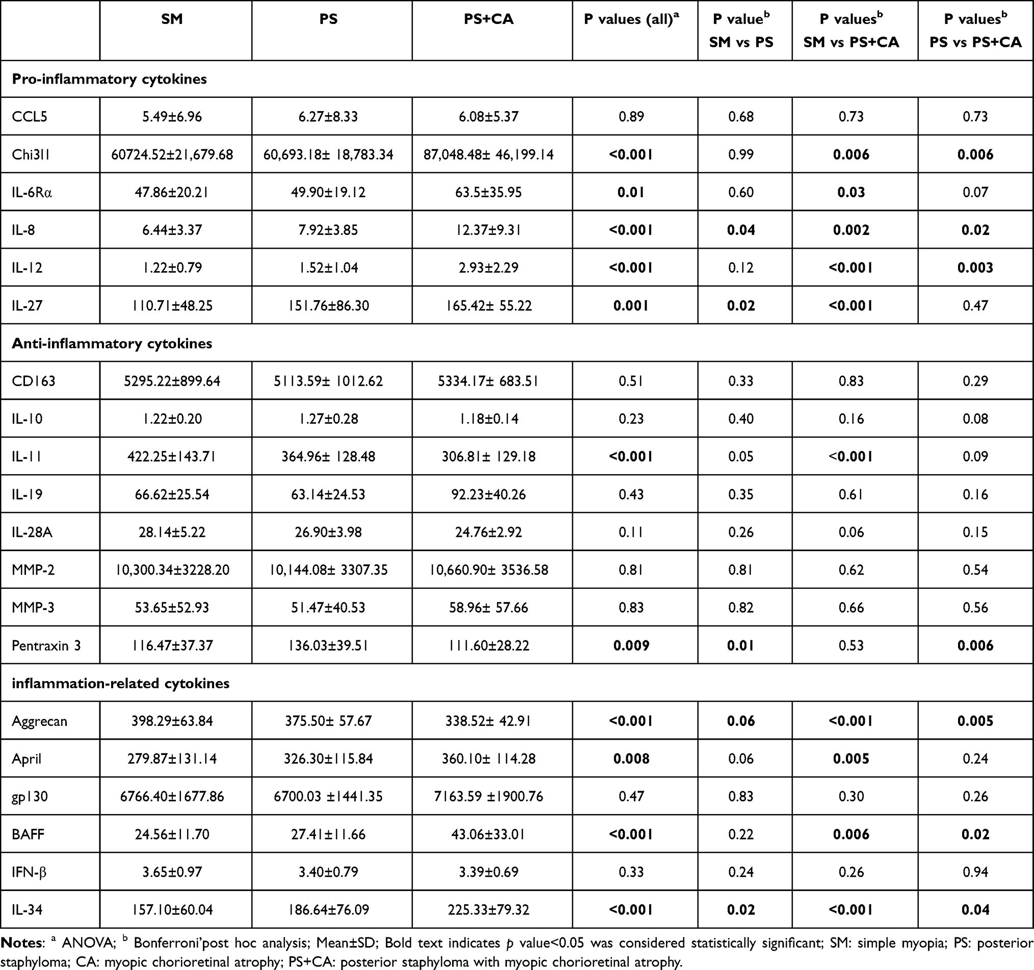 |
Table 2 Inflammatory Cytokines Levels in the Aqueous Humor of Patients from Different Degrees of Pathological Myopia Patients (pg/mL) |
The Relationship Between Intraocular Inflammatory Factors and Axial Length
To explore the link between intraocular inflammatory factors and myopia-mediated ocular pathologies including axial length elongation and retinal vascular and neuronal degeneration, we further analyzed the correlation between the inflammatory cytokine levels and myopia-related ocular parameters. Our results show that the pro-inflammatory factors, including Chi3l1, IL-6Rα, IL-8, IL-12 and IL-27, inflammation-related cytokines including April and BAFF positively correlated with axial length, with the highest being IL-27, Chi3l1, and IL-12 (β=0.62, 0.41, 0.37, respectively, p<0.001, Supplementary Table 2). Other cytokines including IL-11 (β=−0.57, p<0.001), IL-28A (β=−0.59, p<0.001), Aggrecan (β=−0.41, p<0.001, Supplementary Table 2), and IFN-β (β=−0.21, p<0.05, Supplementary Table 2) negatively correlated with axial length. Further subgroup analysis revealed that the PS group had the highest level of correlation (Supplementary Table 2).
The Relationship Between Intraocular Inflammatory Factors and Retinal Vascular Density in Myopic Eyes
We previously reported a progressive loss of macular zone blood vessels and peripapillary vascular degeneration (VD) in myopic retinopathy.14 Next, we examined the relationship between intraocular cytokines and the optic nerve head vascular density (VD). Chi3l1, IL-6Rα, IL-8, IL-27, MMP3, Aggrecan, and BAFF negatively correlated with peripapillary VD (all vessels) with the highest correlation being IL-27 followed by IL-6Rα, and IL-8 (β = −0.64, −0.39, −0.36, respectively, p<0.01, Supplementary Table 3). The anti-inflammatory factors IL-28A and IL-11 positively correlated with peripapillary VD (all vessels).
Regarding macular zone vessels, Chi3l1, IL-6Rα, IL-8 and IL-27, and BAFF negatively correlated with DRVD and SRVD, with the highest values being IL-27 followed by IL-8 (β = −0.32 and −0.32 for DRVD and −0.49 and −0.32 for SRVD, respectively, p<0.01, Supplementary Table 4 and 5). No other correlations were observed.
The Relationship Between Intraocular Inflammatory Factors and Retinal Thickness in Myopic Eyes
Myopic retinopathy is a progressive thinning of macular zone neurons.14 Therefore, we investigated the link between intraocular cytokines and retinal neuronal degeneration. IL-6α and IL-27 negatively correlated with retinal nerve fibre layer (RNFL) thickness (Supplementary Table 6). Chi3l1, IL-6Rα, IL8, IL-12 and BAFF negatively correlated with the superficial retinal thickness (SRT) and deep retinal thickness (DRT), which the highest correlation is Chi3l1 (β = −0.40 and −0.38, respectively, p<0.001, Supplementary Table 7 and 8). However, Aggrecan showed a positive correlation with all retinal neuronal thicknesses, including RNFL, SRT and DRT (β =0.24, 0.27 and 0.31, p<0.05, Supplementary Table 6, 7 and 8). Further subgroup analysis showed that most of the correlations lie in the PS+CA group (Supplementary Tables 7 and 8).
In summary, our results suggest that higher intraocular inflammatory factors are related to myopia-mediated retinal vascular and neuronal degeneration.
The Relationship Between Intraocular Inflammatory Factors and Age
Furthermore, we investigated the relationship between inflammatory factors and age. In our study, the mean age of the subjects was 26.02 ± 5.86 years. Among them, 119 had myopia progression for ≥10 years, 25 had progression lasting from 5 to less than 10 years, and 3 had progression lasting for less than 5 years. There were no significant age differences among groups with varying degrees of myopia, including low to high myopia and different levels of pathological myopia. The normal distribution plot of age was shown in Supplementary Figure 1. Our findings indicated a positive correlation between age and the levels of inflammatory factors, including Chi3l1, IL-6Rα, IL-8, MMP-3, Aggrecan, BAFF, and gp130. Remarkably, the correlation between these inflammatory factors was more pronounced in subjects with higher degrees of pathology, particularly for IL-8 and MMP-3 (Supplement Table 9). This further suggests that the expression of inflammatory factors in myopic eyes is positively associated with age, with the strongest correlation observed in the PS+CA group, which exhibited the most severe pathological manifestations.
PPI and Enrichment Analysis of the Targeted Inflammatory Factors
To better understand the pathways associated with high myopia and pathological myopia, we conducted protein–protein interaction (PPI) analysis using 16 cytokines/growth factors, which were either significantly altered in different groups of myopic retinopathies or linked to myopic retinal vascular or neuronal degeneration using the STRING database. We obtained 11 nodes and 29 edges with a confidence score >0.4. Chi3l1 and Aggrecan appear to be the upstream factors that could affect other factors, whereas BAFF, IL-34 and MMP3 are the downstream factors that could be affected by others in the PPI network (Figure 1A). The KEGG analysis identified cytokine–cytokine receptor interaction, JAK-STAT signaling pathway, rheumatoid arthritis, and Chagas disease as the top 4 significantly enriched pathways (adjusted p-value <0.001, Figure 1B).
 |
Figure 1 Pathway analysis of the differentially expressed proteins (DEPs). (A) The image showed the 11 highest degree of aqueous connection DEPs calculated by Cytohubba plugin. Arrows suggest the direction of protein-protein interaction. The darker the color, the higher degree of the connections between the nodes. (B) The top 15 pathways analyzed by KEGG enrichment of aqueous humor’s DEPs. |
Animal Study
We previously developed a long-term form-deprivation-induced mild myopic retinopathy in guinea pigs.15 Using retinal samples from the animal study, we further investigated the immune-related gene expression. Real-time RT-PCR showed that the expression of Chi3l1, IL-6r, IL-27, PTX3, Aggrecan and BAFF in the FDM retinae was significantly higher compared to that in self-control eyes or in eyes from control guinea pigs (Figure 2). No significant difference was observed in CCL5, IL-10, CD163 and MMP2 genes.
 |
Figure 2 The relative gene expression of inflammatory/inflammation-related cytokines in different groups of Guinea pig retina by qRT-PCR. Data presented as fold change of gene expression compared to control groups. Mean ± SEM, n = 9/group. ns – no significant difference, * p < 0.05. **p < 0.01. One-way ANOVA followed by Dunn’s Multiple comparison test. FDM – form-deprivation induced myopia. |
Discussion
In this study, we detected a significant alteration in the intraocular levels of inflammatory factors in different groups of myopic retinopathy. We found that with the progression of myopic retinopathy, the intraocular levels of pro-inflammatory factors increase, and anti-inflammatory factors decrease. The intraocular levels of Chi3l1, IL-6Rα, IL-8, IL-27 and BAFF negatively correlated with the retinal vascular density; the levels of IL-6Rα also negatively correlated with retinal neuronal thickness. Aggrecan was positively correlated with retinal neuronal thickness. Our results suggest that intraocular inflammatory status is related to myopic retinopathy, specifically, activation of the IL-27/IL6R pathway may contribute to myopic retinal microvascular and neuronal degeneration, and Aggrecan appears to be neuroprotective.
Cytokines, chemokines and growth factors in the aqueous humor originate from intraocular cells and their constitutions reflect the states of the intraocular microenvironment, particularly the retina. Alterations in aqueous humor inflammatory mediators have been reported in many retinal disease conditions, including diabetic retinopathy,17 glaucoma,18 and high myopia.19 However, the intraocular inflammation status remains controversial. Zhu et al found higher intraocular levels of MCP-1 together with reduced IL-1ra in high myopic cataract eyes.19 Yuan et al detected higher IL-6 and MMP2 and lower VEGF in high myopia and myopic retinopathy.20 Shchuko et al reported elevated PDGF, IL-2, IL-5, IL-13, IL-15, IL-17A, TNF-α, IL-8, and RANTES and decreased VEGF in myopic choroidal neovascularization (mCNV),21 although Yukimi et al found that the aqueous levels of VEGF were higher in mCNV eyes.22 In the current study, we measured the intraocular inflammation status of myopic subjects unaffected by other ocular diseases such as cataracts, which could better inflect the intraocular inflammation status associated with myopia. We observed elevated levels of inflammatory mediators including April, BAFF, Chi3l1, IL-6Rα, IL-8, IL-12, and IL-27 and reduced levels of immune regulators/inhibitors such as Aggrecan, IL-10, IL-11, IL-28A, and L-34 in high myopic eyes compared to those in low myopic eyes.
Our results highlight a pro-inflammatory intraocular microenvironment in high-myopic eyes. To determine the role of intraocular inflammation in the development of myopic retinopathy, we further categorized the participants into SM, myopia with PS, and PS + CA based on the severity of myopic retinopathy.14 We found that the levels of pro-inflammatory factors, including Chi3l1, IL-6Rα, IL-8, IL-12, and IL-27 progressively increase from SM to PS and PS+CA. The levels of IL-11 and Aggrecan decrease with the progression of myopic retinopathy. We also observed positive correlations between inflammatory mediators (including Chi3l1, IL-6Rα, IL-8, IL-12, IL-27, April, and BAFF) and axial length, particularly in the PS+CA group, suggesting the potential role of intraocular inflammation and myopic progression. It is well known that cataract surgery could initiate intraocular inflammation,23 including inflammation in the retina.24 The pro-inflammatory intraocular microenvironment may put the high-myopic eyes at risk of ocular complications from additional insults such as intraocular surgery. It may also contribute to the accelerated onset of cataracts in eyes that have undergone phakic implantation procedures, and the occurrence of proliferative vitreoretinopathy (PVR) in cases of myopic retinal detachment. We urge the ophthalmology community to carefully consider ICL implantation in high-myopic eyes. If intraocular surgery is unavoidable in these eyes, intraocular inflammation should be carefully monitored and promptly controlled.
We have previously reported progressive retinal microvascular and neuronal degeneration during the progression of myopic retinopathy from SM, PS, to PS+CA.14 Here, we found that the intraocular levels of immune mediators including Chi1l3, IL-6Rα, IL-8, IL-27, and BAFF negatively correlated with retinal vascular density (peripapillary and macular zone). In terms of retinal neuronal degeneration, negative correlations were observed between the levels of IL-6Rα and retinal neuronal thickness (eg, RNFL, SRT and DRT). However, Aggrecan was positively correlated with retinal neuronal thickness. IL-27 had the highest degree of negative correlation with retinal vascular density, and the intraocular level of IL-27 progressively increased from SM, PS, to PS+CA. Our results suggest that intraocular inflammation is related to retinal vascular and neuronal degeneration. However, the causal relationship between intraocular inflammation and myopic retinal degeneration requires further investigation. IL-27 is a member of the IL-12 cytokine family and can be produced by antigen-presenting cells, monocytes, endothelial cells (ECs), and dendritic cells. During inflammation, IL-27 can activate target cells through binding to its receptors including IL-27R, IL-27Ra (WSX-1) and gp130.25 Depending on the microenvironment, IL-27 can be pro-inflammatory (eg, by promoting Th1 cell differentiation) or anti-inflammatory (eg, by suppressing IL-17 production).26 In the retina, IL-27 together with IL-10 are known to play a role in maintaining the immune suppressive intraocular microenvironment.27 Apart from the immune-related roles, IL-27 can reduce angiogenesis in multiple myeloma.28 Despite the close relationship between IL-27 and retinal vascular and neuronal degeneration, PPI analysis did not reveal a significant interaction of IL-27 with other cytokines, indicating that IL-27 may function alone and the role of IL-27 in myopic retinal vascular and neuronal degeneration warrants further investigation.
Interestingly, we found Chi3l1, IL-12, IL-11, IL-28A, MMP-2, Aggrecan, and April were correlated with AL and the β values (cytokine vs AL) were 0.55, 0.32, −0.28, −0.33, 0.17, −0.58, and 0.38 (these results not shown), respectively, larger or smaller than that in cytokine vs blood vessels/retinal thickness. Indeed, as myopia progresses, the axial length of the eye elongates, stretching the retina and consequently reducing the density of retinal blood vessels and the thickness of the retina. As a result, the retina degenerates gradually. The increased intraocular inflammatory response could be a response to remove the dead cells/debris, but some of the inflammatory mediators may also promote retinal degeneration. It is possible that retinal degeneration and inflammatory cytokine production both result from abnormal AL elongation. Furthermore, we also found that the intraocular levels of IL-8 and MMP-3 demonstrated a strong correlation with age in our study, especially in more severe pathologic myopia. Previous studies reported that aging induced para-inflammation in the retina.29 As aging occurs, the risk of developing various retinal degenerative conditions, like age-related macular degeneration, diabetic retinopathy, and glaucomatous retinopathy increases.30 Some inflammatory markers like IL-6, CRP and TNF-R1 increase during aging.31 For patients with myopia, the onset and progression of myopia are also closely linked to the age.3,32 This highlights the potential use of specific inflammatory biomarkers in the future to reflect the degree of myopic retinopathy and for the prevention and control of high myopia and pathological myopia.
PPI analysis showed that Aggrecan and Chi3l1 were the upstream proteins and MMP-3, BAFF, and IL-34 were the down-stream proteins. It should be mentioned that Aggrecan was reduced in eyes with myopic retinopathy and positively correlated with retinal neuronal thickness, indicating that Aggrecan may be protective in myopia. Aggrecan is a major extracellular matrix protein. Previous studies have shown that Aggrecan could modulate pro-inflammatory cytokine production by T cells in Rheumatoid Arthritis.33 Aggrecan accumulation has been observed in the chick model of myopia.34 Chil3l1, a member of the glycoside hydrolase family 18, is synthesized and secreted by macrophages, neutrophils, chondrocytes and vascular smooth muscle cells.35 Chi3l1 is involved in a variety of biological processes including oxidant injury, inflammasome activation, Th1/Th2 inflammatory balance, macrophage differentiation, ECM regulation and angiogenesis.36 In chronic inflammatory diseases, Chi3l1 was elevated and correlated with the severity of the disease.37,38 In our study, Chi3l1 was significantly higher in PS + CA group, positively correlated with axial length and negatively correlated with retinal vascular density and neuronal thickness. Our results suggest that Aggrecan and Chi3l1 may affect myopic retinopathy indirectly through other down-stream mediators such as IL-8 and MMP-3.
There are several limitations in our study. Firstly, we were unable to acquire aqueous humor from the emmetropic controls. Secondly, all subjects in our study were grouped into simple myopia, posterior staphyloma, and posterior staphyloma with myopic retinopathy. Subjects with pure myopic retinopathy were not present in our study, and therefore we were unable to identify the inflammation status in myopic retinopathy. Thirdly, all pathological lesions were at early and mild stages. Further studies will be needed to understand the inflammation status in more severe myopic maculopathy such as myopic CNV and macular hemorrhage. Fourthly, we did not measure the circulating levels of inflammatory factors. In this study, we could not determine if the status of intraocular inflammation is due to systemic inflammatory factor changes. Finally, aqueous humor samples at multiple time points in this clinical study were not available, and therefore we could only assess the alteration of inflammatory factors at the early stage of pathological myopia. Further functional studies will be needed to prove the causal link between altered inflammatory factors and myopia-related retinal degeneration.
Conclusion
The production of inflammatory factors in the eyes of individuals with high myopia and pathological myopia was altered, and the elevated levels of intraocular pro-inflammatory factors such as Chi3l1, IL-6Rα, and IL-8 were closely associated with myopia-related retinal microvascular and neurodegeneration.
Abbreviations
Average RNFL, average thickness of retinal nerve fiber layers; AL, axial length; BCVA, best corrected visual acuity; CA, myopic chorioretinal atrophy; DRVD, deep retinal vessels density; DRT, deep retinal thickness; FAZ, Foveal avascular zone; FD, foveal density; GCC, ganglion cell complex; ICL, implantable collamer Lens; IDVD-all vessels, inside disc all vessel density; IDVD-small vessels, inside disc small vessel density; OCT, optical coherence tomography; OCTA, OCT angiography; ONHVD-all vessels, optic nerve head all vessel density; ONHVD-small vessels, optic nerve head small vessel density; PERIM, FAZ perimeter; PS, posterior staphyloma; PS+CA, posterior staphyloma with myopic chorioretinal atrophy; SLO, scanning laser ophthalmoscope; SE, spherical equivalent; SM, simple myopia; SRT, superficial retinal thickness; SRVD, superficial retinal vessels density; Peripapillary VD-all vessels, peripapillary all vessel density; Peripapillary VD-small vessels, peripapillary small vessel density.
Data Sharing Statement
The authors declare that all data supporting the findings are available within this paper.
Ethics Approval and Informed Consent
All procedures concerning the collection of aqueous humor were performed following the principles of the Declaration of Helsinki. Informed consent was obtained from all participants and the Institutional Review Board (IRB) at the Aier Eye Hospital Group approved the study (IRB number: AIER 2019IRB03).
Consent for Publication
All authors of this paper have read and consented to publish this version of the manuscript. The authors declare that all data supporting the findings are available within this paper.
Acknowledgments
The authors thank the staff at the Changsha Aier Eye Hospital, Aier Institute of Optometry and Vision Science and Aier Eye Institute for their assistance in this research.
Author Contributions
All authors made a significant contribution to the work reported, whether that is in the conception, study design, execution, acquisition of data, analysis and interpretation, or in all these areas; took part in drafting, revising or critically reviewing the article; gave final approval of the version to be published; have agreed on the journal to which the article has been submitted; and agree to be accountable for all aspects of the work.
Funding
This work was supported by Hunan Provincial Natural Foundation (2023JJ70031 and 2023JJ70036), the Research Fund Project of AIER Eye Hospital Group (AM2201D02, AMK2303D05 and AMF2403D02), the Innovation Platform and Talents Project of Hunan Province (2022WZ1023) and the Key Research and Development Project of Science and Technology Department of Hubei Province (2020BCB013). The sponsor or funding organization had no role in the design or conduct of this research.
Disclosure
The abstract of this paper was presented as, “Altered Levels of Intraocular inflammatory factors Are Related to Retinal Vascular and Retinal Neurodegeneration in Myopic Retinopathy” at the 2024 Annual Meeting of the Association for Research in Vision and Ophthalmology (ARVO) Annual Meeting as a poster presentation with interim findings. The poster’s abstract was published in “Poster Abstract” in ARVO journal name Investigative Ophthalmology & Visual Science, June 2024, Vol.65, 6628: https://iovs.arvojournals.org/article.aspx?articleid=2799770. The authors declare that the research was conducted in the absence of any commercial or financial relationships that could be construed as a potential conflict of interest.
References
1. Mutti DO, Hayes JR, Mitchell GL. et al. Refractive error, axial length, and relative peripheral refractive error before and after the onset of myopia. Invest Ophthalmol Visual Sci. 2007;48(6):2510–2519. doi:10.1167/iovs.06-0562
2. Ohno-Matsui K, Lai TYY, Lai CC, et al. Updates of pathologic myopia. Prog Retinal Eye Res. 2016;52:156–187. doi:10.1016/j.preteyeres.2015.12.001
3. Ohno-Matsui K, Wu PC, Yamashiro KJ, et al. IMI pathologic myopia. Invest Ophthalmol Visual Sci. 2021;62(5):5.
4. Saw SM, Gazzard G, Shih-Yen EC, Chua WH. Myopia and associated pathological complications. Ophthalmic Physiol Opt. 2005;25(5):381–391. doi:10.1111/j.1475-1313.2005.00298.x
5. Kawai T, Akira SZ. Innate immune recognition of viral infection. Nat Immunol. 2006;7(2):131–137. doi:10.1038/ni1303
6. Wei CC, Kung YJ, Chen CS, et al. Allergic conjunctivitis-induced retinal inflammation promotes myopia progression. EBioMedicine. 2018;28:274–286. doi:10.1016/j.ebiom.2018.01.024
7. Lin HJ, Wei CC, Chang CY, et al. Role of chronic inflammation in myopia progression: clinical evidence and experimental validation. EBioMedicine. 2016;10:269–281. doi:10.1016/j.ebiom.2016.07.021
8. Xue M, Ke YF, Ren XJ, et al. Proteomic analysis of aqueous humor in patients with pathologic myopia. J Proteomics. 2021;234:104088. doi:10.1016/j.jprot.2020.104088
9. Giummarra L, Crewther SG, Riddell N, et al. Pathway analysis identifies altered mitochondrial metabolism, neurotransmission, structural pathways and complement cascade in retina/RPE/choroid in chick model of form-deprivation myopia. PeerJ. 2018;6:e5048.
10. Gao TT, Long Q, Yang X. Complement factors C1q, C3 and C5b-9 in the posterior sclera of Guinea pigs with negative lens-defocused myopia. International J Ophthalmol. 2015;8(4):675. doi:10.3980/j.issn.2222-3959.2015.04.06
11. Herbort CP, Papadia M, Neri P. Myopia and inflammation. J Ophthalmic Vis Res. 2011;6(4):270–283.
12. Neelam K, Cheung CMJ, Ohno-Matsui K, et al. Choroidal neovascularization in pathological myopia. Prog Retinal Eye Res. 2012;31(5):495–525. doi:10.1016/j.preteyeres.2012.04.001
13. Kung YJ, Wei CC, Chen LA, et al. Kawasaki disease increases the incidence of myopia. Biomed Res. Int. 2017;2017:1–6. doi:10.1155/2017/2657913
14. Zeng L, Li XN, Pan W, et al. Intraocular complement activation is related to retinal vascular and neuronal degeneration in myopic retinopathy. Front Cell Neurosci. 2023;17.
15. Zeng L, Li XN, Liu J, et al. RNA-seq analysis reveals an essential role of the tyrosine metabolic pathway and inflammation in myopia-induced retinal degeneration in guinea pigs. Int J Mol Sci. 2021;22(22):12598. doi:10.3390/ijms222212598
16. Livak KJ, Schmittgen TD. Analysis of relative gene expression data using real-time quantitative PCR and the 2− ΔΔCT method. Methods. 2001;25(4):402–408. doi:10.1006/meth.2001.1262
17. Dong N, Xu B, Wang B, et al. Study of 27 aqueous humor cytokines in patients with type 2 diabetes with or without retinopathy. Mol Vision. 2013;19:1734.
18. Tane N, Dhar S, Roy S, et al. Effect of excess synthesis of extracellular matrix components by trabecular meshwork cells: possible consequence on aqueous outflow. Exp. Eye Res. 2007;84(5):832–842. doi:10.1016/j.exer.2007.01.002
19. Zhu XT, Zhang KK, He WW, et al. Proinflammatory status in the aqueous humor of high myopic cataract eyes. Exp. Eye Res. 2016;142:13–18. doi:10.1016/j.exer.2015.03.017
20. Yuan IS, Wu SJ, Wang YW, et al. Inflammatory cytokines in highly myopic eyes. Sci Rep. 2019;9(1):3517. doi:10.1038/s41598-019-39652-x
21. Shchuko AG, Zaitseva NV, Yurieva TN, et al. Intraocular cytokines and their correlations with clinical parameters in patients with myopic choroidal neovascularization. Ophthalmologica. 2017;237(2):96–104. doi:10.1159/000455271
22. Yukimi Y, Dai M, Shin-ichi S, et al. Associations of inflammatory cytokines with choroidal neovascularization in highly myopic eyes. Retina. 2015;35(2):344–350. doi:10.1097/IAE.0000000000000311
23. Grzybowski A, Sidaraite A, Zemaitiene R. Management of inflammation after the cataract surgery. Curr Op Ophthalmol. 2023;34(1):9–20. doi:10.1097/ICU.0000000000000912
24. Xu H, Chen M, Forrester JV, et al. Cataract surgery induces retinal pro-inflammatory gene expression and protein secretion. Invest Ophthalmol Visual Sci. 2011;52(1):249–255. doi:10.1167/iovs.10-6001
25. Pflanz S, Timans JC, Cheung J, et al. IL-27, a heterodimeric cytokine composed of EBI3 and p28 protein, induces proliferation of naive CD4+ T cells. Immunity. 2002;16(6):779–790. doi:10.1016/S1074-7613(02)00324-2
26. Yoshimura T, Takeda A, Hamano S, et al. Two-sided roles of IL-27: induction of Th1 differentiation on naive CD4+ T cells versus suppression of pro-inflammatory cytokine production including IL-23-induced IL-17 on activated CD4+ T cells partially through STAT3-dependent mechanism. J Immunol. 2006;177(8):5377–5385. doi:10.4049/jimmunol.177.8.5377
27. Lee YS, Amadi-Obi A, Yu CR, et al. Retinal cells suppress intraocular inflammation (uveitis) through production of interleukin‐27 and interleukin‐10. Immunology. 2011;132(4):492–502. doi:10.1111/j.1365-2567.2010.03379.x
28. Cocco C, Giuliani N, Carlo ED, et al. Interleukin-27 acts as multifunctional antitumor agent in multiple myeloma. Clin Cancer Res. 2010;16(16):4188–4197. doi:10.1158/1078-0432.CCR-10-0173
29. Xu H, Chen M, Forrester JV. Para-inflammation in the aging retina. Prog Retinal Eye Res. 2009;28(5):348–368. doi:10.1016/j.preteyeres.2009.06.001
30. Ardeljan D, Chan CC. Aging is not a disease: distinguishing age-related macular degeneration from aging. Prog Retinal Eye Res. 2013;37:68–89. doi:10.1016/j.preteyeres.2013.07.003
31. Wyczalkowska-Tomasik A, Czarkowska-Paczek B, Zielenkiewicz M, et al. Inflammatory markers change with age, but do not fall beyond reported normal ranges. Arch Immunol et therapiae experimentalis. 2016;64(3):249–254. doi:10.1007/s00005-015-0357-7
32. Chang L, Pan CW, Ohno-Matsui K, et al. Myopia-related fundus changes in Singapore adults with high myopia. Am J Ophthalmol. 2013;155(6):991–999. doi:10.1016/j.ajo.2013.01.016
33. Buzás EI, Végvári A, Murad YM, et al. T-cell recognition of differentially tolerated epitopes of cartilage proteoglycan aggrecan in arthritis. Cell Immunol. 2005;235(2):98–108. doi:10.1016/j.cellimm.2004.08.006
34. Rada JA, Thoft RA, Hassell JR. Increased aggrecan (cartilage proteoglycan) production in the sclera of myopic chicks. Dev. Biol. 1991;147(2):303–312. doi:10.1016/0012-1606(91)90288-E
35. Lee CG, Hartl D, Lee GR, et al. Role of breast regression protein 39 (BRP-39)/chitinase 3-like-1 in Th2 and IL-13–induced tissue responses and apoptosis. J Exp Med. 2009;206(5):1149–1166. doi:10.1084/jem.20081271
36. Zhao T, Su ZP, Li YC, et al. Chitinase-3 like-protein-1 function and its role in diseases. Signal Transduct Target Ther. 2020;5(1):201. doi:10.1038/s41392-020-00303-7
37. Lee CG, Elias JA. Role of breast regression protein-39/YKL-40 in asthma and allergic responses. Allergy Asthma Immunol Res. 2010;2(1):20–27. doi:10.4168/aair.2010.2.1.20
38. Zhou Y, He CH, Herzog EL, et al. Chitinase 3–like–1 and its receptors in Hermansky-Pudlak syndrome–associated lung disease. J Clin Invest. 2015;125(8):3178–3192. doi:10.1172/JCI79792
 © 2024 The Author(s). This work is published and licensed by Dove Medical Press Limited. The
full terms of this license are available at https://www.dovepress.com/terms.php
and incorporate the Creative Commons Attribution
- Non Commercial (unported, 3.0) License.
By accessing the work you hereby accept the Terms. Non-commercial uses of the work are permitted
without any further permission from Dove Medical Press Limited, provided the work is properly
attributed. For permission for commercial use of this work, please see paragraphs 4.2 and 5 of our Terms.
© 2024 The Author(s). This work is published and licensed by Dove Medical Press Limited. The
full terms of this license are available at https://www.dovepress.com/terms.php
and incorporate the Creative Commons Attribution
- Non Commercial (unported, 3.0) License.
By accessing the work you hereby accept the Terms. Non-commercial uses of the work are permitted
without any further permission from Dove Medical Press Limited, provided the work is properly
attributed. For permission for commercial use of this work, please see paragraphs 4.2 and 5 of our Terms.