")
Back to Journals » Journal of Inflammation Research » Volume 17
Hsa_circ_0001359 in Serum Exosomes: A Promising Marker to Predict Bronchopulmonary Dysplasia in Premature Infants
Authors Guo Y, Pan JJ, Zhu W, Wang MZ, Liu TY, Wang XX, Wu QQ, Cheng YX, Qian YS, Zhou XG, Yang Y
Received 18 April 2024
Accepted for publication 16 July 2024
Published 25 July 2024 Volume 2024:17 Pages 5025—5037
DOI https://doi.org/10.2147/JIR.S463330
Checked for plagiarism Yes
Review by Single anonymous peer review
Peer reviewer comments 2
Editor who approved publication: Dr Tara Strutt
Yan Guo,1,* Jing-Jing Pan,2,* Wen Zhu,1 Mu-Zi Wang,3 Tian-Yu Liu,4 Xiao-Xin Wang,5 Qian-Qian Wu,1 Yi-Xin Cheng,1 Yi-Sen Qian,1 Xiao-Guang Zhou,1 Yang Yang1
1Department of Neonatology, Children’s Hospital of Nanjing Medical University, Nanjing, People’s Republic of China; 2Department of Neonatology, The First Affiliated Hospital, Nanjing Medical University, Nanjing, People’s Republic of China; 3Department of Neonatology, The Affiliated Suzhou Hospital of Nanjing Medical University, Suzhou, People’s Republic of China; 4Department of Neonatology, The Eighth Affiliated Hospital, Sun Yat-Sen University, Shenzhen, People’s Republic of China; 5Department of Pediatrics, Shandong Tumor Hospital, Jinan, People’s Republic of China
*These authors contributed equally to this work
Correspondence: Xiao-Guang Zhou; Yang Yang, Department of Neonatology, Children’s Hospital of Nanjing Medical University, 72 Guangzhou Road, Nanjing, Jiangsu, 210008, People’s Republic of China, Email [email protected]; [email protected]
Objective: This prospective study is to explore the role of specific circRNAs in predicting the development of bronchopulmonary dysplasia (BPD).
Methods: From July 1, 2021 to December 1, 2021, peripheral blood samples were collected from 62 premature infants with gestational age (GA) ≤ 32 weeks on the 7th, 14th, and 28th day after birth. Then, on the 28th day, the included infants were divided into the BPD group and the non-BPD group according to the definition of BPD. Serum exosomal circRNAs from peripheral blood were identified, sequenced, and compared between the BPD and non-BPD groups at different time points. Specific differentially expressed circRNAs were further verified from another 42 enrolled premature infants (GA ≤ 32 weeks). The classical lung biological markers in serum were also measured simultaneously.
Results: Hsa_circ_0001359 in serum exosomes showed continuous differential expression between the BPD group and the non-BPD group on the 7th, 14th, and 28th day. Compared with that, classical lung biological markers like IL-6, IL-33, KL-6, and ET-1 did not exhibit continuous differences. Moreover, the expression of hsa_circ_0001359 on day 7 had a higher predictive value in predicting BPD (area under curve:0.853, 95% CI:0.738– 0.968; adjusted odds ratio:6.033, 95% CI:2.373– 13.326). The calibration curve further showed the mean absolute error = 0.033, mean squared error = 0.00231, and quantile of absolute error = 0.058.
Conclusion: Hsa_circ_0001359 in serum exosomes is a promising marker for predicting BPD in preterm infants with gestational age ≤ 32 weeks.
Keywords: bronchopulmonary dysplasia, circular RNAs, high-throughput sequencing, preterm infants
Graphical Abstract:

Introduction
With the advancement of perinatal medicine, more extremely low birth weight infants have survived. However, the incidence of bronchopulmonary dysplasia (BPD) has not been significantly decreased. This disease can lead to long-term physical and intellectual development disorders in premature infants and has become an important risk factor affecting the quality of life.1 However, the early clinical manifestations of BPD are often non-specific.2 Though several inflammatory factors (such as IL-1β, IL-6, and IL-8) have been reported to be associated with BPD,3,4 these markers lack tissue and disease specificity. Meanwhile, some clinical prediction models have also been developed, but these models contain too many variables, which limits their usability in clinical practice. From the current perspective, there has still been a lack of biomarkers for the simple and accurate prediction of BPD.
Circular RNAs (circRNAs) are a special class of noncoding RNAs which exhibit a closed circular structure. And compared with other small RNAs like lncRNAs, circRNAs exhibit high conservatism and tissue specificity.5 In addition, they have significant regulatory effects in various biological processes such as cell development, cell proliferation, stem cell differentiation, and tumor progression.6 Consequently, circRNAs have been natural potential biomarkers for early diagnosis and prediction of major human diseases such as cancers and BPD.7,8 Exosomes are nanoscale vesicles secreted by most tissue cells, containing various active molecules such as proteins, mRNAs, circRNAs, etc.9 Exosomes can bind with receptors to regulate different biological processes between cells, and have functions such as anti-inflammatory, anti-apoptotic, antioxidant, inhibiting pulmonary fibrosis, and promoting damaged lung tissue regeneration.10 For instance, Willis GR et al found mesenchymal stem/stromal cell derived-exosomes treatment could alleviate hyperoxia-induced BPD by improving lung function, pulmonary vascular remodeling, and pulmonary hypertension.11 Furthermore, a microarray analysis from Wang Y et al reported that 317 exosomal circRNAs showed differential expression between BPD group and non-BPD group using umbilical cord blood from 8 infants (4 newborns in the BPD group and 4 in the non-BPD group).12 This indicates that exosomal circRNAs have important prospects as biomarkers for BPD.Based on the above, in this study, we explored specific circRNAs in serum exosomes that continuously showed differential expression within 28 days after birth in BPD infants and compared them with classical lung biological markers, including interleukin 6 (IL-6), interleukin 33 (IL-33), Klebs von den Lungen-6 (KL-6), and endothelin-1 (ET-1) to obtain the promising “circRNA marker” of BPD.
Patients and Methods
Inclusion, Exclusion, and Diagnostic Criteria
- Inclusion criteria: Premature infants with gestational age ≤32 weeks admitted to Children’s Hospital of Nanjing Medical University from July 1, 2021 to December 1, 2021 (period 1) and May 1, 2022 to December 1, 2022 (period 2) were enrolled.
- Exclusion criteria: Infants with severe congenital malformations, metabolic diseases, severe asphyxia, and severe infections were excluded. Newborns who were discharged without following the doctor’s advice or died during hospitalization were also excluded.
- Diagnostic criteria: BPD is defined as oxygen needed for 28 days after birth and graded according to respiratory support at 36 weeks of postmenstrual age (PMA).13
Estimation of Sample Size
As previous retrospective research reported, the incidence of BPD was around 23.5% in premature infants with gestational age ≤32 weeks.14 The incidence rate of our center in a similar population was around 40% in the past five years. Therefore, considering α=0.05 and β=0.20, it is calculated that at least 56 patients are needed by R 4.3.1.
Grouping
- After being screened by inclusion and exclusion criteria, from July 1, 2021 to December 1, 2021 (period 1), 62 infants were finally included for high-throughput sequencing (Figure 1). The diagnosis of BPD in these infants was evaluated on the 28th day after birth. Among them, 27 infants developed BPD and were accordingly set as the BPD group. The remaining 35 infants were set as the non-BPD group.
- For further validation by real-time PCR, from May 1, 2022 to December 1, 2022 (period 2), another 42 infants were enrolled (Figure 1). Among them, 22 infants developed BPD and were accordingly set as the BPD group. The remaining 20 infants were set as the non-BPD group.
 |
Figure 1 Flowchart of this study. |
Sample Collection
- To screen the circRNA profiles in serum exosomes, each 1.5 mL peripheral venous blood sample was collected from 62 premature infants (period 1) on the 7th, 14th, and 28th day of life and stored in a −80°C freezer. After grouping on the 28th day after birth, blood samples were pooled into two independent tubes (one tube for the BPD group and the other tube for the non-BPD group) for high-throughput sequencing on the 7th, 14th, and 28th day.
- Similarly, on the 1st, 7th, 14th, 21st, and 28th day, each 2 mL peripheral venous blood of another 42 premature infants (period 2) with gestational age ≤32 weeks is preserved for further validation. After grouping on the 28th day after birth, the expression of the selected circRNAs on the 7th, 14th, and 28th day was detected through real-time PCR. In addition, the serum levels of IL-6, IL-33, KL-6, and ET-1 were measured on the 1st, 7th, 14th, 21st, and 28th day from the above 42 premature infants.
Isolation and Identification of Exosomes
After centrifugation, the serum samples were separated using the GSTM exosome isolation reagent kit (Nanjing, China) according to the instructions. The exosomes of the serum samples were identified using the electron microscope of FEI Tecnai G2 F20 (Portland, US) for the ultrastructure of exosomes. In addition, use the Marvin Nanosight NS300 nanoparticle tracking analyzer (Marvin, UK) for exosomes particle size detection.
Construction of circRNA Expression by High-Throughput Sequencing
The total RNAs of the serum exosomes were extracted using Trizol after the exosomes were isolated from serum. The library was constructed using the Clontech SMARTer Stranded Total RNA seq kit V2 (San Jose, US). On the 7th, 14th, and 28th day after birth, circRNAs showing continuous differential expression between the BPD and non-BPD groups were selected. Quantile normalization and subsequent data processing were performed using the R software limma package. Differentially expressed circRNAs were identified through fold-change filtering. Hierarchical clustering was performed to show the distinguishable circRNA expression pattern. Differentially expressed circRNAs with statistical significance between two groups were identified through scatter plot filtering. In this study, the criteria for screening the differential expression of circRNAs between two groups were defined as absolute Fold Change > 1.5, P < 0.05, and False Discovery Rate <1.
Bioinformatics Analysis
To identify the functional categories of differentially expressed circRNAs between groups, Gene Ontology (GO; http://www.geneontology.org/) and Kyoto Encyclopedia of Genes and Genomes (KEGG; http://www.genome.jp/) were also used.
Real-Time PCR of Specifically Expressed circRNAs
After the exosomes were isolated from peripheral serum, the total RNA was extracted from the peripheral blood on the 7th, 14th, and 28th day using Trizol. Subsequently, single-stranded cDNA was synthesized. cDNA was subjected to multiple amplification cycles using the Rotor Gene 3000 real-time PCR system (Sydney, Australia). Throughout the entire reverse transcription and PCR process, U6 snRNA was used as the internal reference (Supplementary-Table 1). The PCR results were calculated using a threshold cycle (Ct). The relative expression level of each circRNA was compared using 2−ΔΔCt method.
Enzyme Linked Immunosorbent Assay (ELISA) for IL-6, IL-33, ET-1, and KL-6
The peripheral blood of premature infants (period 2) on the 1st, 7th, 14th, 21st, and 28th day was centrifuged and separated. The serum levels of IL-6, IL-33, ET-1, and KL-6 were measured strictly according to the operating instructions of the test kit using double antibody ELISA (The ET-1 and KL-6 test kits were purchased from Wuhan Yilairuite Biotechnology Co., Ltd. The IL-6 and IL-33 test kits were purchased from Wuhan Doctoral Biotechnology Co., Ltd).
Statistical Analysis
In the present study, when the clinical characteristics and circRNA expression were analyzed, quantitative data were presented as Mean ± Standard deviation. ANOVA was performed to establish equal variance, and a 2-tailed Student’s t test with Bonferroni correction was applied to determine statistical significance. The qualitative data were expressed in frequency and percentage (%), and compared using chi square test. In addition to the above univariate analysis, binary logistic regression analysis was used to conduct multivariate analysis. We assessed the potential confounding variables in the statistical modeling based on the results of univariate analysis of clinical characteristics. Adjusted odds ratio (aOR) with 95% confidence interval (CI) were then collected. In addition, the receiver operating characteristic curve (ROC) was adopted to compare the prediction value of markers. The calibration curve was also drawn to reflect the difference between predicted and actual values. All statistical analyses were performed using R 4.3.1 software. P value < 0.05 was considered statistically significant.
Results
Clinical Characteristics of Included Infants for High-Throughput Sequencing
The clinical characteristics of the BPD infants and non-BPD infants for high-throughput sequencing are presented in Table 1. The BPD group had lower gestational age and birth weight. The incidences of early-onset sepsis and respiratory distress syndrome were also more common in the BPD group (P<0.05).
 |
Table 1 Clinical Characteristics of the Infants for High-Throughput Sequencing |
Identification of Serum Exosomes
Transmission electron microscopy revealed circular structures in blood samples. In addition, through the detection of the nanoparticle tracking analyzer, the main peak values of the separated particles were in the range of 30–150 nm, indicating that the particles were exosomes (Supplementary-Figure 1).
High-Throughput Sequencing and Screening of circRNAs
- Between the BPD group and the non-BPD group, there were 1783 up-regulated and 2384 down-regulated differentially expressed circRNAs on day 7, 2545 up-regulated and 2413 down-regulated differentially expressed circRNAs on day 14, and 807 up-regulated and 864 down-regulated differentially expressed circRNAs on day 28 (Figure 2A–C).

Figure 2 CircRNA expression profile by high-throughput sequencing. (A) Comparison between the BPD group and the non-BPD group on day 7. (B) Comparison between the BPD group and the non-BPD group on day 14. (C) Comparison between the BPD group and the non-BPD group on day 28. (D) Comparison between day 14 and day 7 in the BPD group. (E) Comparison between day 28 and day 14 in the BPD group.
- In BPD infants, there were 2131 up-regulated and 1999 down-regulated differentially expressed circRNAs between day 14 and day 7, and 1050 up-regulated and 773 down-regulated differentially expressed circRNAs between day 28 and day 14 (Figure 2D and E).
- Furthermore, 20 circRNAs showed continuous differential expression (|logFC| >1) on the 7th, 14th, and 28th day between the BPD group and the non-BPD group (Supplementary-Table 2).

Bioinformatics Analysis of Differentially Expressed circRNAs
Based on the correspondence between circRNAs and their parental genes, GO and KEGG enrichment analysis was performed to determine the function of the differentially expressed circRNAs. GO enrichment analysis showed that it is enriched in multiple biological pathways such as regulation of cell development and regeneration (Supplementary-Figure 2). The KEGG enrichment analysis showed the top 10 KEGG pathways such as MicroRNAs in Endocytosis, MAPK, and Cancer (Supplementary-Figure 3).
Expression of Hsa_circ_0001522 and Hsa_circ_0001359 by Real-Time PCR
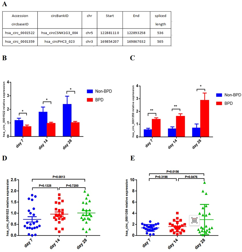
- Among 20 continuously differentially expressed circRNAs (|logFC| >1), four circRNAs [hsa_circCSNK1G3_004 (hsa_circ_0001522), hsa_circPHC3_023 (hsa_circ_0001359), hsa_circCCDC9_003, and hsa_circRNF214_006] showed a consistent expression trend on the 7th, 14th, and 28th day between the BPD group and the non-BPD group (Supplementary-Table 2). Furthermore, because showing higher fold changes, hsa_circ_0001522 and hsa_circ_0001359 were further selected for real-time PCR validation (Figure 3A).
42 infants were enrolled in further real-time PCR validation (Figure 1). Among them, 22 infants developed BPD who were accordingly set as the BPD group. The remaining 20 infants were set as the non-BPD group. The clinical characteristics showed that the gestational age and birth weight were lower in the BPD group. In addition, more infants with intubation in the delivery room were found in the BPD group (Table 2). It turns out that the expression of hsa_circ_0001522 and hsa_circ_0001359 also showed significant differences between the BPD and the non-BPD groups (Figure 3B and C).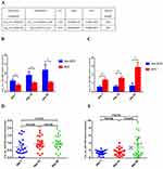
Figure 3 Basic information and real-time PCR of specific circRNAs. (A) Basic information of hsa_circ_0001522 and hsa_circ_0001359. (B) The expression of hsa_circ_0001522 between the BPD group and non-BPD group on the 7th, 14th, and 28th day. (C) The expression of hsa_circ_0001359 between the BPD group and non-BPD group on the 7th, 14th, and 28th day. (D) The expression of hsa_circ_0001522 from day 7 to day 28 in the BPD group. (E) The expression of hsa_circ_0001359 from day 7 to day 28 in the BPD group. *P <0.05, **P < 0.01.

Table 2 Clinical Characteristics of the Infants for Further Validation
- Besides, as far as the BPD group is concerned, the expression level of hsa_circ_0001359 on day 28 was higher than that on day 7 and day 14. In contrast, there was no significant difference in the expression of hsa_circ_0001522 from day 7 to day 28 in BPD patients (Figure 3D and E).

Expression of Serum IL-6, IL-33, ET-1, and KL-6 by ELISA
Compared with non-BPD infants, the BPD group showed a significant increase in serum IL-6 levels on the 1st and 28th day, while there were no significant differences on the 7th, 14th, and 21st day. The IL-33 in BPD infants increased significantly on the 14th and 21st day. As far as ET-1 is concerned, the serum ET-1 levels were higher in BPD infants on the 1st day and 14th day. The difference in serum KL-6 levels between the two groups ultimately occurred only on the 28th day. All four lung biological markers did not show a continuous trend of change over the five time points (1st, 7th, 14th, 21st, and 28th day) (Figure 4).
 |
Figure 4 Comparison of the serum levels of lung biological markers between BPD infants and non-BPD infants on the 1st, 7th, 14th, 21st, and 28th day. (A) Serum IL-6 levels. (B) Serum IL-33 levels. (C) Serum ET-1 levels. (D) Serum KL-6 levels. *P <0.05, **P < 0.01. |
Prediction for BPD
Compared with KL-6 and IL-33, IL-6 and ET-1 exhibited intergroup differences in the early postnatal period (day 1). So, IL-6 and ET-1 were selected for ROC analysis along with specific circRNAs (hsa_circ_0001359 and hsa_circ_0001522). The ROC analysis showed that the hsa_circ_0001359 has the highest area under the curve (AUC) than other markers on day 7 (AUC:0.853, 95% CI:0.738–0.968) (Table 3 and Figure 5A).
 |
Table 3 The Predictive Values of IL-6, ET-1, Hsa_circ_0001522, and Hsa_circ_0001359 on Day 7 for Infants with BPD |
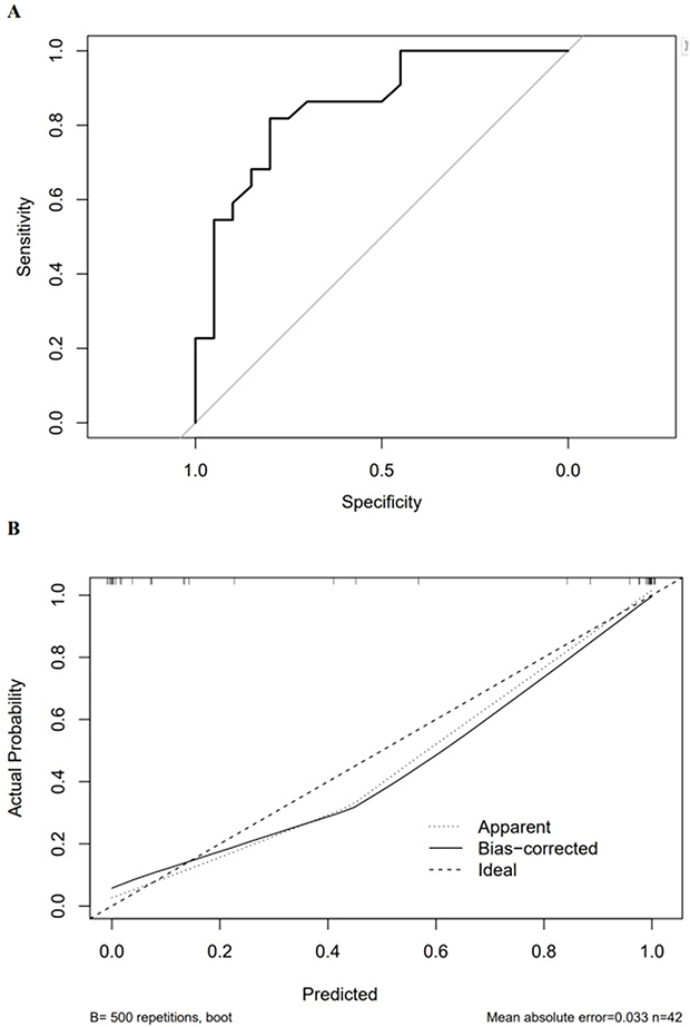 |
Figure 5 ROC and calibration curve. (A) ROC of hsa_circ_0001359 on day 7. (B) Calibration curve of hsa_circ_0001359 on day 7. |
Furthermore, the binary logistic regression showed that the OR of hsa_circ_0001359 is 6.033 (95% CI: 2.373–13.326) when adjusted by gestational age, birth weight, and intubation in the delivery room (Tables 2 and 4). The calibration curve showed that the mean absolute error = 0.033, mean squared error = 0.00231, and quantile of absolute error = 0.058 (Figure 5B).
 |
Table 4 Multivariate Analysis of Hsa_circ_0001359 on Day 7 in Predicting BPD by Binary Logistic Regression |
Discussion
Early identification of newborns at risk of developing BPD would benefit preventive interventions when airway inflammation is reversible. Inflammation is one of the most critical stages in the pathogenesis of BPD.15 Excessive release of inflammatory cytokines can severely damage pulmonary blood vessels and alveoli, which is an important pathological and physiological process in the occurrence and development of BPD16. Some inflammatory cytokines have been reported to early predict the occurrence of BPD.17,18 For example, studies have found that the levels of IL-6 and IL-8 in amniotic fluid or umbilical cord blood have positive clinical significance for early detection and intervention of BPD.19 It was also demonstrated that serum levels of IL-1β, IL-6, IL-8, IL-33, and TNF-α were significantly increased in the early postnatal period of BPD infants.3,4,20 However, the above classic markers are mostly non-specific indicators such as inflammatory factors, making it difficult to distinguish between primary disease and BPD in infants with acute respiratory distress syndrome.2 Therefore, serum IL-6 and IL-3321,22 as inflammatory cytokines, serum ET-1 and KL-623,24 as lung injury markers were measured, and compared between BPD and non-BPD infants. And, the four biomarkers did not exhibit continuous changes during the development of BPD (Figure 4), which cannot explain the pathological mechanism of BPD well.
In order to help neonatologists estimate the probability of future BPD occurrence and provide information for decision-making, many clinical models for predicting BPD have been established in recent years. Most prediction models used only clinical indicators including prenatal, perinatal, and postnatal factors to develop BPD prediction models. However, various types of variables and indicators need to be collected, with a longer period and higher operational difficulty.
CircRNAs are covalently closed cyclic molecules, mainly produced by mRNA precursors through variable splicing processing.25 They are abundant in the eukaryotic transcriptome. Due to its structural characteristics, circRNAs have good biological stability.26 And they also have disease and tissue specificity, which makes them significant advantages as clinical biomarkers. In fact, circRNAs have been found to show differential expression in lung infectious diseases, lung development, and lung cancer,27–29 indicating that circRNAs have potential as biomarkers of respiratory diseases. Exosomes are bilayer vesicles released from cells, with a diameter of 30–150 nm, and exist in human blood, urine, and saliva.30 CircRNAs could be secreted from tissues and organs into the peripheral blood through exocytosis in the form of exosomes, thereby participating in the expression and regulation of important mediators such as inflammatory factors.31 It has been found that some serum circRNAs were differentially expressed in BPD infants. However, there have been few reports on the prediction of BPD through serum exosomal circRNAs. In this study, we first identified four serum exosomal circRNAs differentially expressed between BPD and non-BPD infants through high-throughput sequencing. The KEGG enrichment analysis results further indicated that the parental genes of these circRNAs were mainly enriched in the MAPK, TGF-β signaling pathways, etc. And MAPK and TGF-β signaling pathways have been demonstrated closely related to the progression of BPD.32,33
Among the differentially expressed circRNAs, exosomal hsa_circ_0001359 in serum showed higher fold changes on the 7th, 14th, and 28th day, which suggested it could be a potential marker. The 7th day of life is a proper time to assess the risk of BPD and initiate preventive treatment for BPD, after which lung injury will persist and lung lesions may be irreversible.34,35 In our study, the expression of hsa_circ_0001359 on day 7 showed a high OR (6.033) even adjusted by gestational age, birth weight, and intubation in the delivery room (Table 4). ROC analysis further indicated that hsa_circ_0001359 had a better AUC with good sensitivity and specificity (Table 3). Therefore, hsa_circ_0001359 in serum exosomes could be a good indicator for early prediction of BPD.
As a competitor of endogenous RNAs, circRNAs contain miRNA target sites, which can act as miRNA sponges, thereby relieving the inhibitory effect of miRNAs on target genes and upregulating the expression level of target genes.36,37 In the present study, exosomal hsa_circ_0001359 in the serum of BPD infants was significantly expressed, the change was more significant on the 28th day (Figure 3C). Through the TargetScan database, it is predicted that there is a potential binding site between the hsa_circ_0001359 and let-7 family (Supplementary-Figure 4). In BPD animal models, overexpression of let-7 can alleviate the disease. This may be related to the downregulation of the TGF-β signaling pathway.38 Therefore, hsa_circ_0001359 may act as a let-7 sponge through TGF-β signal pathways regulating lung development and affecting the process of BPD. However, further research is needed to verify the targeted relationship between hsa_circ_0001359 and let-7.
Limitations
Due to limitations on blood collection volume, only serum samples from the 7th, 14th, and 28th day of life were selected for exosomal circRNA sequencing and PCR detection. And the sample size is relatively small with only a single center. Consequently, internal validation was not performed. In addition, this study did not randomize the study subjects at the onset. Although some statistical methods like logistic regression were used to adjust the possible confounding factors, it may affect the reliability of the research findings.
Conclusion
In summary, on the 7th, 14th, and 28th day, differentially expressed circRNAs were found in serum exosomes between BPD infants and non-BPD infants. Especially hsa_circ_0001359, which could serve as a promising and easily obtainable biological marker for predicting BPD as early as 7th day after birth.
Data Sharing Statement
The dataset used during this study is available from the corresponding author upon reasonable request.
Ethics
This study was approved by the ethics committee of the Children’s Hospital of Nanjing Medical University (Number: 202301030-1). Written informed consent was obtained from the parents of the infants. All data were fully anonymized before further statistical analysis. All the procedures were followed by the Declaration of Helsinki.
Consent for Publication
All authors listed have read the complete manuscript and approved the submission.
Acknowledgments
Yan Guo and Jing-Jing Pan are the co-first authors for this study. The authors would like to thank the nurses for helping collect the blood sample.
Author Contributions
All authors made a significant contribution to the work reported, whether that is in the conception, study design, execution, acquisition of data, analysis and interpretation, or in all these areas; took part in drafting, revising or critically reviewing the article; gave final approval of the version to be published; have agreed on the journal to which the article has been submitted; and agree to be accountable for all aspects of the work.
Funding
This study has no funding.
Disclosure
The authors report no conflicts of interest in this work.
References
1. Abiramalatha T, Ramaswamy VV, Bandyopadhyay T, et al. Interventions to prevent bronchopulmonary dysplasia in preterm neonates: an umbrella review of systematic reviews and meta-analyses. JAMA Pediatr. 2022;176(5):502–516. doi:10.1001/jamapediatrics.2021.6619
2. Collaco JM, SA McGrath-Morrow, Griffiths M, et al. Perinatal Inflammatory Biomarkers and Respiratory Disease in Preterm Infants. J Pediatr. 2022;246:34–39.e3. doi:10.1016/j.jpeds.2022.04.028
3. Sahni M, Yeboah B, Das P, et al. Novel biomarkers of bronchopulmonary dysplasia and bronchopulmonary dysplasia-associated pulmonary hypertension. J Perinatol. 2020;40(11):1634–1643. doi:10.1038/s41372-020-00788-8
4. Tao X, Mo L, Zeng L. Hyperoxia induced bronchopulmonary Dysplasia-Like Inflammation via miR34a-TNIP2-IL-1lmonary Dys. Front Pediatr. 2022;10:805860. doi:10.3389/fped.2022.805860
5. Mahmoudi E, Cairns MJ. CircRNA and Ageing. Subcell Biochem. 2023;102:249–270.
6. Sun M, Yang Y. Biological functions and applications of circRNAs-next generation of RNA-based therapy. J Mol Cell Biol. 2023;15(5):mjad031. doi:10.1093/jmcb/mjad031
7. Tang M, Kui L, Lu G, et al. Disease-associated circular RNAs: From biology to computational identification. Biomed Res Int;2020. 6798590. doi:10.1155/2020/6798590
8. Wang J, Yin J, Wang X, et al. Changing expression profiles of mRNA, lncRNA, circRNA, and miRNA in lung tissue reveal the pathophysiological of bronchopulmonary dysplasia (BPD) in mouse model. J Cell Biochem. 2019;120(6):9369–9380. doi:10.1002/jcb.28212
9. Ma X, Chen Z, Chen W, Chen Z, Meng X. Exosome subpopulations: The isolation and the functions in diseases. Gene. 2024;893:147905. doi:10.1016/j.gene.2023.147905
10. Sun H, Gao W, Chen R, et al. CircRNAs in BALF exosomes and plasma as diagnostic biomarkers in patients with acute respiratory distress syndrome caused by severe pneumonia. Front Cell Infect Microbiol. 2023;13:1194495. doi:10.3389/fcimb.2023.1194495
11. Willis GR, Fernandez-Gonzalez A, Anastas J, et al. Mesenchymal stromal cell exosomes ameliorate experimental bronchopulmonary dysplasia and restore lung function through macrophage immunomodulation. Am J Respir Crit Care Med. 2018;197(1):104–116. doi:10.1164/rccm.201705-0925OC
12. Wang Y, Wang X, Xu Q, et al. CircRNA, lncRNA, and mRNA profiles of umbilical cord blood exosomes from preterm newborns showing bronchopulmonary dysplasia. Eur J Pediatr. 2022;181(9):3345–3365. doi:10.1007/s00431-022-04544-2
13. SUPPORT Study Group of the, Kennedy Shriver NICHD Neonatal Research Network E, Finer NN, Carlo WA, et al. Early CPAP versus surfactant in extremely preterm infants [published correction appears inN Engl J Med. N Engl J Med. 2010;362(21):1970–1979.
14. Lee SM, Sie L, Liu J, Profit J, Lee HC. Evaluation of trends in bronchopulmonary dysplasia and respiratory support practice for very low birth weight infants: a population-based cohort study. J Pediatr. 2022;243:47–52.e2. doi:10.1016/j.jpeds.2021.11.049
15. Dankhara N, Holla I, Ramarao S, Kalikkot Thekkeveedu R. Bronchopulmonary Dysplasia: Pathogenesis and pathophysiology. J Clin Med. 2023;12(13):4207. doi:10.3390/jcm12134207
16. Hara T, Shimbo T, Masuda T, et al. High-mobility group box-1 peptide ameliorates bronchopulmonary dysplasia by suppressing inflammation and fibrosis in a mouse model. . Biochem Biophys Res Com. 2023;671:357–365. doi:10.1016/j.bbrc.2023.06.032
17. Hirani D, Alvira CM, Danopoulos S, et al. Macrophage-derived IL-6 trans-signalling as a novel target in the pathogenesis of bronchopulmonary dysplasia. Eur Respir J. 2022;59(2):2002248. doi:10.1183/13993003.02248-2020
18. Su TY, Chen IL, Yeh TF, et al. Salivary cytokine - A non-invasive predictor for bronchopulmonary dysplasia in premature neonates. Cytokine. 2021;148:155616. doi:10.1016/j.cyto.2021.155616
19. McCartney SA, Kapur R, Liggitt HD, et al. Amniotic fluid interleukin 6 and interleukin 8 are superior predictors of fetal lung injury compared with maternal or fetal plasma cytokines or placental histopathology in a nonhuman primate model. Am J Obstet Gynecol. 2021;225(1):89.e1–89.e16. doi:10.1016/j.ajog.2020.12.1214
20. Zhu Y, Yao HC, Lu HY, et al. IL-33-ST2 pathway regulates AECII transdifferentiation by targeting alveolar macrophage in a bronchopulmonary dysplasia mouse model. J Cell Mol Med. 2023;27(2):304–308. doi:10.1111/jcmm.17654
21. Witkowski SM, de Castro EM, Nagashima S, et al. Analysis of interleukins 6, 8, 10 and 17 in the lungs of premature neonates with bronchopulmonary dysplasia. Cytokine. 2020;131:155118. doi:10.1016/j.cyto.2020.155118
22. Jin R, Xu J, Gao Q, et al. IL-33-induced neutrophil extracellular traps degrade fibronectin in a murine model of bronchopulmonary dysplasia. Cell Death Discov. 2020;6:33. doi:10.1038/s41420-020-0267-2
23. Gerull R, Neumann RP, Atkinson A, Bernasconi L, Schulzke SM, Wellmann S. Respiratory morbidity in preterm infants predicted by natriuretic peptide (MR-proANP) and endothelin-1 (CT-proET-1). Pediatr Res. 2022;91(6):1478–1484. doi:10.1038/s41390-021-01493-8
24. He Q, Tang Y, Huang J, et al. The value of KL-6 in the diagnosis and assessment of interstitial lung disease. Am J Transl Res. 2021;13(8):9216–9223.
25. Liang D, Wilusz JE. Short intronic repeat sequences facilitate circular RNA production. Genes Dev. 2014;28(20):2233–2247. doi:10.1101/gad.251926.114
26. Liu CX, Chen LL. Circular RNAs: characterization, cellular roles, and applications [published correction appears in Cell. Cell. 2022;185(12):2016–2034. doi:10.1016/j.cell.2022.04.021
27. Li B, Zhu L, Lu C, et al. circNDUFB2 inhibits non-small cell lung cancer progression via destabilizing IGF2BPs and activating anti-tumor immunity. Nat Commun. 2021;12(1):295. doi:10.1038/s41467-020-20527-z
28. Liu Z, Guo Y, Zhao L, et al. Analysis of the circRNAs expression profile in mouse lung with H7N9 influenza A virus infection. Genomics. 2021;113(1):. doi:10.1016/j.ygeno.2020.10.002
29. Li H, Ma K, Dou H, et al. CircABPD1 alleviates oxidative lung injury of bronchopulmonary dysplasia through regulating miR-330-3p/HIF1α axis. Int J Biochem Cell Biol. 2023;163:106464. doi:10.1016/j.biocel.2023.106464
30. Liu X, Zong Z, Liu X, et al. Stimuli-mediated specific isolation of exosomes from blood plasma for high-throughput profiling of cancer biomarkers. Small Methods. 2022;6(2):e2101234. doi:10.1002/smtd.202101234
31. Si J, Li W, Li X, et al. Heparanase confers temozolomide resistance by regulation of exosome secretion and circular RNA composition in glioma. Cancer Sci. 2021;112(9):3491–3506. doi:10.1111/cas.14984
32. Yuan W, Liu X, Zeng L, et al. Silencing of Long Non-Coding RNA X Inactive Specific Transcript (Xist) Contributes to Suppression of Bronchopulmonary Dysplasia Induced by Hyperoxia in Newborn Mice via microRNA-101-3p and the transforming growth factor-beta 1 (TGF-β1)/Smad3 Axis. Med Sci Monit. 2020;26:e922424. doi:10.12659/MSM.922424
33. Huang LT, Chou HC, Chen CM. Inhibition of FABP4 attenuates hyperoxia-induced lung injury and fibrosis via inhibiting TGF-β signaling in neonatal rats. J Cell Physiol. 2022;237(2):1509–1520. doi:10.1002/jcp.30622
34. Higgins RD, Jobe AH, Koso-Thomas M, et al. Bronchopulmonary Dysplasia: Executive summary of a workshop. J Pediatr. 2018;197:300–308. doi:10.1016/j.jpeds.2018.01.043
35. Alvarez-FuenteI M, Moreno L, Lopez-Ortego P, et al. Exploring clinical, echocardiographic and molecular biomarkers to predict bronchopulmonary dysplasia. PLoS One. 2019;14(3):e0213210. doi:10.1371/journal.pone.0213210
36. Alkan AH, Akgül B. Endogenous miRNA Sponges. Methods Mol Biol. 2022;2257:91–104.
37. Yang Y, Qiu J, Kan Q, Zhou XG, Zhou XY. MicroRNA expression profiling studies on bronchopulmonary dysplasia: a systematic review and meta-analysis. Genet Mol Res. 2013;12(4):5195–5206. doi:10.4238/2013.October.30.4
38. Chen XQ, Wu SH, Luo YY, et al. Lipoxin A4 attenuates bronchopulmonary dysplasia via upregulation of Let-7c and downregulation of TGF-beta1 signaling pathway. Inflammation. 2017;40(6):2094–2108. doi:10.1007/s10753-017-0649-7
 © 2024 The Author(s). This work is published and licensed by Dove Medical Press Limited. The
full terms of this license are available at https://www.dovepress.com/terms.php
and incorporate the Creative Commons Attribution
- Non Commercial (unported, 3.0) License.
By accessing the work you hereby accept the Terms. Non-commercial uses of the work are permitted
without any further permission from Dove Medical Press Limited, provided the work is properly
attributed. For permission for commercial use of this work, please see paragraphs 4.2 and 5 of our Terms.
© 2024 The Author(s). This work is published and licensed by Dove Medical Press Limited. The
full terms of this license are available at https://www.dovepress.com/terms.php
and incorporate the Creative Commons Attribution
- Non Commercial (unported, 3.0) License.
By accessing the work you hereby accept the Terms. Non-commercial uses of the work are permitted
without any further permission from Dove Medical Press Limited, provided the work is properly
attributed. For permission for commercial use of this work, please see paragraphs 4.2 and 5 of our Terms.