")
Back to Journals » Clinical Ophthalmology » Volume 19
Modelling of the Soft Shell Technique Using Computational Fluid Dynamics
Authors Watanabe I , Mukuno H
Received 30 January 2025
Accepted for publication 23 April 2025
Published 28 April 2025 Volume 2025:19 Pages 1427—1433
DOI https://doi.org/10.2147/OPTH.S520105
Checked for plagiarism Yes
Review by Single anonymous peer review
Peer reviewer comments 2
Editor who approved publication: Dr Scott Fraser
Ippei Watanabe,1 Hirokazu Mukuno2
1Medical Affairs, Seikagaku Corporation, Chiyoda-ku, Tokyo, Japan; 2Shinnagata Eye Hospital, Kobe, Hyogo, Japan
Correspondence: Ippei Watanabe, Medical Affairs, Seikagaku Corporation, 1-6-1 Marunouchi, Chiyoda-ku, Tokyo, 100-0005, Japan, Tel +81 3 352208595, Fax +81 3 352208975, Email [email protected]
Purpose: To clarify how ophthalmic viscosurgical devices (OVDs) behave during the soft shell technique.
Methods: We simulated the fluids dynamics of a Dispersive-OVD [Combination of 3% hyaluronic acid (HA) and 4% chondroitin sulfate] and Cohesive-OVD (1% HA with a high molecular weight) during the soft shell technique using the software program Fluent2023R2 and ICEM CFD. During the simulation, 0.1 mL of Dispersive-OVD was injected into the eye model for 9 sec, followed by 0.1 mL of Cohesive-OVD for 5 sec. The mass fraction, static pressure, velocity, shear rate and apparent viscosity of each OVD were evaluated during intraocular injection.
Results: Initially, the Dispersive-OVD was injected upward into the anterior chamber. Although the pressure applied to the cannula tip during injection was high, almost no pressure rise occurred inside the eye. Subsequently, injection of Cohesive-OVD pushed the Dispersive-OVD up, forming a thin layer of Dispersive-OVD covering the entire corneal endothelium. Injection of Cohesive-OVD increased the velocity magnitude towards the eye incision; once the OVD filled the eye, it overflowed from the incision. The shear rate of Cohesive-OVD was higher than that of Dispersive-OVD when injected through the cannula lumen and out of the tip. Therefore, the apparent viscosity of Cohesive-OVD near the cannula tip was about one-third lower than that of Dispersive-OVD. The shear rate of the Dispersive-OVD at the corneal endothelial surface during Cohesive-OVD injection was less than 10− 1– 10 s− 1, and the apparent viscosity was less than 30– 100 Pa∙s.
Conclusion: Fluid dynamics simulation demonstrated how the soft shell technique works using two agents with different physical properties. The parameters obtained in this study provide useful metrics for understanding and predicting OVD behavior during eye surgery.
Keywords: cataract surgery, chondroitin sulfate, hyaluronic acid, ophthalmic viscosurgical devices
Graphical Abstract:

Introduction
The corneal endothelium is a tissue that does not regenerate once damaged. Among ophthalmic surgical procedures, it has been reported that bullous keratopathy occurs most frequently after cataract surgery.1 Ophthalmic viscosurgical devices (OVDs) are used to protect the corneal endothelium from contact with lens fragments and air bubbles that arise during phacoemulsification and aspiration (PEA) in cataract surgery. Hyaluronic acid (HA) products or the combination of HA and chondroitin sulfate (CS) are used as OVDs for ophthalmic surgery. OVDs containing HA exhibit non-Newtonian fluid properties. Although the dominant rheological properties of OVDs vary for each product, they are broadly classified into two types: cohesive and dispersive.2–4 Cohesive OVDs containing high molecular weight HA protect intraocular tissues from invasion by surgical instruments and intraocular lenses during surgery by maintaining a deep anterior chamber due to their high viscoelasticity.5,6 Compared with Cohesive OVDs, Dispersive OVDs containing 3% HA and 4% CS tend to adhere to tissue, resulting in greater corneal endothelial protection during PEA.7–9 Therefore, the soft shell technique, a procedure that takes advantage of the properties of dispersive and Cohesive OVDs, has been widely used in cataract surgery. First, a Dispersive OVD is injected into the anterior chamber. Next, the Cohesive OVD is injected under the Dispersive OVD, pushing the Dispersive OVD up against the corneal endothelial surface. As a result, a thin layer of dispersed OVD is formed on the corneal endothelial surface.10
However, it is difficult to observe the colorless and transparent two-component fluid with varying rheological properties during the soft shell technique in the eye. Initially, we injected a Dispersive OVD colored with fluorescein into porcine eyes, followed by a colorless Cohesive OVD, but were unable to observe the behavior of the two agents. In this study, we used computational fluid dynamics to evaluate the behavior of two OVDs with different rheological properties during the soft shell technique.
Material and Methods
Computational Fluid Dynamics Simulation
The fluid dynamics of Dispersive-OVD and Cohesive-OVD in the anterior chamber of the eye was analyzed using the software program Fluent2023R2 and ICEM CFD (ANSYS Japan K.K., Tokyo, Japan). Shellgan® (Santen Pharmaceutical Co., Ltd., Osaka, Japan) containing 3% HA and 4% CS was used as the Dispersive-OVD, and Sodium Hyaluronate Ophthalmic Viscoelastic Preparation 1% “SEIKAGAKU”® (Santen Pharmaceutical Co., Ltd) containing 1% HA with a high molecular weight was used as the Cohesive-OVD.
For the simulation, three-dimensional eye models were created based on an anterior chamber depth of 3 mm and an angle-to-angle distance of 11 mm,11,12 resulting in an anterior chamber volume of 196 mm3. The simulation began with the 27-gauge cannula (blunt tip) already inserted through a 2 mm incision in an eye model filled with water. The inner diameter of the cannula for Dispersive-OVD injection was set to 240 µm, and that for Cohesive-OVD injection was set to 209 µm. Their outer diameters were both set to 403 µm. The inner and outer diameters of the cannula are standard sizes. The cannula tip was positioned in the center of the anterior chamber, and 0.1 mL of Dispersive-OVD was injected for 9 sec, followed by 0.1 mL of Cohesive-OVD for 5 sec. The injection times of the OVDs in the simulation were based on the time required to inject 0.1 mL of each OVD into isolated porcine eyes (Tokyo Shibaura Zoki K.K., Tokyo, Japan). Each OVD was injected in a lateral orientation (Figure 1). The densities (kg/m3) of water, Dispersive-OVD and Cohesive-OVD were set to 1000, 1040 and 1010, respectively. For the simulation, Cross model based on the determined apparent viscosity of each OVD under different shear rates was used.3
 |
Figure 1 Coordinates and cross-sections of the model eye. The OVD behavior is mainly evaluated from the Y-axis direction. |
Cross model is expressed by the following equation: 
η (Pa∙s): viscosity
η0 (Pa∙s): zero shear rate viscosity, Dispersive-OVD; 83.5, Cohesive-OVD; 200
τ (sec): relaxation time, Dispersive-OVD; 0.14, Cohesive-OVD; 3.0

n: exponent, Dispersive-OVD; 0.875, Cohesive-OVD; 0.883
The following parameters were analyzed: mass fraction, static pressure, velocity, shear rate and apparent viscosity of each OVD during intraocular injection. The following conditions were not considered in this study: expansion of the anterior chamber volume due to filling of OVD in the eye, and inserting or removing the cannula when injecting OVD. The simulation was displayed at half speed of the set conditions. The observation point of the OVD behavior is mainly from the Y-axis direction, which is perpendicular to the injection direction (Figure 1).
Results and Discussion
Figure 2 shows the mass fraction, static pressure, and velocity magnitude when Dispersive-OVD was injected. At the beginning of injection (0.5 sec), the Dispersive-OVD was injected into the upper part of the anterior chamber at an angle of approximately 45°, taking on a linear shape. At this time, static pressure and velocity magnitude in the eye temporarily increased because the OVD was forcefully injected into the water. After 4.5 seconds of the injection, the Dispersive-OVD spread not only to the angle but also to the upper part of the anterior chamber. Although the pressure applied to the cannula tip during injection of Dispersive-OVD was about 400 Pa, almost no pressure was applied to the intraocular tissues.
 |
Figure 2 Mass fraction, static pressure and velocity magnitude during Dispersive-OVD injection. The images show the results for 0.5, 4.5 and 8.5 sec when 0.1 mL of Dispersive-OVD was injected over 9 sec. In a simulation at half-speed, these points are 1, 9, and 17 sec, respectively. The Mass fraction of Dispersive-OVD is shown in red, that of water is shown in blue. The scale ranges indicated for Mass fraction, Static pressure (Pa), and Velocity magnitude (m/s) are as follows: from 0 to 1, from 0 to 400 (Pa), and from 0.0e0 to 4.0e−3 (m/s). |
Figure 3 shows the mass fraction, static pressure, and velocity magnitude when Cohesive-OVD was injected. The Cohesive-OVD injected into the eye had a spherical shape at the start of injection (9.5 sec). As the Cohesive-OVD mass grew larger, the Dispersive-OVD was pushed into the upper part of the anterior chamber, forming a thin layer on the corneal endothelium (after 11.5 seconds). The layer of Dispersive-OVD on the corneal endothelial surface was observed not only from the observation point in the Y-axis direction, but also from the X-axis (Figure S1). In the simulation, the entire corneal endothelial surface was covered with Dispersive-OVD using the soft shell technique. As the Cohesive-OVD filled the eye, the pressure gradually increased in the eye (Figure 3). Finally, the pressure temporarily attained 4000 Pa or more, eventually dropping to around 3000 Pa because the velocity toward the incision increased, and Cohesive-OVD leaked out from the incision (Figures S1 and S2).
 |
Figure 3 Mass fraction, static pressure and velocity magnitude during Cohesive-OVD injection. The images show the results for 9.5, 11.5 and 13.5 sec when 0.1 mL of Cohesive-OVD was injected over 5 sec, after Dispersive-OVD was injected. In a simulation slowed to half-speed, these points are at 19, 23, and 27 sec, respectively. The Mass fraction of Dispersive-OVD is shown in red, with Cohesive-OVD or water shown in blue. The scale ranges indicated for Mass fraction, Static pressure (Pa), and Velocity magnitude (m/s) are as follows: from 0 to 1, from 0 to 400 (Pa), and from 0.0e0 to 4.0e−3 (m/s). |
The relationship between the shear rate and apparent viscosity of OVDs in the eye has been unclear. As shown in Figure 4A and B, the shear rate and apparent viscosity of each OVD during intraocular injection were calculated based on computational simulation. Regardless of the injection time, the shear rate of Dispersive-OVD as it passed through the cannula lumen and was injected from the cannula tip was 102–104 s−1, and the apparent viscosity was 0.1–3 Pa∙s. The viscosity of Dispersive-OVD was mostly less than 30–100 Pa∙s at a low shear rate in the range of 10−2–1 s−1 in the eye. At the beginning of Dispersive-OVD injection (0.5 sec), the apparent viscosity of the Dispersive-OVD below the cannula tip (lens surface) was slightly higher than that above the cannula tip. This may be because the Dispersive-OVD injected below the cannula tip came into contact with the lens surface, resulting in a lower shear rate. As shown in Figure 2, Dispersive-OVD was injected upwards at the beginning of injection. The findings may be due to a slight difference in apparent viscosity above and below the cannula tip.
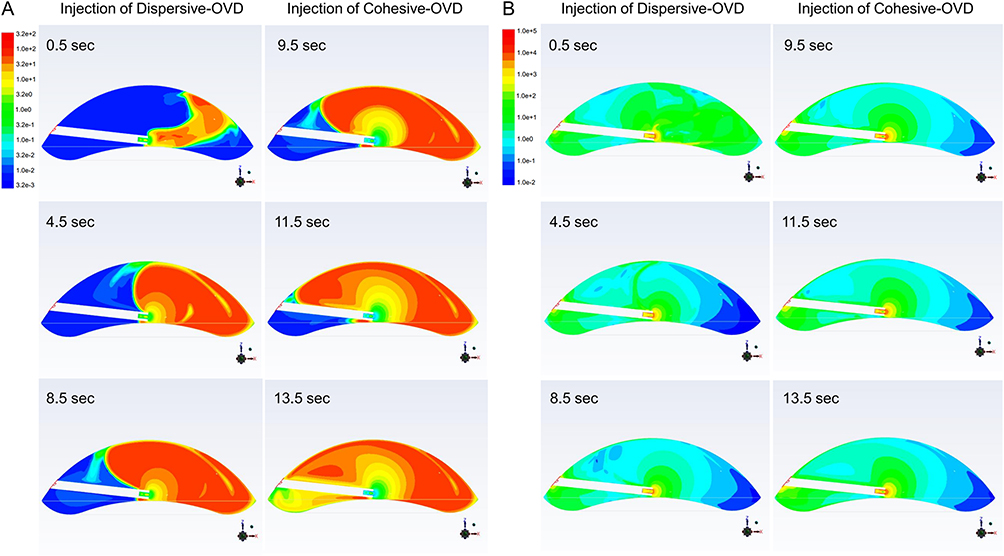 |
Figure 4 Apparent viscosity and shear rates during OVD injection. (A) The images show the apparent viscosity at 0.5, 4.5 and 8.5 sec when 0.1 mL of Dispersive-OVD was injected over 9 sec, and at 9.5, 11.5 and 13.5 sec when 0.1 mL of Cohesive-OVD was injected over 5 sec, after injection of Dispersive-OVD. (B) Shear rate corresponding to the apparent viscosity described above. The scale ranges indicated for apparent viscosity (Pa∙s), and Shear rate (s−1) are as follows: from 3.2e−3 to 3.2e2 (Pa∙s), and from 1.0e−2 to 1.0e5 (s−1). |
The shear rate of Cohesive-OVD passing through the cannula lumen was 103–104 s−1, and the apparent viscosity was approximately 0.03–0.3 Pa∙s. As the distance from the cannula tip increased, the shear rate decreased and the viscosity of Cohesive-OVD increased concentrically. The area of low apparent viscosity (less than 10 Pa∙s) near the cannula tip was larger with Cohesive-OVD than with Dispersive-OVD. The zero shear viscosity of Dispersive-OVD was 83.5 Pa∙s, while that of Cohesive-OVD was 200 Pa∙s. At low shear rates where flow is almost absent, the apparent viscosity of Cohesive-OVD is higher than that of Dispersive-OVD. On the other hand, at shear rates of 1 s−1 or higher, the apparent viscosity of Cohesive OVDs decreases dramatically, and the viscosity of these OVDs is reversed.3 It was suggested that the apparent viscosity of Cohesive-OVD was lower than that of Dispersive-OVD due to the high shear rate in the cannula lumen and tip. This is also related to the fact that Cohesive-OVD can be injected in a shorter time than Dispersive-OVD, when injecting the same amount of agent. The shear rate of the Dispersive-OVD on the corneal endothelial surface during the soft shell technique was less than 10−1–10 s−1, and the apparent viscosity was less than 30–100 Pa∙s.
This study has several limitations. Each OVD product has specific rheological properties. Although not evaluated in this study, simulations for each OVD might provide useful information to support the decision of surgeons to select an appropriate OVD according to the patient’s condition and surgical procedure. As the options for OVDs used in surgery increase, a proper understanding of the properties of each OVD is important to maximize their benefits. Moreover, we were unable to evaluate the behavior of the OVD under a variety of conditions, including the effects of varying the cannula tip position and injection speed. In actual clinical practice, it is expected that the techniques and methods used during OVD injection will vary slightly depending on the surgeon. In future, by evaluating the relationship between such injection conditions and the behavior of the OVD, we may be able to present an optimal approach to the soft shell technique.
In conclusion, we successfully used computational fluid dynamics to simulate the behavior of Dispersive-OVD and Cohesive-OVD, different non-Newtonian fluids used in the soft shell technique. Simulations demonstrated that the Dispersive-OVD was pushed upward into the anterior chamber by the Cohesive-OVD, forming a layer on the corneal endothelial surface. It is difficult to discuss the behavior of an OVD based on surgeons’ perceptions alone, but by expressing perceptions as a quantified parameter, it can be used as a metric. The parameters obtained in this study provide useful information for a greater understanding and prediction of the behavior of OVDs in the eye during the soft shell technique.
Acknowledgments
We thank Research Center of Computational Mechanics, Inc. (Kazuhisa Sugiyama and Moeki Tanaka) for their contribution of expertise.
Funding
This study was funded by Seikagaku Corporation.
Disclosure
The authors report no conflicts of interest in this work.
References
1. Shimazaki J, Amano S, Uno T, et al. National survey on bullous keratopathy in Japan. Cornea. 2007;26(3):274–278. doi:10.1097/ICO.0b013e31802c9e19
2. Watanabe I, Mirumachi H, Konno H, et al. Evaluation of Rheological Properties of Cohesive Ophthalmic Viscosurgical Devices Composed of Sodium Hyaluronate with High Molecular Weight-2019. YakugakuZasshi. 2019;139:1121–1128. doi:10.1248/yakushi.19-00084
3. Watanabe I, Hoshi H, Sato M, et al. Rheological and Adhesive Properties to Identify Cohesive and Dispersive Ophthalmic Viscosurgical Devices. Chem Pharm Bull. 2019;67(3):277–283. doi:10.1248/cpb.c18-00890
4. Arshinoff SA, Jafari M. New classification of ophthalmic viscosurgical devices—2005. J Cataract Refract Surg. 2005;31(11):2167–2171. doi:10.1016/j.jcrs.2005.08.056
5. Hütz WW, Eckhardt HB, Kohnen T. Comparison of viscoelastic substances used in phacoemulsification. J Cataract Refract Surg. 1996;22(7):955–959. doi:10.1016/S0886-3350(96)80198-2
6. Caporossi A, Baiocchi S, Storzi C, et al. Healon GV versus Healon in demanding cataract surgery. J Cataract Refract Surg. 1995;21:710–713. doi:10.1016/S0886-3350(13)80572-X
7. Watanabe I, Yoshioka K, Takahashi K, et al. Advances in Understanding the Mechanism of Ophthalmic Viscosurgical Device Retention in the Anterior Chamber or on the Corneal Surface during Ocular Surgery. Chem Pharm Bull. 2021;69(6):595–599. doi:10.1248/cpb.c21-00116
8. Bissen-Miyajima H. In vitro behavior of ophthalmic viscosurgical devices during phacoemulsification. J Cataract Refract Surg. 2006;32(6):1026–1031. doi:10.1016/j.jcrs.2006.02.039
9. Hsiao CW, Cheng H, Ghafouri R, et al. Corneal Outcomes Following Cataract Surgery Using Ophthalmic Viscosurgical Devices Composed of Chondroitin Sulfate-Hyaluronic Acid: a Systematic Review and Meta-Analysis. Clin Ophthalmol. 2023;17:2083–2096. doi:10.2147/OPTH.S419863
10. Arshinoff SA. Dispersive-cohesive viscoelastic soft shell technique. J Cataract Refract Surg. 1999;25(2):167–173. doi:10.1016/S0886-3350(99)80121-7
11. Ogi S, Nishida T, Kataoka T, et al. Relationship between parameters derived from anterior segment optical coherence tomography and the sulcus-to-sulcus distance by ultrasound bio microscopy. Jpn Orthoptic J. 2018;47:181–189. doi:10.4263/jorthoptic.047F118
12. Mizuno Y, Yamada M, Kataoka M, et al. Intra-and Inter-examiner Reproducibility of Measurements for the Parameters in the Corneal Map of the Anterior Segment OCT (CASIA2). Jpn Orthoptic J. 2018;47:173–179. doi:10.4263/jorthoptic.047F117
 © 2025 The Author(s). This work is published and licensed by Dove Medical Press Limited. The
full terms of this license are available at https://www.dovepress.com/terms.php
and incorporate the Creative Commons Attribution
- Non Commercial (unported, 4.0) License.
By accessing the work you hereby accept the Terms. Non-commercial uses of the work are permitted
without any further permission from Dove Medical Press Limited, provided the work is properly
attributed. For permission for commercial use of this work, please see paragraphs 4.2 and 5 of our Terms.
© 2025 The Author(s). This work is published and licensed by Dove Medical Press Limited. The
full terms of this license are available at https://www.dovepress.com/terms.php
and incorporate the Creative Commons Attribution
- Non Commercial (unported, 4.0) License.
By accessing the work you hereby accept the Terms. Non-commercial uses of the work are permitted
without any further permission from Dove Medical Press Limited, provided the work is properly
attributed. For permission for commercial use of this work, please see paragraphs 4.2 and 5 of our Terms.