")
Back to Journals » International Journal of Nanomedicine » Volume 20
Nanozymes for Accelerating the Foot Wound Healing: A Review
Authors Lv K, Zhang S, Yao Z, Zhou C, Xu J
Received 15 January 2025
Accepted for publication 3 May 2025
Published 10 July 2025 Volume 2025:20 Pages 8907—8934
DOI https://doi.org/10.2147/IJN.S517671
Checked for plagiarism Yes
Review by Single anonymous peer review
Peer reviewer comments 2
Editor who approved publication: Prof. Dr. RDK Misra
Ke Lv,1,* Shumin Zhang,2,* Zimo Yao,3,* Chao Zhou,2 Jianda Xu1
1Department of Orthopaedics, Changzhou Hospital Affiliated to Nanjing University of Chinese Medicine, Changzhou, Jiangsu Province, People’s Republic of China; 2School of Medical and Health Engineering, Changzhou University, Changzhou, Jiangsu Province, People’s Republic of China; 3The Fourth Clinical School of Nanjing Medical University, Nanjing City, Jiangsu Province, People’s Republic of China
*These authors contributed equally to this work
Correspondence: Jianda Xu, Department of Orthopaedics, Changzhou hospital affiliated to Nanjing University of Chinese Medicine, 25 North Heping Road, Changzhou, Jiangsu Province, 213003, People’s Republic of China, Email [email protected] Chao Zhou, School of Medical and Health Engineering, Changzhou University, Changzhou, Jiangsu Province, 213164, People’s Republic of China, Email [email protected]
Abstract: Diabetic foot is a most common and life-threatening complication of diabetes. It often results in a protracted course of wound healing. Currently, treatments for diabetic foot include wound dressings, antibiotics, and debridement. However, these treatments are often inadequate due to the complexity of diabetic foot. Various adjuvant therapies are currently under investigation to accelerate wound healing. In recent years, the emergence of nanozymes, nanomaterial with natural enzyme-like characteristics and activities, have attracted much attention. As a promising candidate in biomedicine, the breadth of application is growing fast, with different mechanisms for accelerating wound healing, such as antioxidant effects, antibacterial effects, promotion of angiogenesis, and regulation of the wound microenvironment. Faced with the inherent defects of nanozyme, new multifunctional nanozymes composite materials have also been continuously developed to achieve synergistic effects with various types of enzymatic activities. This is a new strategy for diabetic foot wound healing in the future. This review summarizes current knowledge about nanozymes and their potential mechanisms in diabetic wound healing. An overview of progress in the applied research on these nanozymes, current challenges and future perspectives of nanozymes in related fields of diabetic wound healing is also discussed to stimulate new strategies and preclinical translations.
Keywords: nanozymes, diabetic foot, wound, healing, therapy
As a most common and serious complication of diabetes, diabetic foot often results in a protracted wound healing course with a relatively high recurrence rate due to peripheral vascular neuropathy. Diabetic foot is usually accompanied by serious consequences, such as infection, necrosis and even amputation, which significantly affect the quality of life of diabetic patients.1 The number of diabetic foot patients worldwide will reach 366 million in 2030, and the incidence of diabetic foot in diabetic patients will reach 25%.2 Diabetic foot accounts for 40% to 60% of nontraumatic foot amputations.3 Given its difficulty in treatment and high cost of treatment, more urgent attention is needed to reduce the socioeconomic burden on family and society.
The importance of local wound treatment in the management of diabetic foot is extremely crucial. Recently, a series of advanced techniques, such as debridement, the application of moist dressings, vacuum-assisted closure (VAC) therapy, stem cell transplantation and growth factor therapy, have been employed in diabetic foot treatment, aiming to directly improve the wound environment, promote granulation tissue growth, and accelerate the healing process. By thoroughly removing necrotic and infected tissues, debridement reduces barriers to new tissue regeneration and creates favorable conditions for healing. However, traditional therapies have certain application deficiencies in clinical practice, and repeated surgical debridement results in great pain for patients.4 The effect of traditional textile dressings occur slowly and therefore are not immediately obvious, and frequent dressing exchange may worsen healing and even pose an anaphylaxis.5 In vacuum-assisted closure therapy, continuous negative pressure suction is used to effectively remove exudates and necrotic tissues from the wound, but blood clots from bleeding may increase the degree of infection.6 Stem cell transplantation and growth factor therapy are currently associated with problems such as poor targeting ability and a short half-life.7 Therefore, an inexpensive and simple treatment method for patients is urgently needed. However, the exploration of new local wound treatment strategies to accelerate wound healing is still an enormous challenge.
Nanozymes, which are emerging nanomaterials, were first reported in 2007.8 Nanozymes can simulate the efficient catalytic activity of natural enzymes through their unique properties. These nanozymes can regulate redox levels in cells and tissues, thereby eliminating or generating reactive oxygen species, playing an anti-inflammatory and anti-bactericidal role. Nanozymes have unique advantages and application potential in the biomedical, chemical, industrial, food, agricultural, and environmental fields.9–12 In diabetic foot, nanozymes have been shown not only to kill pathogenic bacteria directly and eliminate inflammatory factors but also to promote the repair and regeneration of wound tissues.13 By mimicking the activity of natural enzymes, nanozymes facilitate biological processes such as cell proliferation, migration, and differentiation, thereby accelerating wound healing and skin regeneration. This advantage of promoting tissue repair and regeneration makes nanozyme therapy more comprehensive and effective in the treatment of diabetic foot wounds. Nonetheless, to the best of our knowledge, few articles have focused on advances in comprehensive classifying nanozymes for physicochemical properties related with diabetic foot wound healing.
In the present review, we provide a brief perspective on the underlying mechanisms of nanozymes used in diabetic wound healing. Furthermore, we also discuss the progress in the applied research on these nanozymes, as well as current challenges and future perspectives of nanozymes in related fields of diabetic wound healing.
Basic Characteristics of Nanozymes
Nanozymes are a type of simulated enzyme with a nanoscale size (1–100 nm) that not only retain the unique properties of nanomaterials but also possess the catalytic function of natural enzymes.14 With the rapid development of nanotechnology, the use of nanozymes, as a new therapeutic approach, has emerged in the field of biomedical applications (Figure 1). These enzymes exhibit redox-mimicking enzyme activities similar to those of superoxide dismutase (SOD), catalase (CAT), peroxidase (POD), and oxidase (OD).15 Therefore, they can regulate the level of reactive oxygen species (ROS) in the body. ROS upregulation can be used to inhibit the proliferation of bacteria and other pathogens, preventing and controlling infection sources, while downregulation of ROS can alleviate oxidative stress. In addition, the large specific surface area and numerous surface activation sites of ROS enable them to fully realize multiple enzyme cascade catalysis, significantly increasing their catalytic efficiency. With strong structural stability, nanozymes can maintain catalytic activity as mimic enzymes under extreme conditions such as high temperatures and different pH values. Unlike natural enzymes, nanozymes can avoid protein denaturation or digestion by proteases.
 |
Figure 1 Schematic diagram of catalytic activities and types of nanozymes. |
Nanozymes inevitably have some inherent defects. By adjusting the size, morphology, and specific surface area of nanozymes, their physicochemical properties can be regulated, improving their interaction with biomolecules, increasing targeted internalization of drugs at specific locations, achieving sustainable drug release from nanomaterials, and reducing incompatibility and adverse reactions with the body to increase therapeutic effects.16,17 Therefore, new composite materials with synergistic effects have emerged to compensate for these defects.
Mechanism of Nanozymes in Diabetic Foot Wound Healing
The basic process of diabetic foot wound healing usually involves local ischemia and nutritional barriers to necrosis, debridement treatment, and finally entry into the repair and healing stage.18 Diabetic foot wound healing is characterized by a significantly prolonged healing process, a tendency for the development of complications such as difficult-to-control infections, weak tissue repair ability, and a tendency for the development of chronic recurrent ulcers.19,20 Rescue of stagnant wound healing in diabetic foot has remained a great challenge till now. The pathological wound microenvironment is usually associated with local hyperglycemia, excessive oxidative stress, hypoxia and biofilm infection. The strategy of improving the local microenvironment has become the theoretical basis and practical direction for chronic diabetic wound repair.21
Recent progress in nanobiotechnology has made the development of nano-artificial enzymes possible. In the study of diabetic wound healing, nanozymes can simulate the catalysis of natural enzymes or exert their own multienzyme activity to meet various therapeutic needs. Five mainstream mechanisms (Figure 2) for accelerating wound healing have been described, namely, antioxidant effects, antibacterial effects, promotion of angiogenesis, wound blood glucose regulation, and pH monitoring.22,23
 |
Figure 2 Mechanisms and potential therapeutic strategies of nanozymes in diabetic foot. |
Antioxidant Effects
In diabetic foot wounds, high levels of superoxide anions and H2O2 abnormally increase the level of ROS in the wound, which in turn exacerbates oxidative stress and inflammation in the wound and ultimately makes wound healing difficult.24 Antioxidant nanozymes, such as SOD and CAT, can reduce the concentrations of superoxide anions and H2O2 in wounds by catalyzing the degradation of ROS. This process reduces oxidative stress damage at the wound surface and promotes wound healing.25 SOD has strong antioxidant properties and can effectively convert superoxide anions into H2O2 and O2, thereby reducing the ROS level and the hypoxic state in wounds. Therefore, the application of SOD to wounds is considered an effective means to shorten the inflammatory cycle and promote wound healing.26
It is generally believed that the highest level of superoxide anions is reached in the early stage of wound healing and that the presence of these ROS can induce oxidative stress, thereby exacerbating wound inflammation, hindering normal cell proliferation and migration, and significantly prolonging the wound healing cycle.27 Therefore, reasonable early regulation of wound ROS levels can effectively promote the healing of diabetic wounds. SOD-like nanozymes have been applied to protect against oxidative stress in diabetic wounds, convert superoxide anions in wounds into H2O2 and O2, reduce ROS levels in diabetic wounds, relieve the hypoxic status of diabetic wounds, and thus improve wound health and healing.28 Deng et al developed an amorphous cobalt selenide-based amorphous biocatalyst, Ru@CoSe, which has superior ROS scavenging ability and can effectively resolve the unbalanced multielectron reactions during ROS catalysis (Figure 3a). Ru@CoSe can also protect the proliferation ability of mesenchymal stem cells (MSCs) as well as angiogenesis ability as much as possible.29
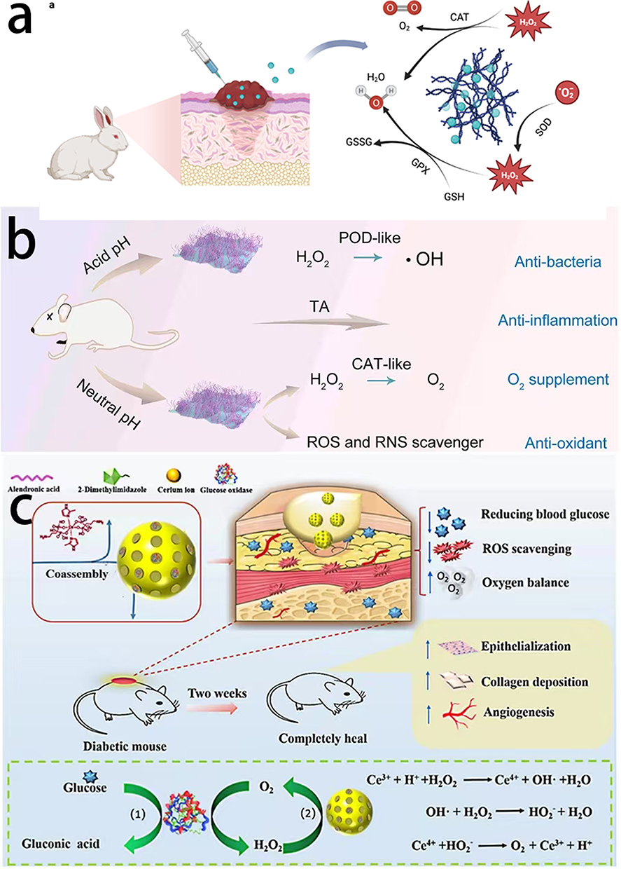 |
Figure 3 Antioxidant effects of nanozymes. (a) Anti-oxidation and anti-inflammation of amorphous Ru@CoSe nanolayer. (b) CAT-like activity and POD-like activity of MoS2@TA/Fe NSs. Reprinted from Li Y, Fu R, Duan Z, et al. Construction of multifunctional hydrogel based on the tannic acid-metal coating decorated MoS2 dual nanozyme for bacteria-infected wound healing. Bioact Mater. 2022;9:461–474. Creative Common.30 (c) Multi-enzymatic activities (Gox-, SOD- and CAT-like) of CHA@Gox. Reprinted from Yu X, Fu X, Yang J, et al. Glucose/ROS cascade-responsive ceria nanozymes for diabetic wound healing. Mater Today Bio. 2022;15:100308. Creative Commons.31 |
During the wound repair stage, CAT removes a large amount of H2O2 present in infected tissues to relieve oxidative stress damage to the tissues. Moreover, the oxygen generated during the decomposition process can ameliorate the local hypoxic environment in the wound, thereby promoting angiogenesis and wound healing.8 Similarly, Li et al fabricated a multifunctional hydrogel loaded with tannic acid-chelated Fe-decorated molybdenum disulfide nanosheets (MoS2@TA/Fe NSs), which exhibited excellent antibacterial ability and CAT-like activity (Figure 3b). MoS2@TA/Fe NS could provide adequate O2 to accelerate wound healing by decomposing H2O2 into O2 in a neutral environment. In addition, MoS2@TA/Fe NSs also had POD-like activity and could catalyze H2O2 into •OH in an acidic environment.30 Zhao Y et al designed a biomimetic nanozyme-decorated hydrogel system with H2O2-activated oxygenation (MnCoO@PDA/CPH). This system could effectively modulate the immune microenvironment, reduce proinflammatory conditions, increase re-epithelialization, and promote the orderly deposition of collagen, thereby improving the healing of diabetic foot wounds.32 In addition, surface modification can also improve the antioxidant effects. Yao et al modified gold nanozymes with interleukin-4 (IL-4) and noted that IL-4-modified gold nanozymes improved the local oxidative environment of wounds in diabetic mice and promoted the polarization of macrophages from the M1 phenotype to the M2 phenotype, thus effectively achieving effective skin regeneration and healing of wounds in diabetic mice.33
To address the possible deficiencies of a single antioxidant nanozyme, nanozymes with multiple antioxidant effects have gradually been designed and used in the healing of diabetic wounds. Yu et al used Ce-driven coassembly of alendronic acid and 2-methylimidazole to form CHA nanoparticles. They then combined CHA with GOx to create a CHA@GOx nanocomposite material, which was eventually used for the treatment of diabetic wounds. The dynamic equilibrium of cerium ions (Ce3+ and Ce4+) on the surface of CHA@GOx endows CHA with increased SOD-like activity (Figure 3c). The loaded GOx could reduce the blood glucose level at the wound surface. The SOD-like activity of CHA@GOx can convert superoxide anions in wounds to H2O2 and O2. In addition, the CAT-like activity of CHA@GOx converted H2O2 into water and oxygen to relieve wound hypoxia. Therefore, CHA@GOx can regulate the ROS level in wounds and relieve wound hypoxia, thereby promoting wound healing.31
Oxidative stress impairs cellular function, disrupts tissue integrity, and promotes inflammation, all of which hinder the healing process. Currently, a reasonable quantitative definition of oxidative stress still cannot be obtained, which also introduces the problem of controlling the intensity of antioxidant activity. Studies on the optimal timing and dosage of antioxidant therapy and its combination with other therapeutic modalities could further advance the field and improve patient outcomes. In addition, we also need to pay more attention to balancing the requirements of high activity, persistence, dispersion, and biocompatibility of new biomaterials or composite materials.
Antibacterial Effects
Owing to the protracted course of diabetic foot wounds, adjacent soft tissues are exposed to pathogens and have a clear tendency to be infected. If not promptly detected and treated, tissue infection may further spread to the surrounding bone tissue and even cause osteomyelitis.34 Antibacterial nanozymes, such as POD-like and nitric oxide synthase-like (NOS-like) nanozymes, can precisely catalyze and produce substances (such as hydrogen peroxide and nitric oxide) with antibacterial activity, thereby inhibiting the growth of bacteria or killing bacteria. For example, a nanozyme with POD-like activity catalyzes the conversion of H2O2 into a hydroxyl radical (•OH), which can rapidly increase the intracellular ROS level to kill bacteria, thereby eliminating infectious pathogens in wounds and promoting wound healing.35 Cong et al reported a Platinum (Pt)-based nanozyme (Figure 4a) with both GOx-like and POD-like activities for the first time. Pt nanoparticles with GOx-like activity can convert glucose to H2O2 under aerobic conditions. POD-like activity catalyzes the decomposition of H2O2 to generate large amounts of toxic •OH, which inactivates bacteria.36 The methods of coating polymers or doping elements are also used to increase the stability and catalytic activity of some nanozymes. Li et al designed a POD-like Fe 3 C/N-doped graphitic carbon nanomaterial (Fe 3 C/N–C) (Figure 4b) via a one-step mild electrochemical method. This Fe 3 C/N–C could simultaneously decompose H2O2 into •OH to achieve antimicrobial activity and accelerate wound healing.37 Similarly, Yu et al developed chitin nanofibrous microspheres loaded with AgNPs and Fe3O4 nanoparticles (Ag-Fe3O4-NMs) (Figure 4c) with increased synergistic antibacterial activity and wound-healing.38
 |
Figure 4 (a A–E) Enhanced POD-like activity of Pt hollow nanodendrites exhibited broad spectrum antibacterial activity via decomposing H2O2 into •OH.Reprinted from Ge C, Wu R, Chong Y, et al. Synthesis of Pt hollow nanodendrites with enhanced peroxidase-like activity against bacterial infections: implication for wound healing. Adv Funct Mater. 2018. © 2018 WILEY-VCH Verlag GmbH & Co. KGaA, Weinheim.36 (b) POD-like activity of Fe3C/N–C decomposed H2O2 into •OH to achieve antimicrobial activity. Reprinted from Carbon, volume 159, Li Y, Ma W, Sun J, et al. Electrochemical generation of Fe3C/N-doped graphitic carbon nanozyme for efficient wound healing in vivo. 149–160, copyright 2020, with permission from Elsevier.37 (c) Synergistic antibacterial activity of Ag-Fe3O4-NMs: peroxidase-like activity and sustained release of Ag+. Reprinted from Int J Pharm, volume 552(1-2), Yu N, Cai T, Sun Y, et al. A novel antibacterial agent based on AgNPs and Fe3O4 loaded chitin microspheres with peroxidase-like activity for synergistic antibacterial activity and wound-healing. 277–287, copyright 2018, with permission from Elsevier.38 |
The control of local infection through the use of peroxidase alone is sometimes not enough. Therefore, researchers have begun to use antibacterial nanozymes in combination with other bacteriostatic and antibacterial agents to synergistically increase the antibacterial effect and reduce the side effects. Cai Get al combined catalase (CAT)-like nanozyme-loaded microneedles (MNs) with polymyxin B (Figure 5a) to further increase the number of M2 macrophages and reduce the expression of TNF-α under the premise of ensuring effective antibacterial agents. This combination also promoted the regeneration of blood vessels and nerves, and eventually promoted the healing of diabetic foot wounds.39 Moreover, the combination with other antibacterial methods has also gradually attracted increasing attention. Duet al designed a pH-switchable iron oxide nanozyme coated with a shell of glucose oxidase (Fe3O4-GOx) (Figure 5b). This enzyme possessed excellent GOx, CAT, and POD-like activities, and could catalyze pH-switchable glucose-initiated GOx/POD and GOx/CAT cascade reactions in acidic and neutral environments, respectively. Under acidic conditions of bacterial infection, Fe3O4-GOx could oxidize large amounts of glucose in diabetic wounds through GOx activity to produce gluconic acid and H2O2. Furthermore, Fe3O4-GOx could decompose H2O2 through POD-like activity and continuously produce a large amount of •OH, which killed the bacteria at the wound site, thereby shortening the inflammatory period and promoting wound healing.40 Cao et al used charge separation technology to design a new Ag/Bi2MoO6 (Ag/BMO) nanozyme (Figure 5c), which could achieve synergy between photoactivated sustainable peroxidase-mimicking activities and NIR-II photodynamic performance via a solvothermal reaction and photoreduction. Ag/Bi2MoO6 exhibited excellent antibacterial activity and wound healing effects on MRSA-infected wounds.41 Considering the complex biological processes of glucose metabolism, Shan et al developed a near-infrared II (NIR-II)-responsive biocatalytic MN patch, which had dual nanozyme activities (glucose oxidase and catalase-like activities). These Au-Cu2MoS4 nanosheets (Figure 5d) could reduce the glucose level at the wound surface, release oxygen, effectively eliminate bacteria, and then rapidly treated MRSA-infected wounds in diabetic mice.42
 |
Figure 5 Antibacterial nanozymes in combination with other bacteriostatic and antibacterial agents to synergistically increase the antibacterial effect. (a) CAT-(MnSH)x@PMB-MNs: catalase (CAT)-like nanozyme-loaded microneedles (MNs) combined with polymyxin B. Reprinted from J Colloid Interface Sci, volume 667, Cai G, Li R, Chai X, et al. Catalase-templated nanozyme-loaded microneedles integrated with polymyxin B for immunoregulation and antibacterial activity in diabetic wounds. 529–542, copyright 2024, with permission from Elsevier.39 (b) Fe3O4-Gox: pH-switchable iron oxide nanozyme coated with a shell of glucose oxidase. Reprinted from Du X, Jia B, Wang W, et al. pH-switchable nanozyme cascade catalysis: a strategy for spatial-temporal modulation of pathological wound microenvironment to rescue stalled healing in diabetic ulcer. J Nanobiotechnology. 2022;20(1):12. Creative Commons.40 (c) Ag/Bi2MoO6 (Ag/BMO) nanozyme: synergy between photoactivated sustainable peroxidase-mimicking activities and NIR-II photodynamic performance. Reprinted from Cao C, Zhang T, Yang N, et al. POD nanozyme optimized by charge separation engineering for light/pH activated bacteria catalytic/photodynamic therapy. Signal Transduct Target Ther. 2023;8(1):214. doi: 10.1038/s41392-023-01476-7]. Signal Transduct Target Ther. 2022;7(1):86. Creative Commons.41 Copyright 2023 Springer Nature. (d) Au-CMS NSs: a catalytic microneedle patch that encapsulates near-infrared-II responsive and dual-nanozyme active Au-Cu2MoS4 nanosheets. Reprinted from Shan J, Zhang X, Cheng Y, et al. Acta Biomater, volume 157, Glucose metabolism-inspired catalytic patches for NIR-II phototherapy of diabetic wound infection. 200–209, copyright 2023, with permission from Elsevier.42 |
Bacteria have a high propensity for attaching to surfaces and forming three-dimensional bacterial biofilms. Bacterial biofilms are complex, structured communities of bacterial cells that adhere to each other and often to a surface, enclosed in a self-produced polymeric matrix, providing protection and enhancing their survival and resistance to environmental stresses and antimicrobial agents. They are difficult to be eliminated because of their unique protective matrix. Owing to the high-glucose microenvironment of diabetic foot wounds, approximately 90% of wounds have biofilm infections. These biofilms can penetrate deeply damaged tissues, significantly prolonging the inflammatory stage and eventually leading to wound deterioration.43 Moreover, exudate and necrotic tissue from wounds hinder the ability of nanozymes to reach subcutaneous tissue, further reducing their effectiveness against biofilms.44 To address the problem of biofilms, Sun C et al fabricated nanoenzyme microneedles (Fe2C/GOx@MNs) (Figure 6a) to rapidly deliver and release Fe2C nanoparticles (NPs)/GOx in the active area of biofilms. The increased tissue and biofilm permeability eventually resulted in increased biofilm elimination and wound healing.45 T.Huang et al fabricated an intelligent and multifunctional Pd-MOF@PAzo@SNP nanoplatform (Figure 6b) (including palladium, UiO-66, photosensitive polyazobenzene, and sodium nitroprusside), which has light controlled nanozyme activity and UV responsive NO release ability. This nanoplatform exhibited excellent antibacterial and antibiofilm activity to accelerate the healing of biofilm-infected diabetic wounds.46
 |
Figure 6 Nanozymes for combating biofilms. (a) Fe2C/GOx@MNs: deliver and release Fe2C nanoparticles (NPs)/GOx in the active area of biofilms. (b) Pd-MOF@PAzo@SNP nanoplatform (including palladium, UiO-66, photosensitive polyazobenzene, and sodium nitroprusside): light controlled nanozyme activity and UV responsive NO release ability. |
Recently, two-dimensional (2D) nanomaterials have been extensively studied in the biomedical field. These materials have exhibited the advantages of a large surface area and good biocompatibility. Among them, 2D nanomaterials with POD-like activities are particularly favored because of their superior stability and multiple active targets. Li et al designed defect-rich molybdenum disulfide nanosheets loaded with bovine serum albumin-modified gold nanoparticles (MoS2 @Au@BSA NSs) (Figure 7a). They were embedded in injectable hydrogels to accelerate diabetic wound healing by achieving an oxygen self- supplying cascade reaction. Gox-like Au catalyzes the oxidation of local glucose into gluconic acid and H2O2, while peroxidase-like activity decomposes H2O2 into hydroxyl radicals (•OH) to kill bacteria and destroy local biofilms effectively. Under alkalescent conditions, MoS2@Au@BSA could transform and decompose superoxide anions into O2 and H2O2 via superoxide dismutase and catalase-like mechanisms. In summary, the MoS2@Au@BSA nanozyme-anchored injectable hydrogel effectively healed of diabetic wounds by reducing oxidative stress, alleviating hypoxia, and facilitating glucose oxidation.47 Metal-organic frameworks (MOFs) have porous structures with large organic frame surface area for enzyme adsorption.48 Liu et al also designed a nano-delivery system that used ultrathin two-dimensional (2D) MOF (2D Cu-TCPP(Fe)) nanosheet (Figure 7b) as carriers to load GOx through physisorption. This MOF-based hybrid nanocatalyst could convert glucose gluconic acid and H2O2 by GOx, and decompose H2O2 to extremely toxic •OH through POD-like activity. In vitro and in vivo results confirmed the antibacterial effects and ability to promote wound healing.49 Moreover, multifunctional 2D MOF have also gradually attracted increasing attention. Xuan Zhang et al successfully developed a composite antibacterial nanomaterial (Au@Zn-Tcpp/ASO-pVEC NSs) (Figure 7c) that combines a cell-penetrating peptide-conjugated 2D MOFs with an adaptive light-response biosensor, effectively incorporating the sensing of living bacteria via multimodal methods (including chemodynamic therapy (CDT), photodynamic therapy (PDT) and photothermal therapy (PTT)). This multifunctional 2D MOF showed strong promise for clinical treatment and bacterial infection monitoring, with a rich antibacterial strategy.50
 |
Figure 7 Two-dimensional (2D) nanomaterials with antibacterial effects in diabetic foot. (a) MoS2@Au@BSA nanozyme-anchored injectable hydrogel: reduce oxidative stress, alleviate hypoxia, and facilitate glucose oxidation. (b) 2D Cu-TCPP(Fe) nanosheet: Gox- and POD-like activities. (c) Au@Zn-Tcpp/ASO-pVEC NSs: chemodynamic therapy (CDT), photodynamic therapy (PDT) and photothermal therapy (PTT). |
The removal of infectious bacteria and biofilms is very important for diabetic foot antibacterial therapy. Nanozymes have achieved the above objectives well, but their inherent targeting disadvantage may damage surrounding normal tissues during treatment, causing inevitable adverse reactions. Therefore, improving the targeting ability of nanozymes to minimize the occurrence of adverse reactions remains a popular focus of current research, which will likely continue in the future.
Promotion of Angiogenesis
Angiogenesis plays a crucial role in the wound healing process. Angiogenesis provides a functional blood vessel network to reestablish a sufficient blood supply for new connective tissue. Diabetic foot patients often have severe vascular disease, which makes it difficult to ensure sufficient exchange of oxygen for cell proliferation and viability.51,52 Angiogenesis drugs based on biomacromolecules usually cause severe oxidative stress damage and degeneration of the diabetic foot wound microenvironment. Therefore, sufficient blood flow remains an important problem in diabetic foot wound healing.53–55 To address this problem, Wang et al designed a biodegradable, pH-responsive CuPt-GOx-CaP nanoreactor (Figure 8a), which has a core‒shell structure and consumed Gs through an enzymatic reaction to release O2 and simultaneously released Cu2+ to promote angiogenesis.56 CuPt-GOx-CaP also exhibited good antibacterial activity against gram-negative and gram-positive bacterial infections.
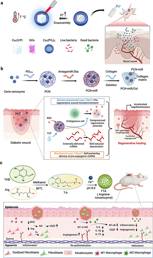 |
Figure 8 Nanozymes for promoting angiogenesis. (a) CuPt-GOx-CaP nanoreactor: release O2 and simultaneously released Cu2+ to promote angiogenesis. (b) PCN miR/Col: reshape the hostile oxidative wound microenvironment, but also ensures the structural integrity of the encapsulated proangiogenic miRNA in the oxidative microenvironment. (c) The arginine-nanozyme to ameliorating angiogenesis by activating the cAMP signaling pathway and reducing inflammation. |
The advantages of hydrogels are their high degree of water absorption, good biocompatibility, tunable physical and chemical properties, excellent biological activity and drug release ability in various applications, which makes them excellent carriers for angiogenesis in diabetic foot.57,58 On the basis of the unique “seed-and-soil” strategy, Wu et al designed a hydrogel with a self-protection effect (PCN miR/Col) by using a miRNA that promotes angiogenesis and a cerium oxide nanozyme with redox regulation, which can not only promote angiogenesis but also effectively remodel the microenvironment of wound healing. Animal experiments also confirmed that PCN-miR/Col (Figure 8b) can effectively promote the growth of new functional blood vessels, thereby accelerating the closure of diabetic foot wounds.23
Some nanozymes, such as Gox-like nanozymes, do not directly promote angiogenesis but can provide a wound with a more conducive angiogenic environment by lowering blood glucose, reducing inflammation, macrophage reprogramming, and relieving hypoxia, thereby promoting wound healing.59 Yang Y et al designed an arginine-nanozyme (Figure 8c) to ameliorating angiogenesis by activating the cAMP signaling pathway and reducing inflammation. This nanozyme removes excess reactive oxygen species at the wound site in situ and converts them to oxygen to ameliorate hypoxia.60 These mechanisms that indirectly promote angiogenesis are also critical for increasing the blood supply and providing nutritional support for diabetic foot wounds.
Wound Blood Glucose Regulation
The hyperglycemic microenvironment of a diabetic wound slow wound healing by reducing angiogenesis and tissue regeneration.61,62 Excessive glucose can decrease the permeability of red blood cells and blood flow for tissue regeneration.63 In addition, local hyperglycemia in the diabetic wound microenvironment provides sufficient nutrients for bacterial infection and the formation of biofilms in infected tissues, thus impeding wound healing.64 Nanozymes with GOx-like activity can convert glucose in wounds to gluconic acid and H2O2, which not only lowers blood glucose but also produces H2O2 as a substrate of nanozymes, which further produces more •OH, increasing the effect of bacterial removal and relieving the hyperglycemia in the wound microenvironment.47 Shi S et al engineered an integrated GOx-GA-Fe nanozyme (GGFzyme) (Figure 9a) to achieve increased reactive oxygen species (ROS) generation. This nanozyme promoted hyperglycemic wound healing via the upregulation of fibroblastic activity and the induction of increased levels of wound-healing cytokines.65 Owing to the low Michaelis constant (Km) and high substrate affinity of GOx-like nanozymes,66 the nanomaterials with GOx-like activity, ie, gold nanoparticles (Au NPs), exhibited good biocompatibility. These nanozymes functions mainly as “hunger agents” in diabetic wound healing. They can consume glucose in wounds, cut off the nutrient source for bacteria and thereby starve bacteria. In addition, H2O2 generated during the glucose oxidation process can be converted to •OH through subsequent Fenton or Fenton-like reactions. Together, these two mechanisms reduce the inflammatory response cycle of wounds and promote the healing of diabetic foot wounds.67
 |
Figure 9 Nanozymes for regulating wound blood glucose. (a) GGFzyme promoted hyperglycemic wound healing via the upregulation of fibroblastic activity and the induction of increased levels of wound-healing cytokines. Reprinted with permission from Shi S, Zhang Q, Sun H, et al. Glucose oxidase-integrated metal-polyphenolic network as a microenvironment-activated cascade nanozyme for hyperglycemic wound disinfection. ACS Biomater Sci Eng. 2022;8(12):5145–5154. Copyright © 2022 American Chemical Society.65 (b) Fe2(MoO4)3@GOx could consume local glucose and produce •OH to efficiently kill bacteria. Reprinted with permission from Zhang Y, Li D, Xu Y, et al. Application of a cascaded nanozyme in infected wound recovery of diabetic mice. ACS Biomater Sci Eng. 2022;8(4):1522–1531. Copyright 2022, American Chemical Society.68 |
To simultaneously obtain GOx activity and intrinsic nanozyme activity, Zhang Y et al coated GOx on POD-like Fe2(MoO4)3 [Fe2(MoO4)3@GOx] (Figure 9b) and fabricated a cascade nanoenzymatic active material. This material could consume local glucose and produce •OH to efficiently kill bacteria.68 In another study, Wu Y et al synthesized cascaded enzyme conjugates with GOx-, SOD- and CAT-like activities. These conjugates could also significantly decrease glucose levels with a prolonged duration.69 These synergistic effects could work well together and regulate the microenvironment to promote diabetic wound healing.
Lowering blood glucose is an important method to promote diabetic wound healing, but it alone is not sufficient to fully achieve healing. Combining nanozymes to lower blood glucose with other enzymes (such as antibacterial agents, which promote angiogenesis) may be extremely efficient.
pH Monitoring
Human tissues generally exhibit alkalinity under physiological conditions, and a pH >8.0 can be found in some chronic wound tissues.70 However, nanozymes require an acidic environment (pH 3–6) to perform peroxidase like activity. In addition, the content of H2O2 under physiological conditions cannot meet needs of chemodynamic therapy.71 Chen L et al constructed an infection-activatable nanozyme (Figure 10) (aptamer-functionalized nanozymes, glucose oxidase, and hyaluronic acid) with a targeting ability for chemodynamic therapy. This nanozyme simultaneously overcame the limitations of local pH and H2O2 under physiological conditions. The increased antibacterial effect improved diabetic wound healing by directly regulating the local microenvironment.35
 |
Figure 10 Nanozymes for monitoring pH. An activatable nanocapsule “APGH” tuned the local pH down through glucose oxidation. Reprinted from Chen L, Xing S, Lei Y, et al. A glucose-powered activatable nanozyme breaking pH and H2 O2 limitations for treating diabetic infections. Angew Chem Int Ed Engl. 2021;60(44):23534–23539. © 2021 Wiley-VCH GmbH.35 |
pH monitoring in diabetic wound healing holds significant value, as it can effectively accelerate wound healing by modulating the microenvironment. Future research directions may include the development of novel biomaterials, such as multifunctional hydrogels, that can intelligently regulate pH levels at different healing stages, providing a dynamic and optimal environment for diabetic wound repair. These advancements have the potential to revolutionize treatment strategies for diabetic foot.
Progress in the Research and Application of Nanozymes in Diabetic Foot Wound Healing
The application prospects of nanozymes in diabetic foot wound healing are very broad. Their unique enzymatic characteristics and biomedical application potential provide a new direction for the treatment of diabetic foot. In recent years, researchers have conducted many studies on the application of nanozymes in diabetic foot wound healing and have made important progress. The progress of nanozymes in clinical applications for diabetic foot ulcers can be summarized as exhibiting promising potential in enhancing wound healing (biocompatibility and safety of nanozymes, and catalytic reaction efficiency) and managements (development of multifunctional nanozymes, combined application of nanozymes, and nanozymes-based dressings).
Biocompatibility and Safety of Nanozymes
Considering the advantages and promising therapeutic potential of nanozymes in diabetic foot, ensuring biosafety and biocompatibility is extremely important before nanozymes can be used clinically. The biocompatibility and safety of nanozymes in biomedical applications are important issues and have been confirmed by some researchers. For example, after nanozymes (such as MOF nanozymes) were injected into healthy SD rats through the tail vein, there was no difference in the weight gain of the rats compared with that of the control group (injected with an equal volume of normal saline). These findings indicated that the nanozyme did not have significant toxicity in vivo. Moreover, the major organs (heart, liver, spleen, lung, and kidney) of the rat did not show severe pathological effects, further confirming the biocompatibility of the nanozymes in the body.72 However, CuO NPs can result in cell death through autophagic and mitochondrial dysfunction.73 Au NPs exhibit good antibacterial activity, but their potential toxicity to human health and the environment remains controversial. Graphene oxide (GO) nanomaterials exhibit good biocompatibility, but still have certain cytotoxic effects on cells and organs. Hu W et al used A549 cells cultured in fetal bovine serum were to evaluate the cytotoxicity of GO nano sheets. They reported that the cytotoxicity of GO nanosheets was significantly concentration dependent. Physical damage to the cell membrane was the main risk factor.74 Metal-based nanozymes exhibit good antibacterial activity, but the released metal ions may induce potential adverse effects. Owing to their strong protein inactivation effects, Fe3+, Zn2+ and Cu2+ can lead to metal poisoning.75 Further research is still essential.76 Long-term study is usually needed to confirm the biosafety of these materials in biological systems.
The toxicity of nanozymes can be modulated by size, morphology, and surface modification.77 To achieve biosafety, several strategies are used to minimize the toxicity of nanozymes. First, ultrasmall sized nanozymes are preferred to achieve rapid renal elimination. Large sized GO can increase the bactericidal ability of nanozymes, but at the same time, it also increases toxicity to normal tissues and cells.78 Second, surface modification can be used to improve the targeting ability of nanozymes and reduce different types of cytotoxicity.79 A previous study showed that the introduction of folic acid (FA) on the surface of a nanozyme could significantly increase its tumor-targeting ability and the endocytosis of tumor cells.80 The targeting ability of nanozymes can also effectively reduce toxic reactions. The targeting ability can be achieved by surface modification or embedding nanozymes into polymeric nanocarriers/hydrogel matrices.79 Xu M et al reported that the cytotoxic effect of polyacrylic acid modified GO had very little cytotoxicity.81 Finally, a reasonable drug dosage and administration method can better ensure safety.82
Some researchers have designed new nanozymes to increase biosafety. Zhao et al reported that pure Au NPs or pure Pt NPs had no antibiotic activity. However, bimetallic nanoparticles (NPs) of AuPt exhibit excellent antibiotic activities by dissipating the membrane potential and increasing ATP levels. This specific non-toxicity to human cells endows it with excellent application prospects.83 Ma et al designed a biomimetic nanozyme (CuxO@EM-K) with minimal immunogenicity, and increased biocompatibility. This nanozyme could effectively reduce Aβ levels in the blood and brain, and simultaneously mitigate Aβ-induced membrane oxidative damage. Moreover, blood biochemical and histological analyses revealed that this nanozyme has no apparent toxicity in 3xTg-AD mice and was biocompatible and safe.84 However, how to synthesize nanozymes with high safety and high enzyme-like catalytic activity through different combinations, structural optimization, and surface modification remains a major challenge.
In conclusion, the activities of nanozymes have been confirmed, but in vivo and human experiments are still in the preliminary stage. Considering the complexity of biological systems, more in vivo and human experiments are urgently needed to better evaluate the biosafety and efficacy of nanozymes.
Development of Multifunctional Nanozymes
Nanozymes have various enzyme activities and can simulate the catalysis of natural enzymes to meet different clinical therapeutic needs. However, nanozymes with a single enzyme-like activity are not sufficient to achieve wound healing because of the constantly changing microenvironment at different stages. Therefore, the development of new multifunctional nanozymes is particularly important and urgent. These multifunctional nanozymes have various types of enzymatic activities. The synergy of various enzymatic activities through cascade reactions can more effectively promote the healing of diabetic foot wounds. Some researchers have partially achieved the comprehensive treatment of diabetic foot by designing nanozymes with various enzymatic activities. Shang et al designed a nanozyme hydrogel spray (Figure 11a), which has five types of enzyme-like activities, including SOD, CAT, GOx, POD, and NOS-like activities, and can form a protective film against diabetic foot wounds. This spray reduces inflammation, ameliorates hypoxia, lowers blood glucose, promotes angiogenesis, eliminates pathogenic bacteria, and comprehensively accelerates diabetic wound healing.85 However, the current multi-nanozyme systems exhibit only moderate activity because the microenvironments required for individual enzyme reactions are usually different or even incompatible. To overcome the shortcomings of traditional assembly strategies, Li G et al designed a fiber-based multifunctional compartment strategy to construct a multinanozyme system that can perform incompatible reactions simultaneously.86 Deng M et al added glucose oxidase (GOx) and quasi-amorphous Fe2O3 to Zn-MOF nanoparticle (F-GZ) (Figure 11b) to achieve cascade enzyme catalytic activities. GOx consumes the local blood glucose, while the released Zn2+ and catalytically produced •OH synergistically control wound infection during the bacterial infection period. In addition, it can also correct the hypoxic environment through the catalysis of H2O2 to produce O2. at the stage of wound recovery. Interestingly, this hydrogel could also improve glucose tolerance and increase insulin secretion via the release of Zn2+, which potentially partially restored pancreatic islet functions.87 Notably, these methods remain to be further studied in the future.
 |
Figure 11 Application of multifunctional nanozymes. (a) Nanozyme hydrogel spray with SOD-, CAT-, GOx-, POD-, and NOS-like activities. (b) F-GZ with Gox activity and ability to release Zn2+. (c) PFOB@PLGA@Pt with oxygen supply capacity and enzyme catalytic performance accompanied by pH self-regulation. (d) MOF/gel system with SOD- and CAT-like dual-enzyme-like activities. (e) Mo,Fe/Cu,I-Ag@GOx with GOx-, POD-, oxidase (OXD)-, CAT- and SOD-like activities. |
In clinical applications, hydrogels have excellent biocompatibility and drug release ability, which makes it possible to meet the multifunctional demand for diabetic foot treatment. Zhou et al functionalized Pt NPs with PLGA and perfluorooctyl bromide (PFOB) to obtain nanoenzyme complexes (PFOB@PLGA@Pt) (Figure 11c), which were further integrated into a methacrylic anhydride-gelatin hydrogel via a Schiff base reaction. In the early stage, the hydrogel used the oxygen loaded by PFOB to promote the GOx-like activity of the Pt NPs to catalyze the production of gluconic acid and hydrogen peroxide from glucose, thus activating the POD-like activity of the Pt NPs in the hydrogel and converting the generated hydrogen peroxide to strongly oxidative ROS to destroy bacteria. In the later stage of wound healing, the SOD and CAT-like activities of the Pt nanozyme are activated, which promotes wound healing by removing excess ROS from the wound, thereby shortening the inflammatory period and accelerating wound healing.88 Chao et al designed a metal‒organic framework (MOF-818) with SOD- and CAT-like dual-enzyme-like activities and embedded it in a thermosensitive gel to form a MOF/gel system (Figure 11d). In infected wounds, this system can catalyze the decomposition of oxygen radicals into H2O2 and O2 through the SOD-like activity of MOF nanozymes. Moreover, CAT-like activity can convert H2O2 into O2. In vivo experiments revealed that MOF/gel could not only reduce the ROS level and relieve oxidative stress in diabetic wounds but also relieve wound hypoxia, thus effectively promoting wound healing.89 Zhang et al designed Au-Pt alloy nanoparticles with GOx and CAT-like activity, entrapped them in antibacterial hydrogels, and obtained a multifunctional hydrogel with ROS scavenging, O2 generation and antibacterial activities. This nanozyme-decorated multifunctional hydrogel effectively promoted chronic diabetic wound healing.90 Furthermore, nanocomposites with multienzyme activity are also emerging. Li Q et al also designed a multienzyme active nanocomposite material (Mo,Fe/Cu,I-Ag@GOx) (Figure 11e) that was embedded in a multifunctional fluorescence hydrogel. This nanozyme gel possessed endogenous GOx, POD, oxidase (OXD), CAT and SOD-like activities, which efficiently promoted pro-angiogenesis and the healing of bacterium-infected wounds in diabetic foot.91
Recently, some advanced nano techniques, such as electrospinning, can provide new routes for the development of multifunctional nanozymes. Gong W et al developed core-shell F2 nanofibers, consisting of polyethylene oxide (PEO), poly(vinyl alcohol-co-ethylene) (PVA-co-PE), and the photo-antibacterial agent vitamin K3 (VK3) using coaxial electrospinning. This composite could improve the process-structure-performance relationship of electrospun nanofibers for potential sunlight-activated antibacterial ability.92 And inspired by natural asymmetry observed in bacterial S-layers and octopus suckers, Chen et al fabricated Janus particles with bifacial structures using one-step side-by-side electrospraying technique. This bioinspired strategy developed multifunctional textiles with applications in protection and bio-contamination resistance.93 These different preparation strategies contribute to the development of multifunctional nanozymes in the future.
In the future, with the continuous development of nanotechnology, more nanozymes and related combinations with specific functions and activities will be developed, providing more options for the treatment of diabetic foot.
Combined Application of Nanozymes
To further increase the therapeutic effect, researchers have explored the combination of nanozymes with other therapeutic strategies. For example, antioxidant nanozymes can be combined with other therapeutic strategies, such as antibacterial nanozymes, to achieve better therapeutic effects. In the wound healing of diabetic foot, the simultaneous use of antioxidant nanozymes and antibacterial nanozymes can have a synergistic effect. First, antioxidant nanozymes can remove ROS from the ulcer site to reduce the tissue damage caused by oxidative stress; then, antibacterial nanozymes can kill bacteria at the ulcer site to reduce the risk of infection. The combined use of these methods can simultaneously solve the two major problems of oxidative stress and bacterial infection and significantly increase the healing rate of DFU and the quality of life of patients. Mao L et al designed a multifunctional composite hydrogel with antibacterial, antioxidant and anti-inflammatory effects (Figure 12a) that could effectively promote the wound repair of full-thickness skin defects in rats.94 The sustainable release profiles of selenium nanoparticles (SeNPs) endow this composite hydrogel with excellent antioxidant and anti-inflammatory capabilities, and outstanding antibacterial activity. Nanozymes can be used in combination with phototherapy and electrotherapy to further improve therapeutic effects. Huang et al fabricated an intelligent and multifunctional Pd-MOF@PAzo@SNP nanoplatform (Figure 12b), that combines light-controlled nanozymes with NO therapy to accelerate the healing of diabetic wounds by dispersing biofilms, reducing the bacterial burden, and promoting angiogenesis and collagen deposition.46 This method provided a reliable and highly efficient strategy for developing an intelligent nanozyme synergistic therapeutic strategy for chronic wound management.
 |
Figure 12 Combined application of nanozymes. (a A and B) A multifunctional composite hydrogel with antibacterial, antioxidant and anti-inflammatory effects. Reprinted from Mao L, Wang L, Zhang M, et al. In situ synthesized selenium nanoparticles-decorated bacterial cellulose/gelatin hydrogel with enhanced antibacterial, antioxidant, and anti-inflammatory capabilities for facilitating skin wound healing. Adv Healthc Mater. 2021;10(14):e2100402. © 2021 Wiley-VCH GmbH.94 (b A and B) Pd-MOF@PAzo@SNP nanoplatform: light-controlled nanozymes with NO therapy. Rprinted from Mater Today Chem, volume 24, Huang T, Yu Z, Yuan B, et al. Synergy of light-controlled Pd nanozymes with NO therapy for biofilm elimination and diabetic wound treatment acceleration. 100831, copyright 2022, with permission from Elsevier.46 (c) An MXene-based bifunctional nanozyme with POD activity and good photothermal properties. Reprinted with permission from Yuan H, Hong X, Ma H, et al. MXene-based dual functional nanocomposite with photothermal nanozyme catalytic activity to fight bacterial infections. ACS Mater Lett. 2023. Copyright 2023, American Chemical Society.95 Copyright 2023, American Chemical Society. (d) ACPCAH with possessed multienzyme activity (including SOD-, CAT-, GOx-, POD-, and NOS-like activities) and effectively accelerated diabetic wound healing by reducing inflammation, relieving hypoxia, lowering blood glucose, promoting angiogenesis, and eliminating pathogenic bacteria. Reprinted with permission from Shang L, Yu Y, Jiang Y, et al. Ultrasound-augmented multienzyme-like nanozyme hydrogel spray for promoting diabetic wound healing. ACS Nano. 2023;17(16):15962–15977. Copyright 2023, American Chemical Society.85 (e) GOX/MPDA/Fe@CDs: an all-in-one glucose-responsive photothermal nanozyme. Reprinted from Acta Biomater, volume 168, Shen Y, Nie C, Pan T, et al. A multifunctional cascade nanoreactor based on Fe-driven carbon nanozymes for synergistic photothermal/chemodynamic antibacterial therapy. 168:580–592, copyright 2023, with permission from Elsevier.96 |
As a class of two-dimensional transition metal carbides, MXenes have garnered significant attention because of their exceptional electrical conductivity and rich surface chemistry. An MXene-based bifunctional nanozyme with POD activity and good photothermal properties (Figure 12c) was fabricated. This nanocomposite generated localized heat under 808 nm laser irradiation. It could accelerate diabetic wound healing when the lesion was infected with methicillin-resistant Staphylococcus aureus (MRSA).95
Similarly, ultrasound can also be used to enhance the activities of nanozymes because of its low energy attenuation and noninvasiveness. Shang L et al fabricated an ultrasound-augmented multienzyme-like nanozyme hydrogel spray, consist of hyaluronic acid encapsulated l-arginine, ultrasmall gold nanoparticles, Cu1.6O nanoparticles coloaded with phosphorus-doped graphitic carbon nitride nanosheets (ACPCAH) (Figure 12d). This ACPCAH possessed multienzyme activity (including SOD-, CAT-, GOx-, POD-, and NOS-like activities) and effectively accelerated diabetic wound healing by reducing inflammation, relieving hypoxia, lowering blood glucose, promoting angiogenesis, and eliminating pathogenic bacteria.85
In addition, nanozymes can be combined with drugs and bioactive agents to form nanodrugs or nanocomposites to increase the bioavailability and improve therapeutic effect of drugs. For example, carbon-based nanozymes are efficient and environmentally friendly antibacterial materials with certain antibacterial effects. These enzymes have a simple structure and good compatibility and can be combined with a variety of antibacterial substances to form a composite antibacterial material, which expands the antibacterial range and increases antibacterial ability.97 Chemodynamic therapy (CDT) and photothermal therapy (PTT) have broad antibacterial spectra and high spatiotemporal controllability. However, these activities can be modulated by the surrounding environment. Shen Y et al developed an all-in-one glucose-responsive photothermal nanozyme (GOX/MPDA/Fe@CDs) (Figure 12e), comprising with glucose oxidase (GOX), Fe-doped carbon dots (Fe@CDs), and mesoporous polydopamine (MPDA). As ideal photothermal conversion agents, these polydopamine (PDA) NPs could convert light energy into heat to damage bacteria under NIR (808 nm) laser irradiation. GOX/MPDA/Fe@CDs could simultaneously achieve self-supply of H2O2 and H+, a cascade that catalyzes •OH production via a subsequent peroxidase-mimetic activity-induced Fenton/Fenton-like reaction. This synergistic effect accelerated diabetic wound healing in infected diabetic wounds.96 Liu S et al engineered a (zeolitic imidazolate framework-8) ZIF-8/(mesoporous polydopamine) MPDA@(deoxyribonuclease I) DNase I ternary nanocomposite system. This nanocomposite system can rapidly degrade in an acidic bacterial environment and release antimicrobial Zn2+ and DNase I. As a special endonuclease, DNase I can kill bacteria by cleaving DNA and destroying bacterial biofilms. Under NIR irradiation, ZIF-8/MPDA@DNase treatment could also achieve effective photothermal antimicrobial therapy due to the photothermal bactericidal effect of MPDA.98
In conclusion, the combined application of nanozymes can exploit the advantages of nanozymes and provide more possibilities for the treatment of diabetic foot. However, key efforts are needed, including exploring innovative strategies for low-toxicity and highly efficient nanozyme complexes, refining the understanding of the catalytic kinetics and mechanisms of nanozymes, and overcoming the complexities of integrating nanozymes into diverse systems while maintaining their stability and functionality.
Nanozyme-Based Dressings
In research on wound healing dressings, adaptive wound dressings utilize the intelligent response and adaptive characteristics of nanozymes to selectively exert therapeutic effects on changes in the microenvironment during wound healing, greatly preventing damage to normal tissues and providing protection for new tissue regeneration. Xu et al embedded Prussian blue nanoparticles (PB NPs) in a PLEL hydrogel to form a PBNP@PLEL wound dressing (Figure 13a), which could slowly release PB NPs. PB NPs convert O2 anions in wounds to H2O2 and O2 through their SOD-like activity and simultaneously convert H2O2 to H2O and O2 through their CAT-like activity. These responses effectively accelerated wound healing in diabetic mice by reducing ROS levels at the wound site, exerting anti-inflammatory effects and promoting angiogenesis.99 Feng Y et al also reported a multifunctional wound dressing that combined a traditional alginate (Alg) hydrogel with a newly developed biodegradable copper phosphate (CuP) nanozyme (Figure 13b), which had good near-infrared (NIR) photothermal conversion ability, sustained copper ion release ability and pH-responsive peroxidase/catalase-mimetic catalytic activity. This dressing highly eradicated bacterial biofilms through the combined action of catalytically generated hydroxyl radicals and the sustained release of Cu ions under low pH conditions.100
 |
Figure 13 (a) PBNP@PLEL wound dressing. Prussian blue nanoparticles (PBNPs) were incorporated into a thermosensitive poly (d,l-lactide)-poly(ethylene glycol)-poly(d,l-lactide) (PDLLA-PEG-PDLLA) hydrogel (PLEL). Reprinted with permission from Xu Z, Liu Y, Ma R, et al. Thermosensitive hydrogel incorporating Prussian blue nanoparticles promotes diabetic wound healing via ROS scavenging and mitochondrial function restoration. ACS Appl Mater Interfaces. 2022;14(12):14059–14071. Copyright 2022, American Chemical Society.99 (b) A multifunctional wound dressing integrating a conventional alginate (Alg) hydrogel with biodegradable copper hydrogen phosphate (CuP) nanozyme, which possesses good near-infrared (NIR) photothermal conversion capabilities, sustained Cu ion release ability, and pH-responsive peroxidase/catalase-mimetic catalytic activity. Reprinted with permission from Feng Y, Su L, Zhang Z, et al. pH-responsive wound dressing based on biodegradable CuP nanozymes for treating infected and diabetic wounds. ACS Appl Mater Interfaces. 2024;16(1):95–110. Copyright 2024, American Chemical Society.100 (c) A membrane that can contemptuously decrease H2O2 and increase O2 levels. The hematite nanozyme particles were integrated into electrospun and cross-linked poly(vinyl alcohol) membranes. Reprinted with permisison from Hu M, Korschelt K, Daniel P, et al. Fibrous nanozyme dressings with catalase-like activity for H2O2 reduction to promote wound healing. ACS Appl Mater Interfaces. 2017;9(43):38024–38031. Copyright 2017, American Chemical Society.101 (d) Nanoenzymatic cryogels with sustained NO-release ability. Reprinted with permission from Li G, Huang Y, Zhao L, et al. Targeting and microenvironment-activated nanoreactor for diabetic chronic wound healing via multienzyme cascade reactions. ACS Appl Mater Interfaces. 2024;16(5):6315–6326. Copyright 2024, American Chemical Society.102 (e) A nanocomposite Pluronic F127 (F127)-based hydrogel dressing incorporated with Ce3+/Tannic Acid/Ulinastatin Nanoparticles (Ce3+/TA/UTI NPs). Reprinted with permission from Wang T, Fang H, Yalikun S, et al. Pluronic F127-lipoic acid adhesive nanohydrogel combining with Ce3+/Tannic acid/ulinastatin nanoparticles for promoting wound healing. Biomacromolecules. 2024;25(2):924–940. Copyright 2024, American Chemical Society.103 |
Moreover, gaseous molecules with therapeutic properties, such as oxygen, can also be combined with nanozymes. This solution can effectively address the shortcomings of using gas therapy alone, such as low tissue permeability, poor solubility and bioavailability. Hu M et al integrated hematite nanozyme particles into electrospun and crosslinked polyvinyl alcohol membranes (Figure 13c). This membrane had high water permeability, which could simultaneously decrease H2O2 and increase O2 levels.101 Nanoenzymatic cryogels with sustained NO-release ability (Figure 13d) have also been found to be useful for treating infected wounds. These cryogels can be used for PDT, PTT, and NO release therapy at the same time. Li G et al developed a nanoreactor with the ability to initiate biocatalytic cascades under acidic conditions. Glucose oxidase (GOx), Fe3O4 nanozymes, and l-arginine (l-Arg) were encapsulated in the nanoreactor, which could convert glucose into gluconic acid (GA) and hydrogen peroxide (H2O2). H2O2 promoted the oxidation of l-Arg under acidic conditions and released nitric oxide (NO) to combat bacterial infections.102
Nanozymes can exhibit different enzymatic catalytic activities in response to different changes in the microenvironment at various stages of wound healing. Hydrogels are good candidates for achieving these functions. Wang T et al developed Ce3+/Tannic Acid/Ulinastatin Nanoparticles (Ce3+/TA/UTI NPs) and embedded them in Pluronic F127 (F127)-based hydrogel (Figure 13e). This dressing exhibited with good injectivity, tissue adhesiveness, and anti-inflammatory performance. Ce3+/TA/UTI NPs scavenged lipopolysaccharide (LPS) and ROS to accelerate LPS-induced wound healing.103 Yang et al constructed a natural NO donor arginine-modified molybdenum disulfide-based nanozyme (MSPA) and embedded it in carboxymethyl chitosan/poly (N-isopropylacrylamide)-based cryogels (CMCS/PNIPAM) with multiple responses (pH, NIR, and temperature). Multiple stimuli-responsive nanozyme-based cryogels efficiently eliminate MRSA bacterial biofilms through NO-assisted photodynamic and photothermal therapy. After the wound infection was eliminated, this cryogel was able to exert SOD-like and CAT-like activities through the adaptive characteristics of the nanozyme, catalyzing the conversion of superoxide radical and endogenous and exogenous hydrogen peroxide into water and oxygen. The relief of excessive oxidative stress, accelerated collagen deposition and angiogenesis work together to promote infected wound healing.104 Owing to their unique enzyme-like catalytic activity and intelligent response characteristics, nanozymes play an important role in the regulation of the wound microenvironment and provide new therapeutic strategies for wound healing.
Catalytic Reaction Efficiency
The demand for increasing the efficiency of nanozyme-catalyzed reactions stems from the need to maximize their catalytic activity and selectivity, enabling them to outperform natural enzymes in terms of stability, reusability, and resistance to environmental factors. Novel nanozymes with high catalytic activity should be carefully designed on the basis of both internal (such as size, morphology, and surface modification) and external factors (pH, temperature, light, and dopant).105,106 Some studies have noted that nanozymes with plasmonic effects can increase the catalytic efficiency of nanozymes. These nanozymes can increase the temperature of the surrounding environment through the hot carrier effect, near-field effect and photothermal effect generated during the surface plasmon resonance decay process, thereby increasing the catalytic efficiency.107 The addition of Pt to the Au NPs significantly increased the catalytic activity of the Au nanoparticles. Zhang et al reported that the GOx-like activity of Au Pt nanoparticles was significantly greater than that of single-metal Au NPs.90 Furthermore, Au NPs surface-modified with BSA can simultaneously have GOx-like and POD-like dual-enzyme activities at the same pH, which can effectively circumvent the different requirements of nanozymes in the external environment and thus improve the clinical prognosis more efficiently.108
Nanozymes exhibit size- and morphology- dependent catalytic activity. Smaller nanozymes (such as ultrasmall gold nanoparticles) can easily pass through the bacterial outer membrane and kill bacteria via releasing highly toxic •OH.109 Moreover, octahedron-shaped magnetite nanoparticles (NPs) exhibit greater POD mimetic activity than sphere-shaped NPs do.110
Unlike traditional natural enzymes, nanozymes can cope with challenges under extreme conditions. Lin et al reported that MoS2 nanosheets had high intrinsic peroxidase-like activity within a range of pH values (2.0 ~ 7.5).111 Different pH values can also change enzyme-like types. Iron oxide nanoparticles exhibit POD like activity under acidic conditions, but CAT-like activity under neutral conditions.112 In addition to pH, temperature and energy sources affect enzyme activity. Lu et al fabricated multifunctional Janus hematite-silica nanoparticles that exhibited different intrinsic peroxidase-like catalytic activities at different temperatures. This nanoparticle exhibited high POD-like catalytic activity at 20–30 °C, but horseradish peroxidase (HRP) activity gradually decreased when the temperature exceeded 30 degrees.113 Light provides a sufficient energy source, and the absorption of photons induces the nanoparticle to be in an excited state with high catalytic activity.114 Xie et al reported that CuS NPs could trigger the release of more ROS and modulate their catalytic activity to promote antibacterial and anti-biofilm therapies.115 Some small molecules, such as adenosine triphosphate (ATP), in nanozymes can also modulate their catalytic activities via electron and energy transfer. ATP can increase the POD-like activity of CeO2 nanocrystals by increasing the affinity between H2O2 and CeO2.116
In conclusion, current research has confirmed the partial catalytic mechanism of nanozymes, but more in-depth mechanistic studies are still needed to better understand other potential catalytic activities and mechanisms of nanozymes. To completely meet the clinical requirements, further enhancement of the catalytic activity of nanozymes is still needed. Relative research on the impact of the complexity of the microenvironment and its impact on nanozyme activity is still lacking. Efficient nanozymes with high catalytic activity are urgently needed to achieve good wound healing.
Perspectives
Diabetic foot often results in a protracted course of wound healing and has a great impact on quality of life. Great progress has been made in the development and application of nanozymes for the treatment of diabetic foot wounds, with favorable prognoses. In this review, we have summarized current knowledge about nanozymes and their potential mechanisms in diabetic wound healing. An overview of progress in the applied research on these nanozymes, current challenges and future perspectives of nanozymes in related fields of diabetic wound healing is also discussed to stimulate new strategies and preclinical translations.
Taking advantage of physicochemical properties, nanozymes have shown significant research progress and great potential for application in diabetic foot wound healing because of their low cost, high stability, and easy preparation. Through the design of nanozymes with various enzymatic activities and their combined application with other treatment strategies, efficient treatment of diabetic foot wounds can be achieved. Despite these encouraging advances, some issues and challenges elaborated below remain to be resolved in current research on nanozymes in diabetic foot wound healing.
Biocompatibility and Safety
Although the current research shows that nanozymes have good biocompatibility and safety, the long-term or massive use of nanozymes may have unknown or unpredictable effects on organisms. In addition, the metabolic pathways, biodistribution and clearance mechanisms of nanozymes in the body are still not fully understood, and there may be potential safety risks. Therefore, how to balance their catalytic activity and biocompatibility is particularly important. In addition, a comprehensive and systematic evaluation method for the biosafety and biocompatibility of nanozymes is particularly needed to ensure minimum adverse effects. Several toxicity testing methods, such as the Cell Counting Kit-8 (CCK-8) and methyl thiazolyl tetrazolium (MTT), can be used to evaluate toxicity.117 However, toxicological evaluations in the human body and long-term toxicity studies are rare. The identification of potential molecular mechanisms is still urgently needed to reduce adverse reactions.
To ensure the safe application of nanozymes in diabetic foot wound healing, the toxicity, biodistribution, and clearance of these nanozymes should be evaluated in vitro and in vivo, which will provide important references for the clinical application of nanozymes. The synthesis and modification of nanozymes on the basis of biomolecules are potential strategies for increasing their biocompatibility and safety. In addition, novel carriers, such as hydrogels, exhibit excellent biological activity and drug release ability in various applications. In the future, with the continuous improvement and optimization of nanozyme preparation technology, their biocompatibility and safety in the field of diabetic foot wound healing will further increase.
Catalytic Reaction Efficiency
Nanozymes have overcome many of the deficiencies of traditional natural enzymes, but their actual catalytic efficiency still cannot satisfy actual needs. Their enzymatic activities can be influenced by both internal (such as size, morphology, and surface modification) and external factors (pH, temperature, light, and dopant). The enzymatic activity of nanozymes can be optimized to meet practical demands by meticulously manipulating their internal structures and external environmental conditions. Therefore, increasing the efficiency of nanozyme-catalyzed reactions is still urgently needed to improve wound healing.
Stability and Targeting Issues
Nanozymes may face stability challenges in some complex biological environments. In organisms, changes in pH, ionic strength and other factors may affect the activity and stability of nanozymes. Moreover, during long-term storage or transportation, the structure and performance of nanozymes may also change and affect their therapeutic effects.
The targeted properties of nanozymes, that is, their ability to target lesion sites, play a critical role in the protection of normal tissues. Compared with natural enzymes, most nanozymes have poor selectivity, and more studies are needed to improve their selectivity. Although nanozymes with multienzyme-like activity have been designed, accurate selectivity is still needed to reduce adverse reactions. Improving the selectivity of nanozymes will still be a popular topic in future research.
Complexity of Treatment Mechanisms
The therapeutic mechanisms of nanozymes in diabetic foot wound healing involve multiple aspects, including antioxidant, antibacterial, and proangiogenic effects. The interaction and synergistic effects among these mechanisms are still not fully understood, which may lead to uncertainty in the prognosis. Fully understanding the specific catalytic mechanism of nanozymes is conducive to the development of novel nanozymes that accelerate wound healing.
The development of nanozyme-based composite materials for diabetic wound healing is still in infancy. To achieve better catalytic activity of nanozymes, we need to determine the optimum internal and external reaction conditions of the enzyme. With this information, we can rationally design structural or composite materials to meet specific application requirements. In addition, more efforts are needed to develop new types of nanozymes for treating diabetic foot in the future.
Nanodrug Delivery
To achieve better delivery efficiency, other new delivery systems, in addition to hydrogels, are still needed. Nanovesicles and plant-derived vesicles are potential promising delivery systems. In addition, plant-derived extracellular vesicles, especially those derived from traditional Chinese medicine, exhibit good antimicrobial, anti-inflammatory, and hypoglycemic effects. These compositions have synergistic effects on improving the healing of diabetic foot.
Challenges to Clinical Application
Before the clinical application of nanozymes, many in vitro and in vivo experiments need to be performed to verify their safety and efficacy, and these experiments may be time-consuming and costly. Factors such as patient individual differences and condition severity also need to be considered in the clinical application of nanozymes. The optimal design of nanozymes (eg, controlling parameters such as size, shape, and surface properties) to achieve accurate functions provide a new option for personalized treatment. The combined application of nanozymes with other therapeutic strategies may involve complex interactions and ultimately synergistic effects. For example, when nanozymes are used in combination with drugs, phototherapy, electrotherapy, etc., interactions and safety may need to be considered. Therefore, how to synthesize nanozymes with high safety and high enzyme-like catalytic activity through different combinations, structural optimization, and surface modification remains a major challenge.
Conclusions
In summary, the overview of nanozymes-mediated diabetic wound healing therapeutic strategies may provide a shortcut for the clinical application. Notably, the above shortcomings are not universal to all nanozymes but vary according to the specific research and application situation. With the continuous development and optimization of nanotechnology, these issues are expected to be gradually resolved. Moreover, in-depth study and analysis of the potential shortcomings of nanozymes in diabetic foot wound healing will help to better promote their clinical application and development. In the future, the design and optimization of nanozymes should be further strengthened, and more therapeutic mechanisms and application fields should be explored to meet the specific needs of diabetic foot wound healing and provide more effective methods for the treatment of diabetic foot.
Disclosure
Ke Lv, Shumin Zhang, Zimo Yao are co-first authors for this study. The authors report no conflicts of interest in this work.
References
1. Dewi F, Hinchliffe RJ. Foot complications in patients with diabetes. Surgery. 2020;38(2):108–113. doi:10.1016/j.mpsur.2019.12.002
2. Wild S, Roglic G, Green A, et al. Global prevalence of diabetes: estimates for the year 2000 and projections for 2030. Diabetes Care. 2004;27(5):1047–1053. doi:10.2337/diacare.27.5.1047
3. Guariguata L, Whiting DR, Hambleton I, et al. Global estimates of diabetes prevalence for 2013 and projections for 2035. Diabet Res Clin Pract. 2014;103(2):137–149. doi:10.1016/j.diabres.2013.11.002
4. Moghaddam Ahmadi M, Ashoobi MT, Darabi Z, et al. Characteristics and outcomes of diabetic foot ulcers treated with surgical debridement and standardized wound care. Int Wound J. 2024;21(4):e14859. doi:10.1111/iwj.14859
5. Mishra A, Kushare A, Gupta MN, et al. Advanced dressings for chronic wound management. ACS Appl Bio Mater. 2024;7(5):2660–2676. doi:10.1021/acsabm.4c00138
6. Yamashiro T, Kushibiki T, Mayumi Y, et al. Negative-pressure wound therapy: what we know and what we need to know. Adv Exp Med Biol. 2023;1436:131–152. doi:10.1007/5584_2023_773
7. Shi H, Yuan X, Fan W, et al. Stem cell therapy for diabetic foot: an umbrella review of systematic reviews and meta-analyses. Adv Wound Care. 2024;13(5):201–216. PMID: 38149885. doi:10.1089/wound.2023.0136
8. Gao L, Zhuang J, Nie L, et al. Intrinsic peroxidase-like activity of ferromagnetic nanoparticles. Nat Nanotechnol. 2007;2(9):577–583. doi:10.1038/nnano.2007.260
9. Yu Z, Lou R, Pan W, et al. Nanoenzymes in disease diagnosis and therapy. Chem Commun. 2020;56(99):15513–15524. doi:10.1039/d0cc05427e
10. Razzaghi M, Homaei A, Vianello F, et al. Industrial applications of immobilized nano-biocatalysts. Bioprocess Biosyst Eng. 2022;45(2):237–256. doi:10.1007/s00449-021-02647-y
11. Sirui L, Yen-Chun H, Jiarui L, et al. Nanozymes in analytical chemistry: from in vitro detection to live bioassays. Biochem Biophys Med. 2018;45(2):129–147. doi:10.16476/j.pibb.2017.0469
12. Jeyachandran S, Srinivasan R, Ramesh T, et al. Recent development and application of “nanozyme” artificial enzymes—a review. Biomimetics. 2023;8(5):446. doi:10.3390/biomimetics8050446
13. Xiao X, Zhao F, DuBois DB, et al. Nanozymes for the therapeutic treatment of diabetic foot ulcers. ACS Biomater Sci Eng. 2024;10(7):4195–4226. doi:10.1021/acsbiomaterials.4c00470
14. Zhang R, Fan K, Yan X. Nanozymes: created by learning from nature. Sci China Life Sci. 2020;63(8):1183–1200. doi:10.1007/s11427-019-1570-7
15. Dong H, Fan Y, Zhang W, et al. Catalytic mechanisms of nanozymes and their applications in biomedicine. Bioconjug Chem. 2019;30(5):1273–1296. doi:10.1021/acs.bioconjchem.9b00171
16. Liang M, Yan X. Nanozymes: from new concepts, mechanisms, and standards to applications. Acc Chem Res. 2019;52(8):2190–2200. doi:10.1021/acs.accounts.9b00140
17. Wu Y, Wen J, Xu W, et al. Defect-engineered nanozyme-linked receptors. Small. 2021;17(33):e2101907. doi:10.1002/smll.202101907
18. Shao Z, Yin T, Jiang J, et al. Wound microenvironment self-adaptive hydrogel with efficient angiogenesis for promoting diabetic wound healing. Bioact Mater. 2023;20:561–573. doi:10.1016/j.bioactmat.2022.06.018
19. Falanga V. Wound healing and its impairment in the diabetic foot. Lancet. 2005;366(9498):1736–1743. doi:10.1016/S0140-6736(05)67700-8
20. Lipsky BA, Senneville É, Abbas ZG, et al. Guidelines on the diagnosis and treatment of foot infection in persons with diabetes (IWGDF 2019 update). Diabetes Metab Res Rev. 2020;36(Suppl S1):e3280. doi:10.1002/dmrr.3280
21. Wang S, Zhang Y, Sun F, et al. Catalase-like nanozymes combined with hydrogel to facilitate wound healing by improving the microenvironment of diabetic ulcers. Mater Des. 2023;225:111557. doi:10.1016/j.matdes.2022.111557
22. Sahu A, Jeon J, Lee MS, et al. Antioxidant and anti-inflammatory activities of Prussian blue nanozyme promotes full-thickness skin wound healing. Mater Sci Eng C Mater Biol Appl. 2021;119:111596. doi:10.1016/j.msec.2020.111596
23. Wu H, Li F, Shao W, et al. Promoting angiogenesis in oxidative diabetic wound microenvironment using a nanozyme-reinforced self-protecting hydrogel. ACS Cent Sci. 2019;5(3):477–485. doi:10.1021/acscentsci.8b00850
24. Kaleci B, Koyuturk M. Efficacy of resveratrol in the wound healing process by reducing oxidative stress and promoting fibroblast cell proliferation and migration. Dermatol Ther. 2020;33(6):e14357. doi:10.1111/dth.14357
25. Jiang J, Li X, Li H, et al. Recent progress in nanozymes for the treatment of diabetic wounds. J Mater Chem B. 2023;11(29):6746–6761. doi:10.1039/d3tb00803g
26. Zhao H, Zhang R, Yan X, et al. Superoxide dismutase nanozymes: an emerging star for anti-oxidation. J Mater Chem B. 2021;9(35):6939–6957. doi:10.1039/d1tb00720c
27. Polaka S, Katare P, Pawar B, et al. Emerging ROS-modulating technologies for augmentation of the wound healing process. ACS Omega. 2022;7(35):30657–30672. doi:10.1021/acsomega.2c02675
28. Du X, Zhang M, Zhou H, et al. Decoy nanozymes enable multitarget blockade of proinflammatory cascades for the treatment of multi-drug-resistant bacterial sepsis. Research. 2022;2022:9767643. doi:10.34133/2022/9767643
29. Deng Y, Gao Y, Li T, et al. Amorphizing metal selenides-based ROS biocatalysts at surface nanolayer toward ultrafast inflammatory diabetic wound healing. ACS Nano. 2023;17(3):2943–2957. doi:10.1021/acsnano.2c11448
30. Li Y, Fu R, Duan Z, et al. Construction of multifunctional hydrogel based on the tannic acid-metal coating decorated MoS2 dual nanozyme for bacteria-infected wound healing. Bioact Mater. 2022;9:461–474. doi:10.1016/j.bioactmat.2021.07.023
31. Yu X, Fu X, Yang J, et al. Glucose/ROS cascade-responsive ceria nanozymes for diabetic wound healing. Mater Today Bio. 2022;15:100308. doi:10.1016/j.mtbio.2022.100308
32. Zhao Y, Wang D, Qian T, et al. Biomimetic nanozyme-decorated hydrogels with H 2 O 2 -activated oxygenation for modulating immune microenvironment in diabetic wound. ACS Nano. 2023;17(17):16854–16869. PMID: 37622922. doi:10.1021/acsnano.3c03761
33. Yao MY, Zhang N, Zhang Q, et al. [Effects of interleukin-4-modified gold nanozymes on the full-thickness skin defects in diabetic mice]. Zhonghua Shao Shang Yu Chuang Mian Xiu Fu Za Zhi. 2023;39(1):15–24. Danish. doi:10.3760/cma.j.cn501225-20220630-00275
34. Lauri C, Leone A, Cavallini M, et al. Diabetic foot infections: the diagnostic challenges. J Clin Med. 2020;9(6):1779. doi:10.3390/jcm9061779
35. Chen L, Xing S, Lei Y, et al. A glucose-powered activatable nanozyme breaking pH and H2 O2 limitations for treating diabetic infections. Angew Chem Int Ed Engl. 2021;60(44):23534–23539. doi:10.1002/anie.202107712
36. Ge C, Wu R, Chong Y, et al. Synthesis of Pt hollow nanodendrites with enhanced peroxidase-like activity against bacterial infections: implication for wound healing. Adv Funct Mater. 2018. doi:10.1002/adfm.201801484
37. Li Y, Ma W, Sun J, et al. Electrochemical generation of Fe3C/N-doped graphitic carbon nanozyme for efficient wound healing in vivo. Carbon. 2020;159:149–160. doi:10.1016/j.carbon.2019.11.093
38. Yu N, Cai T, Sun Y, et al. A novel antibacterial agent based on AgNPs and Fe3O4 loaded chitin microspheres with peroxidase-like activity for synergistic antibacterial activity and wound-healing. Int J Pharm. 2018;552(1–2):277–287. doi:10.1016/j.ijpharm.2018.10.002
39. Cai G, Li R, Chai X, et al. Catalase-templated nanozyme-loaded microneedles integrated with polymyxin B for immunoregulation and antibacterial activity in diabetic wounds. J Colloid Interface Sci. 2024;667:529–542. doi:10.1016/j.jcis.2024.04.121
40. Du X, Jia B, Wang W, et al. pH-switchable nanozyme cascade catalysis: a strategy for spatial-temporal modulation of pathological wound microenvironment to rescue stalled healing in diabetic ulcer. J Nanobiotechnology. 2022;20(1):12. doi:10.1186/s12951-021-01215-6
41. Cao C, Zhang T, Yang N, et al. POD nanozyme optimized by charge separation engineering for light/pH activated bacteria catalytic/photodynamic therapy. Signal Transduct Target Ther. 2023;8(1):214. doi: 10.1038/s41392-023-01476-7]. Signal Transduct Target Ther. 2022;7(1):86. doi:10.1038/s41392-022-00900-8
42. Shan J, Zhang X, Cheng Y, et al. Glucose metabolism-inspired catalytic patches for NIR-II phototherapy of diabetic wound infection. Acta Biomater. 2023;157:200–209. doi:10.1016/j.actbio.2022.12.001
43. Liu Y, Naha PC, Hwang G, et al. Topical ferumoxytol nanoparticles disrupt biofilms and prevent tooth decay in vivo via intrinsic catalytic activity. Nat Commun. 2018;9(1):2920. doi:10.1038/s41467-018-05342-x
44. Zhao F, Su Y, Wang J, et al. A highly efficacious electrical biofilm treatment system for combating chronic wound bacterial infections. Adv Mater. 2023;35(6):e2208069. doi:10.1002/adma.202208069
45. Sun C, Zhou X, Liu C, et al. An integrated therapeutic and preventive nanozyme-based microneedle for biofilm-infected diabetic wound healing. Adv Healthc Mater. 2023;12(30):e2301474. doi:10.1002/adhm.202301474
46. Huang T, Yu Z, Yuan B, et al. Synergy of light-controlled Pd nanozymes with NO therapy for biofilm elimination and diabetic wound treatment acceleration. Mater Today Chem. 2022;24:100831. doi:10.1016/j.mtchem.2022.100831
47. Li Y, Fu R, Duan Z, et al. Injectable hydrogel based on defect-rich multi-nanozymes for diabetic wound healing via an oxygen self-supplying cascade reaction. Small. 2022;18(18):e2200165. doi:10.1002/smll.202200165
48. Huang S, Kou X, Shen J, et al. Armoring the enzymes with metal‐organic frameworks. Angew Chem. 2020;132(23). doi:10.1002/ange.201916474
49. Liu X, Yan Z, Zhang Y, et al. Two-dimensional metal-organic framework/enzyme hybrid nanocatalyst as a benign and self-activated cascade reagent for in vivo wound healing. ACS Nano. 2019;13(5):5222–5230. doi:10.1021/acsnano.8b09501
50. Zhang X, Liu Y, Guo Q, et al. Light-responsive Au@Zn-TCPP nanozyme functionalized with cell-penetrating peptide and antisense oligonucleotide for sensing living bacteria and synergistic therapy of diabetic wounds. Chem Eng J. 2024. doi:10.1016/j.cej.2024.150945
51. Veith AP, Henderson K, Spencer A, et al. Therapeutic strategies for enhancing angiogenesis in wound healing. Adv Drug Deliv Rev. 2019;146:97–125. doi:10.1016/j.addr.2018.09.010
52. Guerra A, Belinha J, Jorge RN. Modelling skin wound healing angiogenesis: a review. J Theor Biol. 2018;459:1–17. doi:10.1016/j.jtbi.2018.09.020
53. Rai V, Moellmer R, Agrawal DK. Stem cells and angiogenesis: implications and limitations in enhancing chronic diabetic foot ulcer healing. Cells. 2022;11(15):2287. doi:10.3390/cells11152287
54. Qiu J, Shu C, Li X, et al. Exosomes from linc00511-overexpressing ADSCs accelerates angiogenesis in diabetic foot ulcers healing by suppressing PAQR3-induced Twist1 degradation. Diabet Res Clin Pract. 2021;180:109032. doi:10.1016/j.diabres.2021.109032
55. Briquez PS, Clegg LE, Martino MM, et al. Design principles for therapeutic angiogenic materials. Nature Rev Mater. 2016;1(1):Article15006. doi:10.1038/natrevmats.2015.6
56. Wang T, Dong D, Chen T, et al. Acidity-responsive cascade nanoreactor based on metal-nanozyme and glucose oxidase combination for starving and photothermal-enhanced chemodynamic antibacterial therapy. Chem Eng J. 2022;446(3):137172. doi:10.1016/j.cej.2022.137172
57. Xing Z, Zhao C, Wu S, et al. Hydrogel-based therapeutic angiogenesis: an alternative treatment strategy for critical limb ischemia. Biomaterials. 2021;274:120872. doi:10.1016/j.biomaterials.2021.120872
58. Kamoun EA, Loutfy SA, Hussein Y, et al. Recent advances in PVA-polysaccharide based hydrogels and electrospun nanofibers in biomedical applications: a review. Int J Biol Macromol. 2021;187:755–768. doi:10.1016/j.ijbiomac.2021.08.002
59. He S, Li Z, Wang L, et al. A nanoenzyme-modified hydrogel targets macrophage reprogramming-angiogenesis crosstalk to boost diabetic wound repair. Bioact Mater. 2024;35:17–30. doi:10.1016/j.bioactmat.2024.01.005
60. Yang Y, Yang Y, Jiang J, et al. Arginine-nanoenzyme with timely angiogenesis for promoting diabetic wound healing. ACS Appl Mater Interfaces. 2024;16(8):9640–9655. doi:10.1021/acsami.3c13072
61. Salazar JJ, Ennis WJ, Koh TJ. Diabetes medications: impact on inflammation and wound healing. J Diabetes Complications. 2016;30(4):746–752. doi:10.1016/j.jdiacomp.2015.12.017
62. Natrus L, Ryzhko I, Blazquez-Navarro A, et al. Correlational analysis of the regulatory interplay between molecules and cellular components mediating angiogenesis in wound healing under normal and hyperglycemic conditions. Clin Hemorheol Microcirc. 2021;78(4):379–390. doi:10.3233/CH-201077
63. Moretto P, Karousou E, Viola M, et al. Regulation of hyaluronan synthesis in vascular diseases and diabetes. J Diabetes Res. 2015;2015:167283. doi:10.1155/2015/167283
64. de Souza A, Santo GE, Amaral GO, et al. Electrospun skin dressings for diabetic wound treatment: a systematic review. J Diabetes Metab Disord. 2023;23(1):49–71. doi:10.1007/s40200-023-01324-z
65. Shi S, Zhang Q, Sun H, et al. Glucose oxidase-integrated metal-polyphenolic network as a microenvironment-activated cascade nanozyme for hyperglycemic wound disinfection. ACS Biomater Sci Eng. 2022;8(12):5145–5154. doi:10.1021/acsbiomaterials.2c00985
66. Chen J, Liu X, Zheng G, et al. Detection of glucose based on noble metal nanozymes: mechanism, activity regulation, and enantioselective recognition. Small. 2023;19(8):e2205924. doi:10.1002/smll.202205924
67. Guo Y, Jia HR, Zhang X, et al. A glucose/oxygen-exhausting nanoreactor for starvation- and hypoxia-activated sustainable and cascade chemo-chemodynamic therapy. Small. 2020. doi:10.1002/smll.202000897
68. Zhang Y, Li D, Xu Y, et al. Application of a cascaded nanozyme in infected wound recovery of diabetic mice. ACS Biomater Sci Eng. 2022;8(4):1522–1531. doi:10.1021/acsbiomaterials.1c01590
69. Wu Y, Lyu Y, Li L, et al. Unimolecular cascaded multienzyme conjugates modulate the microenvironment of diabetic wound to promote healing. Biomacromolecules. 2024;25(1):43–54. doi:10.1021/acs.biomac.3c00698
70. Mai H, Wang Y, Li S, et al. A pH-sensitive near-infrared fluorescent probe with alkaline pKa for chronic wound monitoring in diabetic mice. Chem Commun. 2019;55:7374–7377. doi:10.1039/C9CC02289A
71. Lin LS, Song J, Song L, et al. Simultaneous Fenton-like ion delivery and glutathione depletion by MnO2 - based nanoagent to enhance chemodynamic therapy. Angew Chem Int Ed Engl. 2018;57(18):4902–4906. doi:10.1002/anie.201712027
72. Liu Y, Cheng Y, Zhang H, et al. Integrated cascade nanozyme catalyzes in vivo ROS scavenging for anti-inflammatory therapy. Sci Adv. 2020;6(29):eabb2695. doi:10.1126/sciadv.abb2695
73. Zhang J, Wang B, Wang H, et al. Disruption of the superoxide anions-mitophagy regulation axis mediates copper oxide nanoparticles-induced vascular endothelial cell death. Free Radic Biol Med. 2018;129:268–278. doi:10.1016/j.freeradbiomed.2018.09.032
74. Hu W, Peng C, Lv M, et al. Protein Corona-mediated mitigation of cytotoxicity of graphene oxide. ACS Nano. 2011;5(5):3693–3700. doi:10.1021/nn200021j
75. Halbus AF, Horozov TS, Paunov VN. Colloid particle formulations for antimicrobial applications. Adv Colloid Interface Sci. 2017;249:134–148. doi:10.1016/j.cis.2017.05.012
76. Ginzburg AL, Truong L, Tanguay RL, et al. Synergistic toxicity produced by mixtures of biocompatible gold nanoparticles and widely used surfactants. ACS Nano. 2018;12(6):5312–5322. doi:10.1021/acsnano.8b00036
77. Wang X, Lee JH, Li R, et al. Toxicological profiling of highly purified single-walled carbon nanotubes with different lengths in the rodent lung and Escherichia coli. Small. 2018;14(23):e1703915. doi:10.1002/smll.201703915
78. Li Y, Yuan H, von Dem Bussche A, et al. Graphene microsheets enter cells through spontaneous membrane penetration at edge asperities and corner sites. Proc Natl Acad Sci U S A. 2013;110(30):12295–12300. doi:10.1073/pnas.1222276110
79. Liu B, Liu J. Surface modification of nanozymes. Nano Res. 2017;10(4):1125–1148. doi:10.1007/s12274-017-1420-0
80. Qian L, Xi J, Gao XJ, et al. A metal-free nanozyme-activated prodrug strategy for targeted tumor catalytic therapy. Nano Today. 2020;35:100935. doi:10.1016/j.nantod.2020.100935
81. Xu M, Zhu J, Wang F, et al. Improved in vitro and in vivo biocompatibility of graphene oxide through surface modification: poly(acrylic acid)-functionalization is superior to PEGylation. ACS Nano. 2016;10(2):1021–1030. doi:10.1021/acsnano.6b00539
82. Meng H, Leong W. Leong KW,et al.Walking the line: the fate of nanomaterials at biological barriers. Biomaterials. 2018;174:41–53. doi:10.1016/j.biomaterials.2018.04.056
83. Zhao Y, Ye C, Liu W, et al. Tuning the composition of AuPt bimetallic nanoparticles for antibacterial application. Angew Chem Int Ed Engl. 2014;53(31):8127–8131. doi:10.1002/anie.201401035
84. Ma M, Liu Z, Gao N, et al. Self-protecting biomimetic nanozyme for selective and synergistic clearance of peripheral amyloid-β in an alzheimer’s disease model. J Am Chem Soc. 2020;142(52):21702–21711. doi:10.1021/jacs.0c08395
85. Shang L, Yu Y, Jiang Y, et al. Ultrasound-augmented multienzyme-like nanozyme hydrogel spray for promoting diabetic wound healing. ACS Nano. 2023;17(16):15962–15977. doi:10.1021/acsnano.3c04134
86. Li G, Liu H, Yi J, et al. Integrating incompatible nanozyme-catalyzed reactions for diabetic wound healing. Small. 2023;19(10):e2206707. doi:10.1002/smll.202206707
87. Deng M, Zhang M, Huang R, et al. Diabetes immunity-modulated multifunctional hydrogel with cascade enzyme catalytic activity for bacterial wound treatment. Biomaterials. 2022;289:121790. doi:10.1016/j.biomaterials.2022.121790
88. Zhou Z, Mei X, Hu K, et al. Nanohybrid double network hydrogels based on a platinum nanozyme composite for antimicrobial and diabetic wound healing. ACS Appl Mater Interfaces. 2023;15(14):17612–17626. doi:10.1021/acsami.3c00459
89. Chao D, Dong Q, Yu Z, et al. Specific nanodrug for diabetic chronic wounds based on antioxidase-mimicking MOF-818 nanozymes. J Am Chem Soc. 2022;144(51):23438–23447. doi:10.1021/jacs.2c09663
90. Zhang B, Lv Y, Yu C, et al. Au-Pt nanozyme-based multifunctional hydrogel dressing for diabetic wound healing. Biomater Adv. 2022;137:212869. doi:10.1016/j.bioadv.2022.212869
91. Li Q, Dong M, Han Q, et al. Enhancing diabetic wound healing with a pH-responsive nanozyme hydrogel featuring multi-enzyme-like activities and oxygen self-supply. J Control Release. 2024;365:905–918. doi:10.1016/j.jconrel.2023.12.015
92. Gong W, Wang ML, Liu Y, et al. Shell distribution of Vitamin K3 within reinforced electrospun nanofibers for improved photo-antibacterial performance. Int J Mol Sci. 2024;25(17):9556. doi:10.3390/ijms25179556
93. Chen S, Wu X, Ding X. One-step side-by-side electrospraying of Janus particles for durable multifunctional coatings on cotton textiles. Colloids Surf a Physicochem Eng Asp. 2025;710:136227. doi:10.1016/j.colsurfa.2025.136227
94. Mao L, Wang L, Zhang M, et al. In situ synthesized selenium nanoparticles-decorated bacterial cellulose/gelatin hydrogel with enhanced antibacterial, antioxidant, and anti-inflammatory capabilities for facilitating skin wound healing. Adv Healthc Mater. 2021;10(14):e2100402. doi:10.1002/adhm.202100402
95. Yuan H, Hong X, Ma H, et al. MXene-based dual functional nanocomposite with photothermal nanozyme catalytic activity to fight bacterial infections. ACS Mater Lett. 2023. doi:10.1021/acsmaterialslett.2c00771
96. Shen Y, Nie C, Pan T, et al. A multifunctional cascade nanoreactor based on Fe-driven carbon nanozymes for synergistic photothermal/chemodynamic antibacterial therapy. Acta Biomater. 2023;168:580–592. doi:10.1016/j.actbio.2023.07.006
97. Hu Z, Shan J, Cui Y, et al. Nanozyme-incorporated microneedles for the treatment of chronic wounds. Adv Healthc Mater. 2024;13(20):e2400101. doi:10.1002/adhm.202400101
98. Liu S, Peng D, He S, et al. Ternary low-temperature phototherapy nano-systems for the treatment of diabetic wounds. J Mater Chem B. 2023;12(1):264–274. doi:10.1039/d3tb02224b
99. Xu Z, Liu Y, Ma R, et al. Thermosensitive hydrogel incorporating Prussian blue nanoparticles promotes diabetic wound healing via ROS scavenging and mitochondrial function restoration. ACS Appl Mater Interfaces. 2022;14(12):14059–14071. doi:10.1021/acsami.1c24569
100. Feng Y, Su L, Zhang Z, et al. pH-responsive wound dressing based on biodegradable CuP nanozymes for treating infected and diabetic wounds. ACS Appl Mater Interfaces. 2024;16(1):95–110. doi:10.1021/acsami.3c12997
101. Hu M, Korschelt K, Daniel P, et al. Fibrous nanozyme dressings with catalase-like activity for H2O2 reduction to promote wound healing. ACS Appl Mater Interfaces. 2017;9(43):38024–38031. doi:10.1021/acsami.7b12212
102. Li G, Huang Y, Zhao L, et al. Targeting and microenvironment-activated nanoreactor for diabetic chronic wound healing via multienzyme cascade reactions. ACS Appl Mater Interfaces. 2024;16(5):6315–6326. doi:10.1021/acsami.3c12427
103. Wang T, Fang H, Yalikun S, et al. Pluronic F127-lipoic acid adhesive nanohydrogel combining with Ce3+/Tannic acid/ulinastatin nanoparticles for promoting wound healing. Biomacromolecules. 2024;25(2):924–940. doi:10.1021/acs.biomac.3c01060
104. Yang Y, Li M, Pan G, et al. Multiple stimuli-responsive nanozyme-based cryogels with controlled NO release as self-adaptive wound dressing for infected wound healing. Adv Funct Mater. 2023. doi:10.1002/adfm.202214089
105. Kuah E, Toh S, Yee J, et al. Enzyme mimics: advances and applications. Chemistry. 2016;22(25):8404–8430. doi:10.1002/chem.201504394
106. Wang F, Ju E, Guan Y, et al. Light-mediated reversible modulation of ROS level in living cells by using an activity-controllable nanozyme. Small. 2017;13(25). doi:10.1002/smll.201603051
107. Xu G, Du X, Wang W, et al. Plasmonic nanozymes: leveraging localized surface plasmon resonance to boost the enzyme-mimicking activity of nanomaterials. Small. 2022. doi:10.1002/smll.202204131
108. Lou-Franco J, Das B, Elliott C, et al. Gold nanozymes: from concept to biomedical applications. Nanomicro Lett. 2020;13(1):10. doi:10.1007/s40820-020-00532-z
109. Hu WC, Younis MR, Zhou Y, et al. In situ fabrication of ultrasmall gold nanoparticles/2D MOFs hybrid as nanozyme for antibacterial therapy. Small. 2020;16(23):e2000553. doi:10.1002/smll.202000553
110. Puvvada N, Panigrahi PK, Mandal D, et al. Shape dependent peroxidase mimetic activity towards oxidation of pyrogallol by H2O2. RSC Adv. 2012. doi:10.1039/c2ra01081j
111. Lin T, Zhong L, Guo L, et al. Seeing diabetes: visual detection of glucose based on the intrinsic peroxidase-like activity of MoS2 nanosheets. Nanoscale. 2014;6(20):11856–11862. doi:10.1039/c4nr03393k
112. Chen Z, Yin JJ, Zhou YT, et al. Dual enzyme-like activities of iron oxide nanoparticles and their implication for diminishing cytotoxicity. ACS Nano. 2012;6(5):4001–4012. doi:10.1021/nn300291r
113. Lu C, Liu X, Li Y, et al. Multifunctional Janus hematite-silica nanoparticles: mimicking peroxidase-like activity and sensitive colorimetric detection of glucose. ACS Appl Mater Interfaces. 2015;7(28):15395–15402. doi:10.1021/acsami.5b03423
114. Zhang J, Liu J. Light-activated nanozymes: catalytic mechanisms and applications. Nanoscale. 2020;12(5):2914–2923. doi:10.1039/c9nr10822j
115. Xi J, Zhang J, Qian X, et al. Using a visible light-triggered pH switch to activate nanozymes for antibacterial treatment. RSC Adv. 2020;10(2):909–913. doi:10.1039/c9ra09343e
116. Chishti B, Fouad H, Seo HK, et al. ATP fosters the tuning of nanostructured CeO2 peroxidase-like activity for promising antibacterial performance. New J Chem. 2020;44:11291–11303. doi:10.1039/C9NJ05955E
117. Mei L, Zhu S, Liu Y, et al. An overview of the use of nanozymes in antibacterial applications. Chem Eng J. 2021;418:129431. doi:10.1016/j.cej.2021.129431
 © 2025 The Author(s). This work is published and licensed by Dove Medical Press Limited. The
full terms of this license are available at https://www.dovepress.com/terms.php
and incorporate the Creative Commons Attribution
- Non Commercial (unported, 4.0) License.
By accessing the work you hereby accept the Terms. Non-commercial uses of the work are permitted
without any further permission from Dove Medical Press Limited, provided the work is properly
attributed. For permission for commercial use of this work, please see paragraphs 4.2 and 5 of our Terms.
© 2025 The Author(s). This work is published and licensed by Dove Medical Press Limited. The
full terms of this license are available at https://www.dovepress.com/terms.php
and incorporate the Creative Commons Attribution
- Non Commercial (unported, 4.0) License.
By accessing the work you hereby accept the Terms. Non-commercial uses of the work are permitted
without any further permission from Dove Medical Press Limited, provided the work is properly
attributed. For permission for commercial use of this work, please see paragraphs 4.2 and 5 of our Terms.