Back to Journals » Clinical Ophthalmology » Volume 18
One-Year Results of a Posterior Chamber Toric Phakic Intraocular Lens Implantation in Patients with Keratoconus
Authors Balparda K , Escobar Giraldo M , Valencia Gómez YM, Franco Sánchez I, Herrera Chalarca T
Received 18 June 2024
Accepted for publication 5 September 2024
Published 2 October 2024 Volume 2024:18 Pages 2741—2749
DOI https://doi.org/10.2147/OPTH.S472606
Checked for plagiarism Yes
Review by Single anonymous peer review
Peer reviewer comments 3
Editor who approved publication: Dr Scott Fraser
Kepa Balparda,1– 3 Mariana Escobar Giraldo,4 Yeliana M Valencia Gómez,5 Isabela Franco Sánchez,6 Tatiana Herrera Chalarca2
1Department of Cornea and Refractive Surgery, Clínica de Oftalmología Sandiego, Medellín, Colombia; 2Department of Clinical Research, Black Mammoth Surgical, Medellín, Colombia; 3Private Practice, Medellín, Colombia; 4Department of Ophthalmology, Universidad Pontificia Bolivariana, Medellín, Colombia; 5Department of Ophthalmology, Pontificia Universidad Javeriana, Cali, Colombia; 6School of Medicine, Universidad Pontificia Bolivariana, Medellín, Colombia
Correspondence: Kepa Balparda, Email [email protected]
Purpose: To determine clinical and refractive results after the implantation of EyeCryl Phakic Toric intraocular lens in patients with stable keratoconus.
Methods: The study included all patients diagnosed with keratoconus who underwent implantation of an EyeCryl Phakic Toric intraocular lens (Biotech Healthcare Holding; Ahmedabad, India) in at least one eye and had a follow-up of at least 12 months. Visual and refractive data were collected for all patients, along with corneal tomography measurements using Pentacam, and vault measurement using optical coherence tomography. This retrospective study was conducted at a high-volume private refractive surgery center in Medellín, Colombia.
Results: A total of 83 eyes from 47 patients were included in the study. The majority (71.1%) were female, with a mean age of 31.2 ± 5.1 years. After 12 months of follow-up post-surgery, the spherical equivalent improved significantly from – 8.19 ± 4.04 D to – 0.06 ± 0.48 D (p < 0.001). Furthermore, 77% of eyes had a post-surgical spherical equivalent within ± 0.50 D, while 92% had residual astigmatism ≤ 0.50 D. Twelve months after surgery, mean manifest astigmatism was – 0.28 ± 0.27 D. Uncorrected visual acuity also showed improvement, from 1.11 ± 0.35 LogMAR to 0.14 ± 0.11 LogMAR. Moreover, 52.4% of eyes demonstrated an improvement of at least one line in best-corrected visual acuity. Notably, no intraoperative or postoperative complications were observed in the study population.
Conclusion: The implantation of EyeCryl Phakic Toric intraocular lenses represents a highly effective and safe option for correcting refractive errors in patients with a history of keratoconus. Refractive accuracy is excellent, and a significant proportion of patients experienced an improvement in their best-corrected vision by at least one line.
Keywords: keratoconus, lens implantation, intraocular, refractive error
Introduction
Keratoconus is a common primary corneal ectasia, typically associated with high levels of refractive error, primarily myopia and astigmatism. These defects can lead to a decreased quality of life for patients,1 many of whom seek options to reduce their dependence on glasses or contact lenses. Additionally, there is a subset of patients who, for various reasons, do not tolerate external optical correction, such as those with anisometropia (glasses) or difficult-to-treat dry eye (contact lenses).2 Poor tolerance to contact lenses and glasses severely impacts quality of life, so these may not be optimal options for many patients.
Corneal surgery-based options are not feasible for all patients and usually do not allow for complete correction of the defect in patients with high ametropias. In these patients, especially those with relatively low levels of higher-order aberrations, implantation of phakic intraocular lenses (PIOLs) may be a viable option.3 These lenses offer the advantage of preserving accommodation in patients who are not yet presbyopic, while largely correcting ametropia without compromising corneal anatomy and strength.
Currently, there are few studies evaluating postoperative outcomes of PIOL implantation in patients diagnosed with keratoconus. There are a couple of studies evaluating outcomes using a posterior chamber PIOL based on collamer. Only one study4 evaluated outcomes using a hydrophilic type EyeCryl Phakic Toric lens (Biotech Healthcare Holding; Ahmedabad, India) in keratoconus patients, but it was a report involving few eyes and relatively short follow-up of only six months.
This study was designed to retrospectively evaluate visual and refractive outcomes in keratoconus patients who had undergone EyeCryl Phakic Toric lens implantation in at least one eye. The aim is to provide better information regarding the outcomes achieved with this specific device in this patient population. Providing evidence on the safety of these devices in keratoconus patients could enhance surgeons’ confidence in recommending them to this group. Consequently, it could offer another means of improving their quality of life, particularly when they are unable to tolerate glasses or contact lenses and wish to reduce their dependence on these corrective devices.
Methods
Study Design
This is a retrospective, descriptive study designed to report visual and refractive outcomes 12 months after the implantation of EyeCryl Phakic Toric intraocular lenses in a population of patients diagnosed with keratoconus. Patients who received the lens in at least one eye were included. All interventions were performed by the same experienced refractive surgeon (K. B)., using a standardized surgical technique. Data were collected directly from the medical records and other diagnostic tests of each patient, prior to surgery, at six months, and 12 months after the intervention. When multiple preoperative evaluations were available, the value taken as the preoperative status was the last evaluation performed before the intervention.
Study Population
This study included a total of 83 eyes from 47 patients under 40 years of age, with a confirmed diagnosis of keratoconus by corneal tomography. These were patients who voluntarily sought surgical intervention to reduce their refractive burden, either due to intolerance to glasses or contact lenses, or simply due to a desire to decrease their use. All patients had a completely normal physical examination except for the presence of corneal abnormality and associated refractive defect. Patients with other ocular comorbidities, including glaucoma, cataracts, or retinal detachment, were excluded. Patients with any other ocular conditions, such as pterygia, severe dry eye, or maculopathy, were also excluded. All patients were required to have a visual acuity of at least 20/50 (LogMAR 0.39) or better with the best possible subjective correction using an automated phoropter.4
All patients underwent tomographic, aberrometric, and axial length measurements using the Pentacam AXL Wave (Oculus, Wetzlar, Germany) with software version 6.10r59. Additionally, endothelial cell counts were assessed with a Konan CellCheck specular microscope, and anterior segment optical coherence tomography (OCT) was performed using a Zeiss Cirrus HD-OCT with software version 11.5.2.54532. All tests were conducted without the use of dilating drops to avoid altering the vault measurements, which were taken through the undilated pupil.
Intraocular Lens Calculation
For intraocular lens calculation, data from corneal tomography with Pentacam AXL Wave were primarily used. The manufacturer’s calculator was used for intraocular lens calculation. This calculator is available online at https://biotechcalculators.com/phakic_home.php currently using version V.1.1.2_2018. It utilises data from steep keratometry, flat keratometry, pre-operative sphere, pre-operative cylinder, corneal thickness, back vertex distance, white-to-white distance, and anterior chamber depth. The keratometry readings were primarily obtained using the Equivalent K Reading 65 (EKR Display) in the 4.5mm zone. Refraction was mainly based on subjective manifest refraction, although decision-making for cylinder power and axis partially relied on data from Total Corneal Refractive Power in the 3.0mm zone and objective refraction by aberrometry in the 3.0mm and 4.0mm optical zones. In all patients, the surgical refractive target was emmetropia, so the lens predicting a spherical equivalent closest to 0.00 D was selected.
Surgical Technique
All surgeries were performed by the first author (K. B). In cases where surgery was performed on both eyes, it was done under immediate sequential bilateral manner, operating on both eyes the same day, according to protocols previously discussed by our group.5
All patients initially had biometric evaluation using ARGOS (Alcon; Fort Worth, United States) to compensate for cyclorotation in the supine position. For all surgeries, the surgeon sat at the head of the patient. No surgery was performed with the surgeon seated in a temporal approach. The patient’s face was washed with 10% povidone-iodine on eyelids and 5% on conjunctivas. Sterile surgical fields were placed followed by Tegaderm. The procedure began with a superior paracentesis using a 1.1mm diamond blade, followed by filling the anterior chamber with preservative-free lidocaine. Subsequently, the anterior chamber was filled with 1.4% Sodium Hyaluronate (Biohyalur Plus; Biotech Healthcare Holding; Ahmedabad, India) until complete pressurization was observed. Next, a temporal incision of 2.8mm was made with a diamond blade. Since the surgeon was seated at the top, the incision for the right eye was made with the right hand, while for the left eye, the hand on that same side was used. The intraocular lens was carefully injected into the anterior chamber, and its four haptics were then placed behind the iris. Toric orientation was verified using the VERION system (Alcon) using data previously obtained by ARGOS. Finally, the anterior chamber was filled with Moxifloxacin 0.5% / Dexamethasone 0.1% preservative-free (Oftamox D UD; Tecnoquímicas, Colombia).
The lead author does not usually use Acetylcholine to induce miosis since some cases of anterior segment toxic syndrome have been reported in Colombia in patients in which this compound was used.6
Statistical Analysis
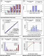
All quantitative data are expressed as measures of central tendency and dispersion. On the other hand, qualitative variables are expressed through absolute and refractive proportions. Data are presented using the six standardized figures for reporting refractive outcomes.7
When comparisons were made between preoperative and 12-month data, the following approach was taken: First, the assumption of normal distribution of data for the different variables was evaluated using the Shapiro–Wilk test. If both variables being evaluated had a normal distribution, a paired Student’s t-test was used for comparison. In cases where there was no normal distribution, a Wilcoxon test was performed. The significance level for achieving statistical significance was set at 0.05.
Bioethics
The protocol of this study was previously reviewed and approved by the ethics committee of the Clínica de Oftalmología Sandiego (Medellín, Colombia). Since it was a retrospective study based solely on the review of medical records, obtaining informed consent was not deemed necessary, patient data privacy and confidentiality was respected. The study adhered to the tenets of the Declaration of Helsinki and was approved by local institutional review boards.
Results
A total of 83 eyes from 47 patients were included in this study. Bilateral surgery was performed in 36 patients (76.6%), while the remaining 11 patients (23.4%) underwent surgery in only one eye. The majority of included patients were female (n = 33, 70.2%), with a mean age of 31.2 ± 5.1 years.
Of the total subjects, 18 (38.3%) had a history of receiving Crosslinking in at least one eye. Similarly, in 8 of the operated eyes (9.6%), there was a history of intra-stromal ring segment implantation, with half performed by the primary author of this study and the other half by previous surgeons. One patient (2.1%) had a history of Artiflex phakic intraocular lens implantation in the contralateral eye to the one undergoing surgery. According to the Amsler-Krumeich classification, 72 eyes (86.7%) were categorized as grade 1, while the rest (n = 11; 13.3%) were grade 2.
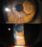
The baseline characteristics of the study population, along with visual and refractive outcomes, can be seen in Table 1 and Figures 1 and 2 depicts the slit-lamp examination photograph of one of the study patients with a history of intra-stromal ring segment implantation in the eye where the intraocular lens was implanted. A Scheimpflug-based image of the phakic lens in a patient with corneal ring can be found in Figure 3.
 |
Table 1 Table Summarizing the Preoperatory Results and Those Achieved 12 Months After Surgery, Along with the p value of the Wilcoxon Test |
All surgeries were straightforward and free of complications. None of the patients experienced elevated intraocular pressure episodes or spontaneous intraocular lens rotations. No complications were observed throughout the study period.
Discussion
Keratoconus is a relatively common corneal disease that can significantly impact not only visual quality but also the quality of life of affected individuals.8 Patients with this condition typically exhibit high levels of associated refractive errors, even after undergoing other surgical interventions, such as intrastromal ring segment implantation. On average, patients with keratoconus managed with glasses or contact lenses may have mean spherical equivalent levels ranging from −3.87 ± 1.61 D to −4.12 ± 1.19 D.9 In patients undergoing intrastromal ring segment implantation, the mean spherical equivalent may be around −7.39 ± 2.42 D.10
Refractive management in patients with keratoconus poses a therapeutic challenge in most situations. Various approaches may help reduce refractive burden to some extent, aiming to decrease the need for glasses or contact lenses. Corneal-based approaches, such as the “Athens Protocol” or the “Crete Protocol”, can help regularize the cornea and decrease refractive burden in well-selected subjects.11 An important study published by Kannellopoulos12 has demonstrated how, 10 years after topography-guided photorefractive keratectomy (PRK) followed by corneal cross-linking (“Athens Protocol”), most patients achieve complete corneal stability and significant visual improvement without correction. It may also be feasible to consider performing PRK or transepithelial PRK as a secondary procedure (after cross-linking) in selected patients.11
Approaches based on corneal laser surgery offer the benefit of better corneal regularization, primarily by reducing higher-order aberrations, a universal characteristic of corneal ectasias.13 However, a limitation of cornea-based approaches is that they require ablation of variable amounts of corneal tissue, producing a decrease in thickness of a primarily weakened tissue, corresponding precisely to the pathophysiology of the disease. Additionally, only a percentage of the patient’s refraction typically can be corrected to allow for greater preservation of corneal tissue. These patients are generally left with variable levels of refractive error.
Hence, some authors have suggested that phakic intraocular lens implantation may enable precise and safe correction of refractive error in some keratoconus patients. This could be particularly applicable in patients with ectasias with low levels of higher-order aberrations, either because their disease is mild, or because they have undergone previous corneal regularization procedures such as intrastromal ring segment implantation or topography-guided excimer laser.14 Therefore, phakic intraocular lens implantation might be especially recommended in patients younger than 40 years with relatively mild keratoconus, achieving relatively good vision with phoropter or glasses.
An additional benefit of intraocular lens implantation is that it allows for essentially complete correction of refractive error. This is important because, as known, corneal-based approaches do not always target complete correction due to the imperative need to conserve most of the corneal tissue.
The present study demonstrates the 12-month follow-up experience in a group of keratoconus patients who underwent EyeCryl Phakic Toric intraocular lens implantation by a high-volume expert surgeon in Colombia (K. B). This same research group had previously published their experience in such situations, with a shorter follow-up (six months) and a smaller number of cases.4
In the current study, visual and refractive outcomes were excellent. The preoperative spherical equivalent of −8.19 ± 4.04 D improved to −0.06 ± 0.48 D after 12 months of postoperative follow-up. This value is similar to that reported previously using the same type of intraocular lenses,4 where an improvement in spherical equivalent from −10.31 D to +0.09 D was found six months after surgical intervention. Previously, Antonios et al15 had found that, after implantation of ICL lenses in keratoconus patients, the spherical equivalent improved from −6.81 ± 3.68 D to −0.83 ± 0.76 D. The fact that our results are slightly closer to emmetropia can probably be explained by the fact that, in Antonios’ study, the steeper keratometry and maximum keratometry (50.49 ± 4.42 and 53.08 ± 5.17, respectively) were higher than those found in the present study, corresponding to patients with more advanced clinical conditions than those reported in the present study.
The improvement in visual quality with and without correction after implantation of this type of device is also significant. In the present study, 20% of patients achieved uncorrected visual acuity of 20/20 (LogMAR 0.00) or better. This is similar to the 40.6% of patients who achieved similar vision after surgery in Fairaq et al’s study.16 In a previous article,15 using ICL lenses, the uncorrected visual acuity achieved 12 months after surgery was 0.17 ± 0.06 LogMAR, similar to the 0.14 ± 0.11 achieved in the present study. Our group had previously reported uncorrected vision of 0.18 LogMAR at six months in a similar population.4
Regarding refraction, the present study demonstrated excellent refractive accuracy, with 77% of eyes having a spherical equivalent within ±0.50 D after twelve months of follow-up. This value is comparable to previous studies such as that of Kamiya et al,17 who found this level of refractive accuracy in 67% of their cohort.
In the studied population, no postoperative complications compromising visual quality occurred. There were no instances of ocular hypertension, glaucoma, unintended lens rotations, or other adverse events. This supports the concept of the safety of such interventions, as emphasized by previous articles.
This article has several limitations that should be acknowledged. First, the retrospective nature of the study may lead to incomplete data for some patients and lacks the higher level of standardization typically achieved in a prospective clinical study. Second, it is important to note that the biometric data for intraocular lens (IOL) calculation was obtained using different equipment (Pentacam AXL Wave) than what was used for toric alignment (Argos/Verion). Some may consider this discrepancy a potential limitation. However, the authors have been using this combination for several years without any issues. While Argos/Verion is primarily employed to assist with the precise placement of toric IOLs, all IOL power calculations were conducted using the Pentacam AXL Wave, which has proven to be reliable across a wide range of clinical scenarios. Another potential limitation is the presence of varied surgical backgrounds among the patients, with some having undergone crosslinking and others having intra-stromal ring implantation, for example. This diversity could introduce some confounding factors into the measurements. However, this variability might also be considered a strength, as it offers evidence on the effectiveness of these phakic lenses in well-selected patients, regardless of their prior surgical history.
Conclusions
The implantation of EyeCryl Phakic Toric intraocular lenses represents a safe, effective, and efficient option for correcting high refractive errors in patients with mild to moderate keratoconus. In the studied population, up to 98% of patients had a spherical equivalent of ±1.00 D. This suggests that, with proper patient selection, individuals with mild keratoconus can achieve excellent visual outcomes with this surgery, with 98% attaining uncorrected functional vision (20/40 or better).
Acknowledgments
The authors would like to express their gratitude for the technical and scientific assistance provided by the staff of Biotech Healthcare Holding and its regional affiliates. Special thanks go to Jennifer Tabares, Zulma Hernández, José Posada, and Luz María Espinosa (Biotech Colombia), as well as Carlos Ríos (Biotech Iberia) for their invaluable support.
We also extend our thanks to Leonardo Orjuela, OD, for capturing the clinical photographs included in this article.
This article is dedicated to the memory of Don Horacio.
Author Contributions
All authors made a significant contribution to the work reported, whether that is in the conception, study design, execution, acquisition of data, analysis and interpretation, or in all these areas; took part in drafting, revising or critically reviewing the article; gave final approval of the version to be published; have agreed on the journal to which the article has been submitted; and agree to be accountable for all aspects of the work.
Disclosure
The lead author (K.B.) is a paid consultant and speaker for Biotech Healthcare and participates in educational and training programs for other surgeons on the use of the brand’s products. However, Biotech Healthcare had no role, influence, or involvement in the design, data collection, statistical analysis, or writing of this article. They also had no role in the decision to conduct or publish the study and did not have access to any individual patient databases. The author received no compensation for conducting this study.
The other co-authors have no conflicts of interest to declare.
References
1. Balparda K, Herrera-Chalarca T, Cano-Bustamante M. Standardizing the measurement and classification of quality of life using the keratoconus end-points assessment questionnaire (KEPAQ): the ABCDEF keratoconus classification. Eye Vis. 2022;9(1):17. doi:10.1186/s40662-022-00288-0
2. Sengor T, Aydin Kurna S. Update on contact lens treatment of keratoconus. Turk J Ophthalmol. 2020;50(4):234–244. doi:10.4274/tjo.galenos.2020.70481
3. Fard AM, Patel SP, Nader ND. The efficacy of 2 different phakic intraocular lens implant in keratoconus as an isolated procedure or combined with collagen crosslinking and intra-stromal corneal ring segments: a systematic review and meta-analysis. Int Ophthalmol. 2023;43(11):4383–4393. doi:10.1007/s10792-023-02813-z
4. Balparda K, Vanegas-Ramirez CM, Herrera-Chalarca T, Silva-Quintero LA. Early results with the eyecryl phakic toric intraocular lens implantation in keratoconus patients. Rom J Ophthalmol. 2021;65(2):163–170. doi:10.22336/rjo.2021.32
5. Balparda K, Silva-Quintero LA, Herrera-Chalarca T. Unilateral toxic anterior segment syndrome after immediate sequential bilateral phakic intraocular lens implantation. JCRS Online Case Rep. 2022;10(2):e00072. doi:10.1097/j.jcro.0000000000000072
6. Balparda K, Vanegas-Ramirez CM, Marquez-Trochez J, Herrera-Chalarca T. Unilateral toxic anterior segment syndrome resulting in cataract and urrets-zavalia syndrome after sequential uneventful implantation of a posterior chamber phakic toric intraocular lens at two different surgical facilities: a series of unfortunate events. Case Rep Ophthalmol Med. 2020;2020:1216578. doi:10.1155/2020/1216578
7. Reinstein DZ, Archer TJ, Randleman JB. JRS standard for reporting astigmatism outcomes of refractive surgery. J Refract Surg. 2014;30(10):654–659. doi:10.3928/1081597X-20140903-01
8. Balparda K, Herrera-Chalarca T, Silva-Quintero LA, Torres-Soto SA, Vanegas-Ramirez CM. Development and validation of the ”keratoconus end-points assessment questionnaire” (KEPAQ), a disease-specific instrument for evaluating subjective emotional distress and visual function through rasch analysis. Clin Ophthalmol. 2020;14:1287–1296. doi:10.2147/OPTH.S254370
9. Barba-Gallardo LF, Jaramillo-Trejos LM, Agudelo-Guevara AM, Galicia-Duran AP, Casillas-Casillas E. Binocular vision parameters and visual performance in bilateral keratoconus corrected with spectacles versus rigid gas-permeable contact lenses. J Optom. 2024;17(3):100514. doi:10.1016/j.optom.2024.100514
10. Ramirez M, Manzanillo C, Estrada A, et al. Early keratometric and refractive results after intra-corneal rings surgery in keratoconus patients. Cir Cir. 2022;90(S1):92–95. doi:10.24875/CIRU.22000018
11. Dupps WJ Jr. Corneal refractive surgery in keratoconus. J Cataract Refract Surg. 2020;46(4):495–496. doi:10.1097/j.jcrs.0000000000000174
12. Kanellopoulos AJ. Ten-year outcomes of progressive keratoconus management with the Athens protocol (Topography-guided partial-refraction PRK combined with CXL). J Refract Surg. 2019;35(8):478–483. doi:10.3928/1081597X-20190627-01
13. Motwani M. A novel procedure for keratoconus/corneal ectasia treating epithelial compensation of higher-order aberrations, topographic guided ablation, and corneal cross linking - the CREATE+CXL protocol. Clin Ophthalmol. 2023;17:1981–1992. doi:10.2147/OPTH.S411472
14. Nowrouzi A, D’Oria F, Del Barrio JL A, Alio JL. Phakic intraocular Lens implantation in keratoconus patients. Eur J Ophthalmol. 2023;11206721231199780.
15. Antonios R, Dirani A, Fadlallah A, et al. Safety and visual outcome of visian toric icl implantation after corneal collagen cross-linking in keratoconus: up to 2 years of follow-up. J Ophthalmol. 2015;2015:514834. doi:10.1155/2015/514834
16. Fairaq R, Almutlak M, Almazyad E, Badawi AH, Ahad MA. Outcomes and complications of implantable collamer lens for mild to advance keratoconus. Int Ophthalmol. 2021;41(7):2609–2618. doi:10.1007/s10792-021-01820-2
17. Kamiya K, Shimizu K, Kobashi H, et al. Three-year follow-up of posterior chamber toric phakic intraocular lens implantation for the correction of high myopic astigmatism in eyes with keratoconus. Br J Ophthalmol. 2015;99(2):177–183. doi:10.1136/bjophthalmol-2014-305612
 © 2024 The Author(s). This work is published and licensed by Dove Medical Press Limited. The
full terms of this license are available at https://www.dovepress.com/terms.php
and incorporate the Creative Commons Attribution
- Non Commercial (unported, 3.0) License.
By accessing the work you hereby accept the Terms. Non-commercial uses of the work are permitted
without any further permission from Dove Medical Press Limited, provided the work is properly
attributed. For permission for commercial use of this work, please see paragraphs 4.2 and 5 of our Terms.


