Back to Journals » Clinical Ophthalmology » Volume 18
Outcomes After Switching to Faricimab for Refractive Macular Edema in Treatment-Experienced Eyes with Neovascular Age-Related Macular Degeneration
Authors Qaseem Y, Hou KK, Pettenkofer MS
Received 20 June 2024
Accepted for publication 20 September 2024
Published 29 October 2024 Volume 2024:18 Pages 3097—3102
DOI https://doi.org/10.2147/OPTH.S483563
Checked for plagiarism Yes
Review by Single anonymous peer review
Peer reviewer comments 2
Editor who approved publication: Dr Scott Fraser
Yaqoob Qaseem,1 Kirk Kohwa Hou,1,2 Moritz S Pettenkofer1
1Retina Division, Stein Eye Institute, University of California Los Angeles, Los Angeles, CA, USA; 2Doheny Eye Institute, Los Angeles, CA, USA
Correspondence: Moritz S Pettenkofer, Retina Division, Stein Eye Institute, University of California Los Angeles, 200 Stein Plaza Driveway, Los Angeles, CA, 90095, USA, Email [email protected]
Purpose: To examine response to faricimab in neovascular age-related macular degeneration (nARMD) refractory to traditional anti-vascular endothelial growth factor (anti-VEGF) agents.
Patients and methods: Retrospective chart review was conducted on eyes with nARMD with persistent subretinal and/or intraretinal fluid despite previously receiving ≥ 15 injections with ≥ 2 different anti-VEGF agents. Best corrected visual acuity (BCVA) and optical coherence tomography (OCT) parameters were collected at baseline, initial post-injection visit, and most recent visit with OCT following last faricimab.
Results: Nineteen eyes were included. Average logMAR BCVA was 0.47 ± 0.60 at baseline, 0.42 ± 0.47 at initial follow-up (p=0.38), and 0.51 ± 0.63 at final visit (p = 0.50). Average central subfield thickness (CST) was 310 ± 92 μm at baseline, 279 ± 88 μm at initial follow-up (p = 0.001), and 274 ± 100 μm at last visit (p < 0.001). 9 eyes (47%) achieved resolution of fluid at both initial and final follow-up visits.
Conclusion: Faricimab mildly decreased CST and reduced fluid in some nARMD eyes refractory to traditional anti-VEGF agents but had minimal effect on BCVA.
Keywords: anti-VEGF, choroidal neovascularization, faricimab, neovascular age-related macular degeneration
Introduction
Age-related macular degeneration (ARMD) is the leading cause of severe vision loss in the developed world for people over 55 years old.1 Anti-vascular endothelial growth factor (anti-VEGF) therapies for macular neovascularization have been pivotal in decreasing the number of individuals visually impaired by neovascular age-related macular degeneration (nARMD).1 Faricimab is a novel antibody for the treatment of nARMD that aims to neutralize not only VEGF-A but also Angiopoietin-2 (Ang-2), thus targeting two distinct pathways involved in nARMD pathogenesis.2
Faricimab was initially shown to be non-inferior to aflibercept in terms of change in best corrected visual acuity (BCVA) at one year for treatment-naïve individuals. Many patients were also able to achieve long dosing intervals of 12 or 16 weeks on faricimab.3
More recently, several real-world studies have examined the role of faricimab in treating eyes with a history of prior injections. These studies generally found that faricimab resulted in improvements in both BCVA and central subfield thickness (CST) in previously treated eyes.4–6 One of these studies specifically focused on patients with persistent subretinal and/or intraretinal fluid despite prior use of aflibercept, while the other studies did not specify baseline fluid as an inclusion criterion.
This study aimed to collect functional and optical coherence tomography (OCT)-based morphological observations after faricimab treatment in a particular subset of eyes that showed persistent subretinal and/or intraretinal fluid despite previous treatment with at least two other anti-VEGF agents. We hypothesized that eyes refractory to treatment with multiple prior anti-VEGF agents may still be able to respond anatomically and/or functionally to faricimab injections based on the novel mechanistic nature of the antibody.
Methods
A retrospective chart review was conducted on patients who received an initial intravitreal injection of faricimab between October and December 2022 within the retina division of a large academic referral center. The inclusion criteria were a diagnosis of nARMD, a history of receiving at least 15 total anti-VEGF injections with at least two different agents prior to faricimab, and persistent subretinal and/or intraretinal fluid on baseline OCT despite prior treatments. Only one eye was included for each patient. This study was approved by the University of California, Los Angeles Institutional Review Board (IRB#11-001350-AM-00012). Informed consent for the study was waived by the University of California, Los Angeles Institutional Review Board due to the study posing no more than minimal risk to patients and the lack of adverse effect on participants’ rights and welfare from waiving consent. Patient data confidentiality was strictly maintained, and the study adhered to the tenets of the Declaration of Helsinki.
Basic demographic information and intravitreal injection history were gathered for each patient. Best corrected visual acuity (BCVA) and optical coherence tomography (OCT) macular imaging parameters were collected at each patient’s baseline visit, initial post-injection visit, and most recent clinical visit with OCT following last faricimab injection as of October 2023. OCT parameters analyzed included retinal atrophy, the presence of subretinal and intraretinal fluid, and the central subfield thickness (CST). Retinal atrophy was assessed by assigning baseline OCT to one of the following categories: complete retinal pigment epithelium (RPE) and outer retinal atrophy (cRORA); incomplete RPE and outer retinal atrophy (iRORA); complete outer retinal atrophy; incomplete outer retinal atrophy; no atrophy.7 The dosing interval between the two faricimab injections prior to the final OCT was also noted. Snellen visual acuities were converted to equivalent logMAR acuities. Wilcoxon signed-rank test was used to compare BCVA and central subfield thickness (CST) at each follow-up to baseline values.
Results
39 eyes received an initial intravitreal injection of faricimab for a diagnosis of nARMD between October and December 2022. 19 of these 39 eyes met the study’s specific inclusion criteria and were analyzed. Of the 19 eyes analyzed, one eye had insufficient quality of OCT imaging at the first follow-up visit and was thus excluded from only the imaging analysis.
The mean age of the patients was 82 ± 8 years. 9 patients (47%) were female, and 10 patients (53%) were male. Four patients (21%) were on ranibizumab treatment prior to faricimab, while 15 patients (79%) were on aflibercept. 17 patients (89%) had an injection interval of 4 weeks prior to switching to faricimab and 2 patients (11%) had an injection interval of 10 weeks. One of these two patients was noted to have recurrent subretinal and intraretinal fluid at an 11-week interval with aflibercept, with a prior history of recurrent fluid at 10 weeks. This patient was injected with aflibercept with the treatment interval shortened to 10 weeks. Due to persistent fluid 10 weeks after aflibercept, the patient was switched to faricimab. The other patient with a baseline interval of 10 weeks had been previously lost to follow-up for 20 weeks with stable OCT and vision and was thus on a less frequent treat-and-extend injection schedule per patient preference (despite some persistent fluid).
Fourteen patients (74%) had received 2 types of anti-VEGF agents before faricimab, while 5 patients (26%) had received 3 types of anti-VEGF agents before faricimab. Average total number of anti-VEGF injections before first faricimab injection was 51 ± 31 (range: 17 to 119). Average duration of prior anti-VEGF therapy was 59 ± 30 months (range: 16 to 104).
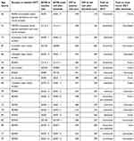
Seventeen eyes (89%) had atrophy on baseline OCT, while 2 eyes (11%) had no atrophy. 1 eye (5%) had cRORA, 12 eyes (63%) had iRORA, 3 eyes (16%) had complete outer retinal atrophy, and 1 eye (5%) had incomplete outer retinal atrophy. Table 1 shows baseline atrophy information for each patient alongside BCVA and CST outcomes.
The mean time elapsed between initial faricimab injection and subsequent BCVA testing was 6.1 ± 3.4 weeks. 1 eye received 3 injections prior to this testing, while the other 18 eyes had received a single faricimab injection.
The mean time elapsed between initial injection and final visual acuity and OCT analysis was 40.1 ± 10.7 weeks. 2 eyes were switched from faricimab to another anti-VEGF agent prior to the most recent OCT, one due to fluid recurrence on faricimab and another due to patient perception that new diagnosis of hypertension and lower extremity edema was related to faricimab. For these patients, the follow-up visit after final faricimab injection was analyzed. Eyes received an average of 7.4 ± 3.2 faricimab injections prior to final analysis, with the two faricimab injections prior to last follow-up separated by an average of 6.2 ± 3.2 weeks (range: 4 to 14). 9 eyes (47%) had a final injection interval of >4 weeks.
Average logMAR BCVA was 0.47±0.60 ≈ Snellen 20/59 at baseline, 0.42 ± 0.47 ≈ Snellen 20/53 at initial follow-up (p = 0.38), and 0.51±0.63 ≈ Snellen 20/64 at final follow-up (p = 0.50) (Figure 1). At initial follow-up, 5 eyes (26%) had an improvement in BCVA, 6 eyes (32%) had no change, and 8 eyes (42%) had a decrease. At final follow-up, 6 eyes (32%) had an improvement in BCVA from baseline, 9 eyes (47%) had no change, and 4 eyes (21%) had a decrease.
 |
Figure 1 Graph depicting the average logMAR visual acuity and average central subfield thickness (CST) at various time points following faricimab injections. |
Average CST was 310 ± 92 μm at baseline, 279±88 μm at initial follow-up (p = 0.001), and 274 ± 100 μm at last follow-up (p < 0.001) (Figure 1). 8 eyes (42%) had intraretinal fluid at baseline, 9 eyes (47%) had subretinal fluid, and 2 eyes (11%) had both. At initial follow-up, 6 eyes (32%) had intraretinal fluid, 3 eyes (16%) had subretinal fluid, and 9 eyes (47%) had neither. At final follow-up, 5 eyes (26%) had intraretinal fluid, 5 eyes (26%) had subretinal fluid, and 9 eyes (47%) had neither.
Discussion
While many eyes with nARMD respond favorably to initial anti-VEGF therapies, there is a significant subset of eyes that show persistent signs of disease activity with non-resolving macular edema despite several routine anti-VEGF treatments and thus may have worse visual outcomes. For this subset of patients, switching anti-VEGF agents is often a consideration.8,9 We hypothesized that faricimab may benefit eyes with previous incomplete response or non-response to two or more traditional anti-VEGF agents due to the novel mechanistic nature of the antibody.
The results of the present study suggest that faricimab may result in anatomic improvement in this subset of patients without evidence of an associated functional benefit in terms of improvement in BCVA. CST, which started at an average of 310 ± 92 μm, decreased to 279 ± 88 μm by the initial follow-up visit, with minimal subsequent decrease to a final average of 274 ± 100 μm at last follow-up. Additionally, 9 eyes (47%) showed resolution of fluid at both initial follow-up and final follow-up visits. One example of a patient who responded favorably to faricimab is presented in Figure 2. However, average BCVA did not show any significant change from baseline at both early and late follow-up visits.
There are a variety of factors that could account for why subjects in this study had minimal change in BCVA despite anatomic improvement on OCT. The large variation in BCVA and small sample size significantly limit detection of changes in BCVA. Additionally, baseline outer retinal atrophy in the study population likely limited improvement in vision, with 17 out of 19 patients having some level of atrophy on baseline OCT as shown in Table 1. Furthermore, the relationship between long-term presence of intraretinal and subretinal fluid in nARMD and vision is uncertain. The presence of intraretinal fluid has a stronger correlation with vision, but the correlation between subretinal fluid and vision is less clear.10 The location of fluid at baseline and on last analyzed OCT is also shown for each patient in Table 1.
While this study suggests that faricimab has the potential to anatomically improve eyes with persistent macular edema despite the use of multiple prior anti-VEGF agents, it is uncertain whether this is a result of the newly addressed Ang-2 pathway or simply because faricimab offers a novel anti-VEGF molecule. Additionally, though functional improvement was not demonstrated during our time of observation, anatomic improvement could still translate to longer treatment intervals and thus improved quality of life for patients with previously refractory macular edema. While only 2 patients out of 19 had an injection interval of >4 weeks prior to faricimab, 9 patients had a final injection interval of >4 weeks. The reduction in exudative fluid may also result in improved preservation of vision over time, which could potentially be demonstrated with longer follow-up.
Other limitations to the present study besides the low sample size were the retrospective nature of the study and the lack of a control group. Additionally, it is possible that some patients noted to have persistent exudation would have shown resolution of fluid if assessed at shorter post-injection intervals; this could imply that such patients may warrant a nontraditional treatment schedule. Nonetheless, we report meaningful outcomes in a homogenous and unique group of treatment-refractory patients.
More research is needed to determine the efficacy of switching intravitreal therapies in patients with prior nonresponse or incomplete response to initial treatments. Future research could explore the comparative efficacy of switching to various agents in patients with fluid despite varying degrees of prior treatment and assess OCT biomarkers for prediction of treatment response. With larger sample sizes, statistical analysis in future studies may also be able to suggest which of these patients may functionally benefit from switching to faricimab.
Conclusions
In a subset of eyes with nARMD that showed persistent intraretinal and/or subretinal fluid refractory to treatment with traditional anti-VEGF agents, faricimab resulted in anatomic improvement in terms of mildly decreased CST and resolution of fluid in several eyes. However, there was no significant associated change in BCVA.
Funding
There is no funding to report.
Disclosure
The authors report no conflicts of interest in this work.
References
1. Fleckenstein M, Keenan TDL, Guymer RH, et al. Age-related macular degeneration. Nature Revi Disea. 2021;7(1):1–25. doi:10.1038/s41572-021-00265-2
2. Panos GD, Lakshmanan A, Dadoukis P, Ripa M, Motta L, Amoaku W. Faricimab: transforming the Future of Macular Diseases Treatment - A Comprehensive Review of Clinical Studies. Drug Des Devel Ther. 2023;17:2861–2873. doi:10.2147/DDDT.S427416
3. Heier JS, Khanani AM, Quezada Ruiz C, et al. Efficacy, durability, and safety of intravitreal faricimab up to every 16 weeks for neovascular age-related macular degeneration (TENAYA and LUCERNE): two randomised, double-masked, Phase 3, non-inferiority trials. Lancet. 2022;399(10326):729–740. doi:10.1016/S0140-6736(22)00010-1
4. Leung EH, Oh DJ, Alderson SE, et al. Initial Real-World Experience with Faricimab in Treatment-Resistant Neovascular Age-Related Macular Degeneration. Clin Ophthalmol. 2023;17:1287. doi:10.2147/OPTH.S409822
5. Rush RB, Rush SW. Intravitreal Faricimab for Aflibercept-Resistant Neovascular Age-Related Macular Degeneration. Clin Ophthalmol. 2022;16:4041. doi:10.2147/OPTH.S395279
6. Khanani AM, Aziz AA, Khan H, et al. The real-world efficacy and safety of faricimab in neovascular age-related macular degeneration: the TRUCKEE study – 6 month results. Eye. 2023:1–8. doi:10.1038/s41433-023-02553-5
7. Sadda SR, Guymer R, Holz FG, et al. Consensus Definition for Atrophy Associated with Age-Related Macular Degeneration on OCT: classification of Atrophy Report 3. Ophthalmology. 2018;125(4):537–548. doi:10.1016/j.ophtha.2017.09.028
8. Amoaku WM, Chakravarthy U, Gale R, et al. Defining response to anti-VEGF therapies in neovascular AMD. Eye. 2015;29(6):721. doi:10.1038/EYE.2015.48
9. Broadhead GK, Hong T, Chang AA. Treating the untreatable patient: current options for the management of treatment-resistant neovascular age-related macular degeneration. Acta Ophthalmol. 2014;92(8):713–723. doi:10.1111/AOS.12463
10. Kaiser PK, Wykoff CC, Singh RP, et al. Retinal Fluid And Thickness As Measures Of Disease Activity In Neovascular Age-Related Macular Degeneration. Retina. 2021;41(8):1579–1586. doi:10.1097/IAE.0000000000003194
 © 2024 The Author(s). This work is published and licensed by Dove Medical Press Limited. The
full terms of this license are available at https://www.dovepress.com/terms.php
and incorporate the Creative Commons Attribution
- Non Commercial (unported, 3.0) License.
By accessing the work you hereby accept the Terms. Non-commercial uses of the work are permitted
without any further permission from Dove Medical Press Limited, provided the work is properly
attributed. For permission for commercial use of this work, please see paragraphs 4.2 and 5 of our Terms.


