Back to Journals » International Medical Case Reports Journal » Volume 18
Ovarian Yolk Sac Tumor in a Young Girl in a Resource-Limited Setting: A Rare Case Report From Somalia
Authors Hussein SA, Hussein AI, Omar AA, Hirsi MO , Mohamed NA, Dalmar AA, Hirei HH, Iyow SN
Received 27 January 2025
Accepted for publication 16 July 2025
Published 17 July 2025 Volume 2025:18 Pages 887—891
DOI https://doi.org/10.2147/IMCRJ.S518785
Checked for plagiarism Yes
Review by Single anonymous peer review
Peer reviewer comments 2
Editor who approved publication: Dr Tanvi Dhere
Safio Ahmed Hussein,1 Ahmed Issak Hussein,1 Abdikarim Ali Omar,1 Mohamed Omar Hirsi,1 Najma Adan Mohamed,1 Abdirashid Abdullahi Dalmar,2 Hanan Hassan Hirei,3 Sowdo Nur Iyow4
1Obstetrics and Gynecology Department, Mogadishu Somalia Turkish Training and Research Hospital, Mogadishu, Somalia; 2General Surgery department, Mogadishu Somalia Turkish Training and Research Hospital, Mogadishu, Somalia; 3Radiology Department, Mogadishu Somalia Turkish Training and Research Hospital, Mogadishu, Somalia; 4Emergency department, Mogadishu Somalia Turkish Training and Research Hospital, Mogadishu, Somalia
Correspondence: Ahmed Issak Hussein, Obstetrics and Gynecology Department, Mogadishu Somalia Turkish Training and Research Hospital, Mogadishu, Somalia, Tel +252615597479, Email [email protected]
Abstract: Ovarian yolk sac tumors, accounting for less than 1% of all malignant ovarian germ cell tumors, primarily affect adolescents and young women. These tumors are typically unilateral, making fertility preservation a critical consideration. Despite their rarity, they generally respond well to chemotherapy, leading to a favorable prognosis. This report describes a 21-year-old woman presenting with abdominal pain and distension. Imaging revealed a substantial pelvic mass, with alpha-fetoprotein (AFP) levels elevated to 2258 mg/mL. She underwent a left salpingo-oophorectomy, and histopathology confirmed the presence of an ovarian yolk sac tumor. Timely diagnosis and treatment play a crucial role in patient outcomes. This case underscores the diagnostic and therapeutic challenges in a resource-constrained setting with limited access to chemotherapy necessitating transfer of most patients to abroad for oncology centers, making difficult for follow up and surveillance of patients, highlighting the pivotal role of surgical management and ongoing monitoring.
Keywords: Yolk sac tumor, malignant, ovary, adolescents, case report
Introduction
Ovarian yolk sac tumors (YSTs), also called endodermal sinus tumors, are rare malignant germ cell tumors that account for less than 1% of ovarian cancers.1 They are the second most common germ cell malignancy after dysgerminomas, characterized by rapid unilateral growth. These tumors mainly affect young women in their teens and twenties.2
Ovarian yolk sac tumors are curable requiring management and follow-up by a specialist multidisciplinary team. Unlike epithelial ovarian cancer where most patients will present with advanced disease, more than half of malignant ovarian germ cell tumors patients (60–70%) will present with FIGO stage I disease, which has an excellent prognosis.3 YSTs are marked by elevated serum AFP levels and often present with symptoms such as abdominal pain or swelling. Due to their sensitivity to chemotherapy, these tumors have a favorable prognosis, and fertility-preserving surgeries have become standard practice.4 Survival rates following fertility-preserving procedures are impressive, with 5-year overall survival reaching 84% and event-free survival at 79%.5 This report describes the diagnosis and treatment of a 21-year-old patient with an ovarian yolk sac tumor in a setting with limited medical resources.
Case Report
A 21-year-old female presented with two months of persistent dull abdominal pain, accompanied by distension and irregular menstrual cycles. On physical examination, the abdomen was distended and tender, with a palpable mass in the abdomen. Her vital signs were stable Abdominal ultrasound revealed a huge adnexal mass about 16 cm. MRI showed a large, solid-cystic adnexal mass with heterogeneity and fluid-filled regions, consistent with a neoplastic etiology.
Laboratory results showed significantly elevated AFP at 2258 ng/mL (normal: 0–20 ng/mL in non-pregnant women), elevated lactate dehydrogenase (523 U/L; normal: 125–220 U/L), and increased CA-125 levels (144 U/mL; normal: <35 U/mL). Advanced diagnostic tools, such as immunohistochemistry, were unavailable, so diagnosis relied on clinical features, laboratory findings, imaging and histopathology.
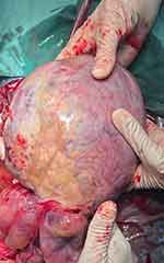
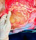
An exploratory laparotomy revealed a large irregular mass arising from the left ovary with adhesions to the omentum and appendix as shown in Figures 1 and 2. The patient underwent left salpingo-oophorectomy, omentectomy, and appendectomy. The uterus, right ovary, and right fallopian tube were normal and preserved for future fertility. Histopathological examination confirmed a yolk sac tumor. Grossly, left ovary integrity capsule ruptured, the tumor appeared as a solid yellow-white mass with areas of necrosis and hemorrhage, measuring 16×8×12 cm. Figure 3 The fallopian tube measured 5×1 cm with gross abnormalities. Microscopically, the tumor showed microcystic and endodermal sinus patterns with Schiller–Duval bodies and tumor cells containing hyaline globules with microscopic extrapelvic peritoneal involvement without positive retroperitoneal lymph nodes; pT3a (IIIA). Figure 4. Histologic Grade; G2, moderately differentiated. The omentum and the appendix was unaffected.
 |
Figure 1 Shows an intraoperative image of the left adnexal mass. |
 |
Figure 2 Illustrates the omentum adherent to the adnexal mass. |
 |
Figure 3 Displays the surgical specimen including the excised left adnexal mass. |
 |
Figure 4 Cross-section of the ovarian mass revealed a heterogeneous cut surface with solid and focal cystic areas, along with hemorrhage and necrosis. |
The patient recovered uneventfully postoperatively. Follow-up included regular AFP monitoring and clinical evaluations. AFP level decreased to 548 ng/mL. Due to the lack of chemotherapy availability, arrangements were made for the patient’s transfer abroad for further treatment. However, further follow-up could not be conducted.
Discussion
Ovarian germ cell tumors comprise 15–20% of ovarian tumors and originate from primordial germ cells. These malignancies are often detected early due to the presence of specific tumor markers, aiding diagnosis before histological confirmation.5 This allows for fertility-preserving procedures instead of radical treatments.6
Malignant ovarian germ cell tumors represent 3–5% of all ovarian cancers, with yolk sac tumors (YSTs) being rare but highly aggressive. YSTs account for approximately 1% of ovarian malignancies and are the second most common subtype of malignant ovarian germ cell tumors after dysgerminomas.7,8 YSTs primarily affect adolescents and young women, typically aged 18–30.5 Common symptoms include abdominal pain, a pelvic mass, and abdominal enlargement. Elevated AFP levels are critical for diagnosis and post-treatment monitoring, with normalization indicating no residual disease.7 Increased CA-125 levels are also occasionally observed in YSTs.2 Histopathological diagnosis is based on the presence of structures resembling early embryonic development, such as Schiller–Duval bodies.9 Standard treatment involves fertility-preserving surgery and combination chemotherapy with BEP (bleomycin, etoposide, and cisplatin).5 Regarding our case study which was in Histologic Grade 2, surgery alone was not sufficient and studies suggest that conservative surgery combined with adjuvant chemotherapy yields high cure rates, even in advanced disease stages.10,11 This case underscores the importance of considering YSTs in young women with abdominal pain and elevated tumor markers, especially in resource-limited settings. Challenges include limited chemotherapy access and the reliance on clinical suspicion and basic diagnostic tools.
Conclusion
This case highlights the importance of early diagnosis and multidisciplinary management of ovarian yolk sac tumors. In resource-limited settings, prompt surgical intervention and rigorous follow-up are vital for improving outcomes. Expanding access to chemotherapy and advanced diagnostic.
Abbreviations
AFP, Alpha Fetoprotein; MRI, Magnetic Resonance Imaging; YST, Yolk Sac Tumor.
Ethical Approval
According to the guidelines of the Ethics Committee of Somali Turkey Recep Tayyip Erdogan Hospital institutional approval was not required to publish the case reports.
Informed Consent
The patient provided written informed consent for publication of the details of her case and the images shown in the figure.
Author Contributions
All authors made a significant contribution to the work reported, whether that is in the conception, Case design, execution, acquisition of data, analysis and interpretation, or in all these areas; took part in drafting, revising or critically reviewing the case; gave final approval of the version to be published; have agreed on the journal to which the article has been submitted; and agree to be accountable for all aspects of the work.
Funding
The authors declare that this study has not received any funding resources.
Disclosure
The authors report no conflicts of interest in this study.
References
1. WHO Classification of Tumors Editorial Board. Female genital tumors. 5th ed. vol. 4. Lyon, France. WHO classification of tumors series. International Agency for Research on Cancer. 2020. Available from: https://publiations.iarc.fr/592.
2. Dallenbach P, Bonnefor H, Pelte MF, Vlastos G. Yolk sac tumours of the ovary: an update. Eur J SurgOncol. 2006;32(10):1063–1075. doi:10.1016/j.ejso.2006.07.010
3. Patterson DM, Murugaesu N, Holden L, Seckl MJ, Rustin GJ. A review of the close surveillance policy for stage I female germ cell tumors of the ovary and other sites. Int J Gynecol Cancer. 2008;18(1):43–50. doi:10.1136/ijgc-00009577-200801000-00007
4. Smith HO, Berwick M, Verschraegen CF, et al. Incidence and survival rates for female malignant germ cell tumors. ObstetGynecol. 2006;107(5):1075–1085.
5. Nawa A, Obata N, Kikkawa F, et al. Prognostic factors of patients with yolk sac tumors of the ovary. Am J Obstet Gynecol. 2001;184(6):
6. Kojimahara T, Nakahar K, Takano T, Yaegashi N, Nishiyama H, Fujimori K. Yolk sac tumor of the ovary: a retrospective multicenter study of 33 Japanese women by Tohoku Gynaecologic Cancer Unit (TGCU. Tohoku J Exp Med. 2013;210:211–217. doi:10.1620/tjem.230.211
7. Eddaoualline H, Sami H, Rais H, et al. Ovarian yolk sac tumor: a case report and literature review. Clin Case Rep Int. 2018;2:1057.
8. Guida M, Pignata S, Palumbo AR, Miele G, Marra ML, Visconti F. Laparoscopic treatment of a yolk sac tumour: case report and literature review. Translational Med. 2013;7(1):1–5.
9. Khoo TK, Donald PC. Embryonal tumour of the ovary with the endodermal sinus pattern. Singapore Med J. 1966;7:1.
10. Tewari K, Cappuccini F, Disaia P, Berman ML, Manetta A, Kohler MF. Malignant germ cell tumors of the ovary. Obstet Gynecol. 2000;95:128. doi:10.1016/s0029-7844(99)00470-6
11. De La Motte Rouge T, Pautier P, Duvillard P, et al. Survival and reproductive function of 52 women treated with surgery and bleomycin, etoposide, cisplatin (BEP) chemotherapy for ovarian yolk sac tumor. Ann Oncol. 2008;19(8):1435–1441. doi:10.1093/annonc/mdn162
 © 2025 The Author(s). This work is published and licensed by Dove Medical Press Limited. The
full terms of this license are available at https://www.dovepress.com/terms.php
and incorporate the Creative Commons Attribution
- Non Commercial (unported, 4.0) License.
By accessing the work you hereby accept the Terms. Non-commercial uses of the work are permitted
without any further permission from Dove Medical Press Limited, provided the work is properly
attributed. For permission for commercial use of this work, please see paragraphs 4.2 and 5 of our Terms.

