Back to Journals » Clinical Ophthalmology » Volume 19
Physiological Intraocular Pressure in Cataract Surgery: A Comparative Consecutive Case Series Study
Authors Sarossy A, Chakrabarti R
Received 18 May 2025
Accepted for publication 9 July 2025
Published 15 July 2025 Volume 2025:19 Pages 2289—2294
DOI https://doi.org/10.2147/OPTH.S532483
Checked for plagiarism Yes
Review by Single anonymous peer review
Peer reviewer comments 2
Editor who approved publication: Dr Scott Fraser
Alexander Sarossy,1,2 Rahul Chakrabarti1– 4
1Essendon Eye Clinic, Essendon, Victoria, Australia; 2Faculty of Medicine, Nursing, Health Sciences, Monash University, Clayton, Victoria, Australia; 3The Royal Victorian Eye and Ear Hospital, Melbourne, Victoria, Australia; 4Department of Ophthalmology, The University of Melbourne, Parkville, Victoria, Australia
Correspondence: Alexander Sarossy, Faculty of Medicine, Nursing, Health Sciences, Monash University, PO Box 960, Moonee Ponds, Victoria, 3039, Australia, Email [email protected]
Introduction
Traditionally, cataract surgery has been conducted at higher intraocular pressure (IOP) ranges of 50–90mmHg to maintain anterior chamber stability. This significantly deviates from the physiological range of 10–21mmHg.1 Higher IOP during routine cataract surgery has shown potential consequences including compromised blood flow to the optic nerve head leading to ischemic damage, increased mechanical stress on retinal ganglion cell axons, and greater risk of visual field defects in vulnerable patients. Additional documented complications include reverse pupillary block, higher stress on the posterior capsule, and greater incidence of anterior hyaloid detachment.2
Post-operative risks of higher IOP include vascular compromise to the optic nerve (non-arteritic anterior ischemic optic neuropathy) and retina (paracentral acute middle maculopathy, retinal artery and vein occlusion).2 There is also a greater risk of ocular inflammation from corneal endothelial decompensation with heightened corneal oedema and post-operative anterior uveitis. This is particularly relevant in patients with glaucoma, compromised endothelium, or high myopias, where elevated IOP may exacerbate existing pathologies.
Technological advancements in surgical equipment have challenged this conventional approach. The Active Sentry™ handpiece (Alcon, Fort Worth, TX, USA), provides real-time IOP sensing with a built-in sensor that constantly monitors pressure within the infusion port to sense occlusion breaks and mitigate surge; allowing dynamic adjustments to maintain a stable anterior chamber at lower infusion pressures. This technology theoretically enables surgeons to operate at more physiological pressure levels without compromising surgical safety or efficiency.3
Methods
We conducted a single-surgeon, retrospective, non-randomised, consecutive case series analysing 258 eyes (129 per group) undergoing routine cataract surgery at 2 private day surgery centres in Melbourne, Victoria, Australia, between January and August 2023. The study was approved by the institutional review board and adhered to the Declaration of Helsinki. Patients with NC2-NC4 age-related cataracts were included (LOCS III classification). We excluded patients with previous ocular surgery, uveitis, advanced glaucoma, pseudoexfoliation, or corneal pathology.
Preoperative assessment included best-corrected visual acuity (BCVA), slit-lamp examination, intraocular pressure measurement, dilated fundus exam, and optical biometry. Automated perimetry was performed for patients with glaucoma risk factors, establishing baseline visual field status.
The physiological IOP group utilised the Alcon Centurion Vision System with Active Sentry handpiece, maintaining pressures at 30mmHg. The high IOP group used as a standard Ozil handpiece with IOP maintained at 65mmHg. All surgeries were performed under topical anaesthesia with standard pupillary dilation.
A temporal approach used two side ports (1.2mm) and main wound (2.4mm). After performing a continuous curvilinear capsulorhexis (5.0–5.5mm), phacoemulsification used a divide and conquer technique. Nuclear disassembly used a standard divide-and-conquer technique in both groups. Bimanual irrigation-aspiration was employed for cortical cleanup, with appropriate fluidic parameters. A foldable acrylic monofocal IOL (Johnson and Johnson PureSee EDOF ™) was implanted in all cases, with standard wound hydration performed at procedure completion.
The Active Sentry system’s pressure sensor continuously monitors IOP and automatically adjusts fluid infusion by squeezing the infusion bag between pressure plates to maintain the target pressure range within milliseconds of detecting fluctuations. When pressure decreases below the threshold, the system rapidly increases fluid inflow to compensate, whereas minor elevations above target pressure result in automatic flow reduction. This dynamic response ensures that anterior chamber depth remains stable throughout the procedure despite the lower baseline pressure settings. As the pressure sensor also responds quickly to post-occlusion breaks by the use of a quick valve, there is an additional safety margin allowing the user to set a lower IOP.
Detailed fluidic parameters for each stage of surgery were recorded, as shown in the Supplementary Table 1. For the high IOP group, IOP was maintained at 60–75mmHg during phacoemulsification stages, with vacuum settings ranging from 110–475 mmHg and aspiration flow rates of 20–40 cc/min depending on the surgical phase. In contrast, the physiological IOP group operated at significantly lower pressures (30–45 mmHg) while maintaining comparable vacuum and slightly reduced aspiration flow rates to ensure chamber stability.
Intraoperative parameters encompassed total ultrasound time, cumulative dissipated energy (CDE), Active Sentry actuations (ASM), aspiration time, total estimated fluid aspirated, surgical complications, and subjective assessment of patient comfort using a standard 1–10 pain scale. The surgeon noted any instances of anterior chamber instability, including shallowing or deepening, iris prolapse or other complications.
Post-operative evaluation at day 1, week 1, and month 1 included visual acuity, corneal clarity assessment (graded as clear, mild oedema, moderate oedema or severe oedema) and anterior chamber inflammation (cell count per high-power field). Corneal clarity and postoperative anterior chamber inflammation were graded through slit-lamp assessment at day 1. Formal specular microscopy was not available in this study. Intraocular pressure was measured at each visit to assess for post-operative hypertension or hypotony using Goldmann applanation tonometry. Statistical analysis was performed using two-tailed t-tests for continuous variables and chi-square tests for categorical data, with p<0.05 considered statistically significant. Sample size calculation determined that 129 eyes per group would provide 90% power to detect a 15% difference in postoperative BCVA.
Results
Surgical time was similar between groups (average 9 mins, range 7.5–18 minutes). Results demonstrated significant advantages in the physiological IOP group (Table 1). First post-operative BCVA was significantly better in the physiological IOP group (0.684 vs 0.617 decimal, p<0.01), a trend that continued to final post-operative BCVA (0.913 vs 0.820 decimal, p<0.01). This represents a clinically meaningful improvement in visual rehabilitation that persisted throughout follow-up.
 |
Table 1 Comparison of Intra-Operative and Post-Operative Metrics Between High IOP and Physiological IOP Groups |
Corneal clarity was markedly better in the physiological IOP group, with 94.6% of patients exhibiting clear corneas compared to only 69.0% in the high IOP group (p<0.01). Remaining physiological IOP patients had more mild oedema resolving by week 1, while high IOP patients had more persistent oedema. This improved corneal status directly contributes to faster visual recovery and potentially fewer post-operative visits.
Patient-reported perioperative pain was significantly reduced in the physiological IOP group (1.67 vs 3.19 on a 10-point scale, p<0.01), representing an important improvement in the patient experience during surgery. Subjective feedback from patients suggested improved comfort in the physiological IOP group, though formal satisfaction scores were not collected.
The physiological IOP group exhibited significantly lower CDE (6.02 vs 9.84, p<0.01) and shorter ultrasound time (40.7 vs 44.0 seconds, p<0.01), indicating decreased energy delivery to ocular tissues. This reduction in energy exposure suggests a potential protective effect on corneal endothelium and other delicate intraocular structures. Total aspiration time was reduced (3.18 vs 4.19 mins, p<0.01), potentially contributing to more efficient surgical workflow. Estimated fluid aspirated was marginally higher in the physiological IOP group (46.0 vs 43.9 mL, p=0.05), though this difference is unlikely to be clinically significant.
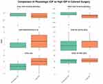
Post-operative uveitis remained comparable between groups (0.733 vs 0.857, p=0.16), indicating lower IOP does not compromise inflammatory control. This is reassuring given theoretical concerns about inflammation from chamber stability fluctuations. The Active Sentry system averaged 11.1 actuations per case, reflecting surge mitigations by the active sentry handpiece, demonstrating regular but not excessive intervention to maintain the target IOP. Figure 1 illustrates the comparative differences in key intraoperative and postoperative metrics between the high IOP (65mmHg) and physiological IOP (30mmHg) groups, highlighting the significant improvements observed across multiple clinical parameters.
 |
Figure 1 Paired boxplot of results of intra-operative and post-operative metrics between high IOP (55mmHg) and phys-IOP (26–30mmHg). |
Discussion
The improved visual outcomes observed in the physiological IOP group may be attributed to better preservation of retinal and optic nerve function. Higher IOP has been shown to compress retinal vessels, potentially causing transient ischemia and subclinical retinal insults that may affect visual recovery. The correlation between reduced CDE in the physiological IOP group and better visual outcomes suggests that combining lower energy levels with more physiological pressure creates an optimised environment for ocular tissues during surgery.
The marked improvement in corneal clarity in the physiological IOP group represents a clinically significant finding with direct impact on visual recovery. Endothelial cells are particularly vulnerable to mechanical stress and ultrasound energy during phacoemulsification. Higher IOP may amplify these detrimental effects through increased corneal strain and reduced endothelial perfusion. The combination of lower IOP and reduced energy (ultrasound) delivery likely contributed to enhanced corneal endothelial preservation and faster visual rehabilitation.
Lower IOPs minimize stress on the corneal endothelium, potentially reducing cell loss—crucial for long-term corneal clarity.4 This protective effect is valuable for patients with compromised endothelium or corneal decompensation risk. Additionally, in patients with pre-existing optic neuropathy (eg, glaucoma), lower IOPs during surgery may minimize further damage to the optic nerve head, potentially preserving visual field and function. The reduced pressure on the retina during the procedure also minimizes retinal stress, which may be especially beneficial for patients with pre-existing retinal pathology.
Reduced perioperative pain has important implications for patient experience. Elevated IOP can directly stimulate trigeminal nerve endings in the limbal area and induce strain on corneal sensory fibers, triggering ocular discomfort. Higher IOP may cause tissue ischemia and inflammatory mediator release, heightening the nociceptive response. This improvement in comfort represents an often-overlooked aspect of surgical quality that directly impacts patient satisfaction.
Specific patient populations may particularly benefit from physiological IOP during cataract surgery. Patients with weakened corneal endothelium (eg, Fuchs’ dystrophy) may experience reduced risk of decompensation. Patients with glaucoma benefit from minimized IOP fluctuations during surgery. Similarly, patients with high myopia, who often have deep and fragile eye structures, may experience reduced strain on the sclera and retina with lower surgical IOP. The consistent benefits observed across our study cohort suggest that physiological IOP may become the standard approach for all routine cataract cases, rather than being reserved for special populations.
From a surgeon’s perspective, performing routine cataract surgery at physiological IOP proved safe without requiring significant modification to standard surgical technique, though specific fluidic adjustments were required. The learning curve for transitioning to this approach was minimal, requiring approximately 15–20 cases for the surgeon to become fully comfortable with the technique. Most surgeons will likely feel comfortable operating at an infusion pressure of 30–36 mmHg with minimal adaptation. The primary adjustment involved trusting the Active Sentry system to maintain chamber stability rather than relying on artificially elevated IOP. Furthermore, there was ease of fragment removal through better followability, contributing to improved overall efficiency (and hence potentially a lower CDE/ultrasound) through comparable surgical times, despite minor differences in fluid volumes used.
For surgeons considering adopting physiological-IOP cataract surgery, we recommend a gradual transition approach. Starting with moderate pressure reduction (30–36mmHg), surgeons can adapt before progressing to physiological ranges (20–26mmHg) This bracketing approach allows for confidence building while maintaining surgical safety. With experience, surgeons typically find that anterior chamber stability can be maintained even at lower pressure settings than previously thought necessary, challenging long-held assumptions about required IOP during phacoemulsification.
Limitations
Our study has several limitations. As a single-surgeon, non-randomized, retrospective analysis, the results may not be fully generalizable across different surgical settings and case complexities. The lack of objective quantification of corneal endothelial cell density changes limits our ability to precisely measure endothelial protection. Additionally, we did not perform macular OCT to assess subclinical retinal changes or evaluate surgically induced astigmatism. Despite these limitations, the consistent trends observed across multiple outcome measures provide strong physiological evidence supporting the benefits of lower IOP during cataract surgery. The findings of this study (i.e the lower CDE/ultrasound) may be explained by factors that are not captured in an audit. However, we believe these findings still show a promising trend, which needs to be further investigated via future Randomised Control Trials.
Economic Considerations
The economic implications of our findings merit consideration. While the Active Sentry technology represents an additional investment, the improved outcomes, faster visual recovery, and potential reduction in follow-up visits may offset these costs through improved clinical efficiency.
Considerations
In conclusion, our findings demonstrate that routine cataract surgery can be safely and effectively performed at near-physiological IOP using modern fluidic control systems. The consistent improvements in corneal clarity, visual acuity, and patient comfort, combined with reduced energy requirements and equivalent surgical safety, challenge long-held assumptions about necessary surgical conditions for cataract extraction. These benefits make a compelling case for the broader adoption of physiological IOP during routine cataract surgery, though larger multi-center randomized control trials across different surgical settings and case complexity are warranted to further validate these findings.
Ethics Statement
The study has been evaluated by the Ethics Committee of Essendon Eye Clinic and deemed not to require ethics approval. The project was conducted as part of routine quality assurance audit and approved as such by the institutional HREC (Human Research Ethics Committee) approval reference 128.21. As this was conducted under quality assurance audit exemption, patient consent to review medical records was not required by the IRB, and patient data confidentiality was maintained throughout the study.
Funding
The authors received no specific funding for this work.
Disclosure
The authors report no conflicts of interest in this work.
References
1. Melancia D, Abegão Pinto L, Marques-Neves C. Cataract surgery and intraocular pressure. Ophthalmic Res. 2015;53(3):141–148. doi:10.1159/000377635
2. Grzybowski A, Kanclerz P. Early postoperative intraocular pressure elevation following cataract surgery. Curr Opin Ophthalmol. 2019;30(1):56–62. doi:10.1097/ICU.0000000000000545
3. Beres H, de Ortueta D, Buehner B, Scharioth GB. Does low infusion pressure microincision cataract surgery (LIPMICS) reduce frequency of post-occlusion breaks? Rom J Ophthalmol. 2022;66(2):135–139. doi:10.22336/rjo.2022.27
4. Wang S, Tao J, Yu X, Diao W, Bai H, Yao L. Safety and prognosis of phacoemulsification using active sentry and active fluidics with different IOP settings - a randomized, controlled study. BMC Ophthalmol. 2024;24(1):350. doi:10.1186/s12886-024-03626-z
 © 2025 The Author(s). This work is published and licensed by Dove Medical Press Limited. The
full terms of this license are available at https://www.dovepress.com/terms.php
and incorporate the Creative Commons Attribution
- Non Commercial (unported, 4.0) License.
By accessing the work you hereby accept the Terms. Non-commercial uses of the work are permitted
without any further permission from Dove Medical Press Limited, provided the work is properly
attributed. For permission for commercial use of this work, please see paragraphs 4.2 and 5 of our Terms.

