")
Back to Journals » International Journal of Nanomedicine » Volume 20
Surface-Modified Carbon Dots for Cancer Therapy: Integrating Diagnostic and Therapeutic Applications
Authors González M, Romero MP
Received 26 November 2024
Accepted for publication 25 April 2025
Published 16 June 2025 Volume 2025:20 Pages 7715—7741
DOI https://doi.org/10.2147/IJN.S508181
Checked for plagiarism Yes
Review by Single anonymous peer review
Peer reviewer comments 2
Editor who approved publication: Professor Jie Huang
Myriam González, Maria P Romero
Escuela Politécnica Nacional, Departamento de Materiales, Quito, 170143, Ecuador
Correspondence: Maria P Romero, Escuela Politécnica Nacional, Departamento de Materiales, Quito, 170143, Ecuador, Email [email protected]
Abstract: Carbon dots (CDs) have become versatile nanomaterials that have found practical applications in cancer therapy due to their small size, tunable photoluminescence, and high biocompatibility. Modified CDs have shown remarkable potential in targeted drug delivery systems, enhancing solubility and specificity in tumor sites while minimizing systemic toxicity. Gene therapy applications take advantage of the ability of CDs to condense and protect genetic material from degradation, thereby facilitating efficient cellular uptake. Furthermore, metal-doped CDs can function as fluorophores and enhance imaging capabilities for tumor detection through fluorescence and MRI. Besides, in phototherapy applications, when combining photodynamic (PDT) and photothermal therapy (PTT), CDs exhibit synergistic effects wherein therapeutic efficacy is increased by the generation of reactive oxygen species (ROS) and heat. This review summarizes recent developments in surface-modified and doped CDs for in vitro and in vivo applications, particularly in drug delivery, gene therapy, multimodal imaging, photodynamic therapy (PDT), photothermal therapy (PTT), chemodynamic therapy (CDT), sonodynamic therapy (SDT) and gas therapy, for cancer therapies. Advances in modalities of surface modification that include ligand binding and metal doping have significantly improved CDs’ biocompatibility and targeting precision. However, limitations such as low drug-loading capacity, complex synthesis processes, and the challenges created by hypoxic tumor environments need to be opened for further research. Future directions will focus on enhancing drug-loading efficiency, establishing long-term biocompatibility, and optimizing multifunctional nanocomposite designs for integrated cancer therapies.
Keywords: carbon dots, cancer therapy, nanocarrier system, photodynamic therapy, photothermal therapy, chemodynamic therapy, sonodynamic therapy
Graphical Abstract:

Introduction
Carbon dots are nanomaterials with sizes typically less than 10 nm, known for their remarkable optical properties and excellent biocompatibility, which make them suitable candidates for applications in cancer therapy, such as drug and gene delivery, bioimaging and biosensing, and photodynamic and photothermal therapy.1 CDs exhibited stable performance in aqueous solutions and have photoluminescence that varies with the excitation wavelength, distinguishing them from traditional organic dyes and semiconductor quantum dots.2 CDs can be synthesized using various techniques, including microwave, hydrothermal, solvothermal, as well as approaches utilizing natural or synthetic organic precursors.3 Other synthesis techniques, such as ultrasonication, electrochemical oxidation, and pyrolysis, have also been explored to optimize their optical and structural properties for biomedical applications.3,4 However, despite their advantages, CDs have limitations such as low quantum yield (QY), reduced biological functionality, and potential inference from non-specific interactions in detection processes.1 Recent advancements in surface modification and doping strategies have been developed to overcome these challenges, enhancing their fluorescence efficiency, biocompatibility, and targeted delivery for improved diagnostic and therapeutic applications.5
Surface modification techniques have been developed to enhance the targeted delivery of CD-based theranostic platforms.5 A common approach involves conjugating CDs with active targeting molecules (eg, hyaluronic acid (HA),5,6 folic acid (FA),6 lysosomes,7 lipids,8 and protein transferrin9), which which enhance the targeting of drugs (eg, doxorubicin (Dox),10 docetaxel (DTX),11 paclitaxel (PTX),12 epirubicin and temozolomide (EPI-TMZ)9) and genes (pDNA and siRNA).13–20
Recent advances have focused on self-targeting strategies to simplify these systems.21 By using photosensitizers (eg, porphyrin, phthalocyanine, pheophytin22–24), and organic molecules (eg, chitosan,22 hyaluronic acid25) with inherent targeting properties as precursors during CDs synthesis, after the synthesis can preserve the targeting functionality on the CDs surface. This inherent targeting eliminates the need for additional targeting agents and improves the precision of tumor-specific delivery.26 These innovations in surface modification significantly enhance the diagnostic and therapeutic potential of CDs, making them more effective for cancer treatment.
Doping CDs with metals (eg, copper,27 iron,14 hafnium,28 gadolinium,29,30 magnesium31,32) or various heteroatoms (eg, nitrogen,33 sulfur,34 phosphorus,35 boron36) is another effective strategy to enhance their optical properties, making them more suitable for applications in bioimaging and cancer therapy.37 The synthesis of doped CDs involves introducing atomic impurities to adjust the material’s electronic band gap (HOMO-LUMO).27 This process directly impacts their optical properties by increasing the efficiency of light absorption and emission.37 Furthermore, co-doping with multiple elements can result in synergistic effects, enhancing their optical and electronic properties.1 These advances have broadened the applicability of CDs in various biomedical fields, including biosensing, bioimaging, and therapeutic platforms.1
Surface-modified and doped CDs in cancer therapy have shown remarkable potential in improving the effectiveness of both PDT and PTT. PDT involves using photosensitizers (PSs) that generate ROS upon activation by light, leading to oxidative stress and apoptosis in cancer cells.38 Doped CDs, especially those modified with metals or heteroatoms such as phosphorus-nitrogen,33–35 copper,27 tin,39 or manganese,40,41 and selenium42 have shown enhanced photosensitization properties, improving ROS generation and increasing the therapeutic efficacy of PDT. These dopants alter the electronic structure of CDs, enhancing their light absorption capabilities and making them more efficient in generating ROS under light irradiation.43 As a result, these doped CDs function as effective photosensitizers that precisely target and destroy cancerous tissues.
PTT leverages the ability of CDs to convert light energy into heat, leading to the localized ablation of tumor cells.14 Surface-modified or doped CDs are photothermal agents (PTAs), absorbing near-infrared (NIR) light and generating sufficient heat to induce cancer cell death.44 The photothermal conversion efficiency (PCE) of these CDs can be further improved through doping with PTAs (eg, indocyanine green (IR825),45 polydopamine,46 and peptides47) and metals or heteroatoms (eg, nitrogen,48,49 sulfur,50 copper-sulfur,51 and selenium-sulfur52 co-doped), making them highly effective in PTT applications. Combining PDT and PTT in a single treatment platform allows for a synergistic effect, where the heat generated by PTT can enhance oxygen diffusion to the tumor, overcoming hypoxia, a common limitation in PDT. Likewise, the ROS produced in PDT can disrupt cancer cell defenses, making them more susceptible to the thermal effects of PTT.
Surface-modified CDs and their integration into nanocomposites significantly enhance the efficiency of PDT and PTT through a synergistic approach.53 By incorporating CDs with other nanoparticles (eg gold nanorods,54 calcium phosphate nanoparticles10), the photothermal conversion efficiency is markedly improved, leading to increased generation of ROS under laser irradiation. This heightened ROS production, coupled with the heat generated during PTT, results in greater apoptosis of cancer cells.55 Additionally, lipid-coated CDs8 and lysosome-targeted CDs7 facilitate enhanced cellular uptake and provide a protective environment for therapeutic agents, ensuring improved stability and targeted delivery to tumor sites. This lipid encapsulation also mitigates plasma protein adsorption, further amplifying the therapeutic effects of both PDT and PTT while maintaining biocompatibility and reducing toxicity.56 Collectively, these advancements pave the way for more effective and targeted cancer treatments.
Other applications such as chemodynamic therapy (CDT), sonodynamic therapy (SDT) and gas therapy have been explored to enhance cancer treatment. In CDT, metal-catalyzed reactions generate ROS within the tumor microenvironment, including oxidative stress and selective cancer cell apoptosis.57 Doped CDs, such as Fe3+-decorated CDs and Cu-doped CDs, have been designed to enhance Fenton and Fenton-like reactions, increasing ROS production and tumor-specific cytotoxicity.58 In SDT, ultrasound energy actives sonosensitizers, leading to ROS production through cavitation effects.59 CDs doped with Ti, Cu or Mn as well as CDs integrated into microbubbles, have demonstrated improved ultrasound absorption, enhanced ROS yield, and deep tissue penetration, making SDT a promising non-invasive therapeutic strategy.60 Gas therapy involves the controlled release of therapeutic gases, such as nitric oxide (NO) or hydrogen sulfide (H2S), which modulate cellular pathways and enhance cancer cell apoptosis.61 Surface-modified CDs, including NO-releasing CDs and CDs combined with catalase-mimicking nanozymes, have been developed to generate and regulate gas-mediated tumor suppression.61
This review aims to comprehensively explore and summarize the advancements in the field of surface-modified CDs for both in vitro and in vivo applications in cancer therapy. It will emphasize the significance of surface functionalization and doping strategies in augmenting the therapeutic potential of CDs. Additionally, the review will highlight key applications, including drug delivery, multimodal imaging, PDT, PTT, CDT, SDT and gas therapy. By reviewing studies conducted to date, this work seeks to assess the limitations and strengths of the approaches taken to improve therapeutic outcomes and optimize the design of CDs within targeted cancer therapy, ensuring an absolute equilibrium between therapeutic efficacy, biocompatibility, and safety.
Surface-Modified CDs as a Nanocarrier System
Surface modification of carbon dots (CDs) plays a crucial role in their application as nanocarriers in cancer therapy. Enhancing drug and gene delivery systems through surface modifications allows CDs to improve targeting specificity, biocompatibility, and therapeutic efficiency.4 This section focuses on how surface-modified CDs function as nanocarriers in drug and gene delivery systems, with a detailed discussion of synthesis methods and in vitro and in vivo applications and compares their strengths and limitations (Table 1).
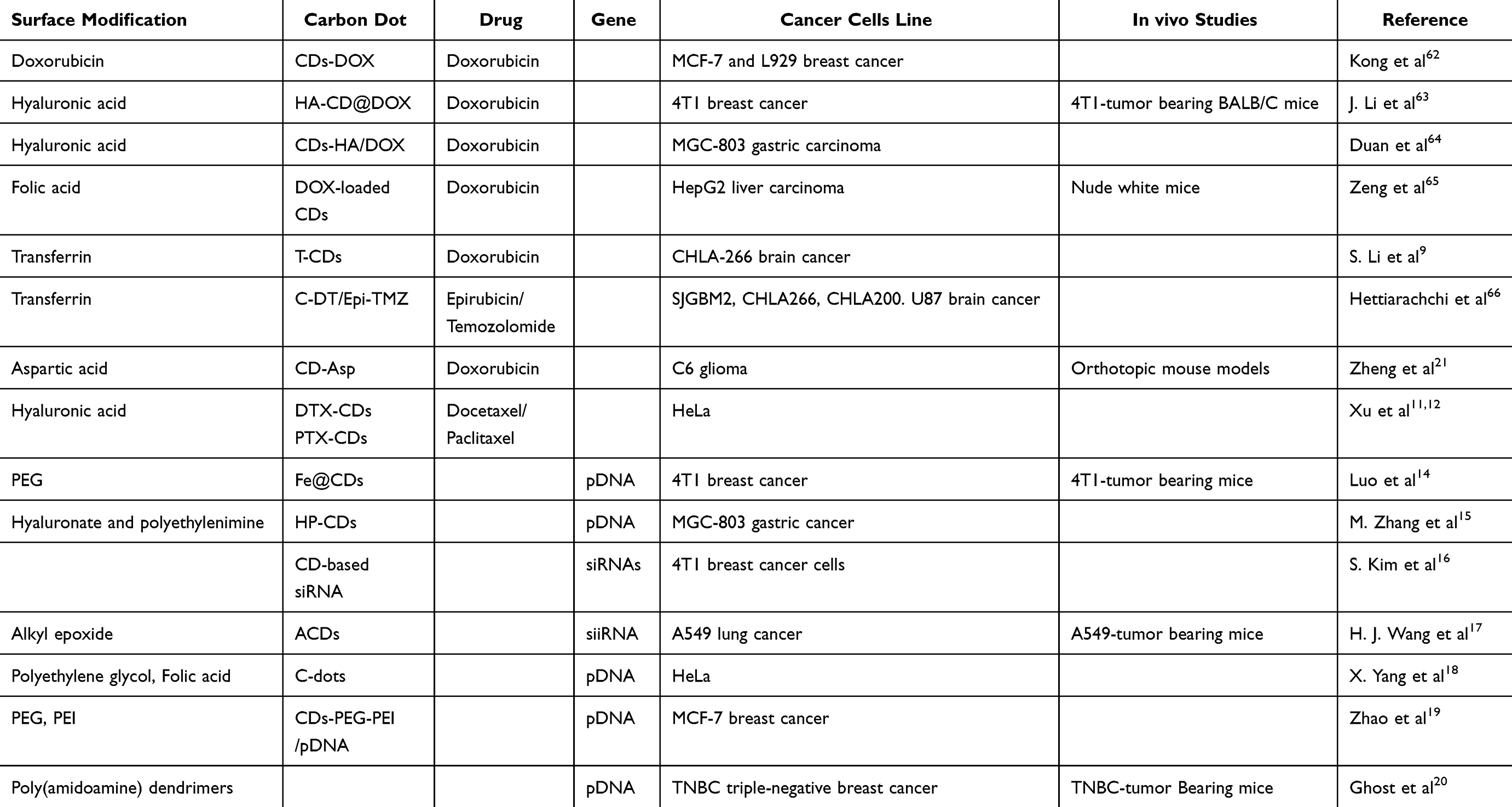 |
Table 1 Surface Modified CDs as a Nanocarrier Applications |
Drug Delivery Systems for Cancer Therapy
Surface-modified CDs have gained attention as ideal drug delivery systems for cancer therapy due to their excellent biocompatibility, small size, and ease of functionalization. These modified CDs can selectively bind to receptors overexpressed on cancer cell surfaces, facilitating the internalization of drugs and improving therapeutic efficacy while minimizing systemic toxicity.67
The drug delivery mechanisms of CDs typically rely on various interactions between the CDs and the drug molecules. The electrostatic interaction is the most common mechanism, where drugs with opposite charges can attach to the surface-modified CDs. For instance, Zeng et al65 synthesized fluorescent CDs with carboxyl-rich surfaces using a hydrothermal method, resulting in a particle size of ≈5 nm that exhibited excellent biocompatibility and low cytotoxicity. Similarly, Yuan et al68 produced CDs from milk hydrothermolysis, achieving an average size of 4 nm and demonstrating high loading efficiency for doxorubicin (DOX) via electrostatic interactions.
Despite the previous results, there are still limitations in targeting drugs and achieving the best interaction between the drug and CDs. CDs are often surface-modified with various ligands, polymers, or targeting agents to enhance this interaction and improve drug delivery. These modifications can improve the selective targeting of cancer cells, enhance solubility, and facilitate drug-controlled release. For instance, J. Li et al63 modified CDs’ surface with a hyaluronic acid (HA) hydrophilic group that helps the electrostatic interaction between CDs and DOX, forming pH-responsive HA-CD@DOX. In a similar study, S. Li et al9 utilized protein transferrin to modify CD’s surface and help the interaction with a cancer drug DOX, enhancing the solubility.
Additionally, the ability of CDs to target the cancer drug docetaxel (DTX) to develop DTX-CDs and paclitaxel (PTX-loaded CDs) and surface modified HA, through covalent bonding; the functional groups, carboxyl and hydroxyl on the surface of CDs can form a covalent bond with drug molecules providing stability in drug loading. Both systems exhibited excellent biocompatibility and enhanced drug delivery with minimized side effects.11,12
The ability to control drug release is a critical feature of CDs-based delivery systems. For instance, S. Wang et al6 reported a pH-responsive release profile with folic acid (FA) to develop an FA-CDs-DOX system, where 79.31% of DOX was released at pH 5.5 compared to just 16.39% at pH 7.4. This differential release mechanism is attributed to the protonation of amino groups in DOX under acidic conditions prevalent in tumor microenvironments, enhancing drug solubility and facilitating targeted therapy. Similarly, Duan et al64 reported modified CDs with HA and DOX, developing a dual-responsive CDs-HA/DOX system that demonstrated a release efficiency of 66% of DOX, showcasing the potential for utilizing both pH and HA responsiveness to enhance therapeutic outcomes. However, Kong et al62 demonstrated 75% of DOX release at pH 7.4 with high accumulation and cytotoxicity of DOX in MCF-7 cancer cells.
In vitro studies have demonstrated the efficacy of surface-modified CDs in drug delivery across various cancer types. Zeng et al65 reported that their DOX-loaded CDs induced a 40% death rate in HepG2 liver carcinoma cells at 2.5 µg/mL concentration while maintaining safety for normal liver cells (HL-7702). In a similar study, Kong et al62 evaluated pharmaceutical activity and cellular toxicity of CDs-DOX on MCF-7 and L929 breast cancer cells with cell viability above 90% at 0.5 μg/, reducing cancer cell growth significantly. The selective targeting capability and cytotoxic effect of CDs improved significantly through surface modification with HA.
J. Li et al63 utilized HA-modified CDs (HA-CDs) to deliver DOX to 4T1 breast cancer cells, achieving high cytotoxicity rates and demonstrating a tumor growth inhibitory rate of 80.23% compared to free DOX. Similarly, Duan et al64 demonstrated the inhibition of growth and migration of CDs-HA-Hep/DOX system dual responsive by HA and pH value in MGC-803 human gastric carcinoma cell line. Additionally, the delivery of drugs for brain cancer has been proved. S. Li et al9 explored transferrin-conjugated CDs (T-CDs) for delivering DOX to brain tumor cells (CHLA-266), achieving greater uptake than non-targeted systems. In a similar study, Hettiarachchi et al66 utilized triple conjugated C-DT with transferrin and drugs (Epirubicin and Temozolomide) to treat pediatric (SJGBM2, CHLA266, CHLA200) and adult (U87) brain tumor cells, and Quiao et al69 proved the high endocytosis rate of C6 brain glioma cells treated with CDs from glucose and L-aspartic acid. This study highlights the importance of functionalizing CDs with targeting ligands to enhance cellular uptake and therapeutic effectiveness in brain tumors.
In vivo studies further validate the efficacy of targeted drug delivery using CDs. Zheng et al21 demonstrated that their CD-Asp system effectively targeted C6 glioma cells in orthotopic models, with the highest fluorescence intensity observed at 15 minutes post-injection. This rapid accumulation indicates efficient targeting and potential for real-time monitoring. Zeng et al65 and S. Wang et al6 evaluated CDs-DOX in liver cancer cells of nude white mice. The HepG2 tumors reduced 50% of the volume in their first 24 hours and 72.13% after 20 days of treatment.
Additionally, J. Li et al63 evaluated the effectiveness of DOX in 4T1 tumor-bearing BALB/C mice, which were intravenously injected with ICG-loaded HA-CD@p-CBA-DOX. The results showed a tumor growth inhibitory rate of 80% compared to free DOX. These studies confirmed that the pH-responsive release mechanism significantly contributed to the selective accumulation of DOX in tumor tissues while minimizing effects on healthy cells.
The targeted drug delivery capabilities of carbon dots are supported by numerous studies demonstrating their effectiveness across different cancer types through tailored synthesis methods and functionalization strategies. The combination of in vitro and in vivo studies highlights the potential of modified carbon dots as ideal carriers for anticancer drugs like DOX, providing a promising avenue for improving therapeutic efficacy while minimizing side effects. Despite their efficiency in drug release, CDs may have a limited capacity for loading large amounts of therapeutic agents. Moreover, the surface modification process, especially with polymers and targeting ligands, can be complex and may affect the stability of the CDs during storage or in biological systems.
Gene Therapy for Cancer Therapy
Gene therapy involves introducing genetic material into cells to correct or inhibit the expression of disease-related genes. This process requires an efficient delivery system to protect the genetic material and ensure its uptake by the target cells. Surface-modified CDs have emerged as promising candidates for gene delivery due to their ability to condense genetic material and protect it from degradation while offering efficient cellular uptake and gene transfection.70
CDs serve as carriers for plasmid DNA (pDNA), small interfering RNA (siRNA), or other nucleic acids in gene delivery for cancer therapy. The main mechanisms through which CDs deliver genes are electrostatic interactions. Luo et al14 synthesized iron-doped carbon dots (Fe@CDs) functionalized with a dendritic lipopeptide (PEG-RLS). This modification improved the stability of CDs and facilitated plasmid DNA (pDNA) targeting gene delivery. The positive charges on surface-modified CDs interact with negatively charged nucleic acids, facilitating efficient binding and condensation of the genetic material. The resulting nanohybrid system exhibited a diameter of 77 nm and demonstrated effective gene transfection in vitro, with fluorescence intensity indicating a two-fold increase in green fluorescent protein (GFP) expression upon laser irradiation compared to controls (see Figure 1A and B).
 |
Figure 1 PEG-RLS/Fe@CDs: (A) schematic mechanism of cancer nanotheranostic platform for gene delivery, PTT and bioimaging applications. Reproduced from Luo T, Nie Y, Lu J, et al. Iron doped carbon dots based nanohybrids as a tetramodal imaging agent for gene delivery promotion and photothermal-chemodynamic cancer synergistic theranostics. Mater Des. 208. © 2021 The Authors. Published by Elsevier Ltd. Creative Commons CC-BY-NC-ND license.14 (B) In vivo photo-enhanced therapeutic effect of PEG-RLS/Fe@CDs under 660 nm (0.5 W/cm2 for 5 min) in 4T1 breast cancer, tumor growth inhibition after different treatments, tumor tissues after treatment. HP-CDs/DNA: (C) Zeta potential of free DNA, HP-CDs and HP-CDs/DNA complex, identification of transfection efficiency of HP-CDs, fluorescent images of gene expression obtained by a confocal laser scanning microscope in HEK-293T cells transfected with DNA/HP-CDs complexes, (D) Schematic synthesis and biomedical applications of HP-CDs. Reproduced from Zhang M, Zhao X, Fang Z, et al. Fabrication of HA/PEI-functionalized carbon dots for tumor targeting, intracellular imaging and gene delivery. RSC Adv. 2017;7:3369–3375. This article is licensed under a Creative Commons Attribution-NonCommercial 3.0 Unported Licence.15 |
Similarly, M. Zhang et al15 developed hyaluronate and polyethylenimine (HA-PEI) functionalized carbon dots (HP-CDs) through a bottom-up synthesis method. These HP-CDs exhibited excellent dispersibility and biocompatibility, allowing efficient internalization into cancer cells via CD44 receptor-mediated endocytosis. After binding on nucleic acids, CDs are internalized by cells through endocytosis, which allows for the delivery of genes into the cytoplasm and eventual gene expression. In vitro transfection experiments using HEK-293T human embryonic kidney cells showed that HP-CDs could effectively condense pDNA, enhancing gene expression in MGC-803 gastric cancer cells (see Figure 1C and D).
Moreover, CDs can be surface-modified to carry interfering RNAs (siRNAs). S. Kim et al16 developed a CD-based siRNA approach and delivered into breast cancer cells for the first time. Similarly, H. J. Wang et al17 constructed amphiphilic carbon dots (ACDs) by conjugating hydrophobic alkyl epoxide to PEI-derived CDs, achieving a high transfection efficiency of approximately 96.7% of siRNA in A549 lung cancer cells. The ACDs exhibited superior cellular uptake compared to traditional transfection agents like Lipofectamine 2000, indicating their potential as effective gene carriers and causing cell apoptosis.
Surface modifications help protect genetic material from degradation and enhance cellular uptake, allowing for effective gene silencing or expression in cancer cells. X. Yang et al18 synthesized positively charged CDs using PEI and FA, facilitating selective imaging and transfection of folate receptor-positive HeLa cells. The C-dots demonstrated a significant ability to target pDNA due to electrostatic interactions, achieving a transfection efficiency of 42.63% in HeLa cells. This study underscores the importance of targeting specific receptors for enhanced gene delivery. In a related study, Zhao et al19 reported the development of CDs modified with PEG and PEI to deliver pDNA, developing CDs-PEG-PEI/pDNA complexes that exhibited pH-responsive charge reversal, enhancing cellular uptake in mildly acidic tumor microenvironments. This system achieved high transfection efficiency in MCF-7 breast cancer cells, with a notable % tumor growth inhibition rate of 70% observed in vivo. The study highlights the potential of pH-responsive systems for targeted gene therapy.
In vivo studies provide critical insights into the therapeutic potential of CDs for gene delivery. Luo et al14 developed Fe-doped CDs (Fe@CDs) and modified the surface with PEG to enhance the biocompatibility and delivery of genes. The delivery system PEG/Fe@CDs demonstrated 80% tumor growth inhibition in 4T1 breast cancer models, with the delivery of green fluorescent protein (EGFP) gene expression that achieves fluorescence intensity signals indicating effective accumulation at tumor sites over time (see Figure 1B). The results suggest that this multifunctional delivery system could be an effective platform for photothermal-responsive gene therapy. According to H. J. Wang et al17 demonstrated that Amphiphilic CDs (ACDs) were developed as vectors to target siRNA. ACDs showed promising results in delivering siRNA to A549 lung cancer cells. The study reported enhanced apoptosis rates due to effective gene silencing, further validating the potential of ACDs as efficient gene delivery vehicles. Ghosh et al20 developed CDs functionalized with poly(amidoamine) (PAMAM) dendrimers for triple-negative breast cancer (TNBC). Their CD-PAMAM system demonstrated low toxicity even at high concentrations and improved gene transfection efficiency, making it a promising candidate for TNBC therapy.
Multimodal-Imaging in Cancer Therapy
Multimodal imaging is a crucial aspect of cancer diagnostics, offering real-time monitoring and visualization of tumor cells. Surface-modified CDs and heteroatoms/metals-doped CDs are pivotal for enhancing imaging capabilities due to their tunable fluorescence, high quantum yield, and biocompatibility (Table 2).19 This section focuses on using surface-modified and doped CDs for tumor detection and bioimaging, discussing their synthesis methods, mechanisms, and performance in vitro and in vivo studies.
 |
Table 2 Multimodal Imaging Applications of Surface-Modified Carbon Dots |
Surface Modified-CDs for Tumor Detection
Fluorescence imaging leverages the photoluminescence (PL) properties of CDs, which originate from quantum confinement effects and surface defect states.74 The reduced size of CDs (<10 nm) restricts the motion of electrons and holes, resulting in size-dependent emission. Additionally, functional groups such as –COOH, –OH, and –NH2 on the CD surface enhance fluorescence by introducing trap states, that influence electron–hole recombination.75
For instance, Wang et al17 synthesized amphiphilic carbon dots (ACDs) by conjugating alkyl epoxide to CDs. The alkyl epoxide reacts with amine (–NH2) or hydroxyl (–OH) groups on the CD surface through epoxide ring-opening reactions, forming covalent bonds. This process produces CDs with hydrophobic alkyl chains and hydrophilic functional groups (–OH, –COOH, –NH2) on the surface. The amphiphilic modification enhances interaction with cell membranes and improves cellular uptake, enabling efficient delivery to the target sites. These ACDs exhibited bright fluorescence in A549 lung cancer cells and demonstrated effective cellular imaging.
Similarly, Yang et al18 developed positively charged carbon dots by functionalizing them with folic acid (FA) and polyethyleneimine (PEI). These CDs showed selective imaging of folate receptor-positive HeLa cells. FA binds specifically to folate receptors (FRα), which are overexpressed on cancer cells, facilitating receptor-mediated endocytosis. Meanwhile, the positive charges on PEI interact electrostatically with negatively charged phospholipids in the cell membrane, promoting cellular uptake. This resulted in efficient intracellular fluorescence signals, demonstrating potential for real-time imaging applications.
In both studies, surface functionalization played a critical role in modulating the electronic bandgap, thereby influencing excitation wavelength and emission intensity.75 Hydrophilic groups improved surface passivation by reducing nonradiative recombination and enhancing fluorescence. Furthermore, the amphiphilic structure of ACDs facilitates penetration through lip bilayers, enabling cytoplasmic localization. Amine (–NH2) groups of PEI and FA contributed to covalent bonding with targeting agents, improved surface passivation, and provided positive charges for electrostatic interactions. Additionally, the cationic charges of PEI enhanced interaction with negatively charged cell membranes, promoting intracellular delivery and localized fluorescence.
In vivo applications of functionalized carbon dots have shown promising results for tumor imaging and monitoring therapeutic efficacy. Zheng et al21 synthesized highly fluorescent CD-Asp from L-aspartic acid, which showed selective imaging of C6 glioma cells in orthotopic models. The highest fluorescence intensity was observed 15 minutes post-injection, indicating rapid tumor accumulation, a critical feature for real-time imaging during therapeutic interventions. Similarly, Li et al9 reported the use of transferrin-conjugated CDs to deliver doxorubicin to brain tumor cells (CHLA-266). The conjugated system demonstrated significantly higher uptake in tumor tissues compared to normal cells, enabling precise monitoring of drug distribution via fluorescence imaging.
Moreover, Zhao et al19 developed a pH-responsive system (CDs-PEG-PEI/pDNA) that exhibited size-shrinkage in response to the acidic tumor microenvironments, promoting enhanced accumulation and penetration into tumor sites. This was confirmed through in vivo imaging of MCF-7 tumor-bearing mice, highlighting the system’s adaptability to physiological conditions. These studies underscore the versatility of CDs, which can be functionalized with ligands such as FA, or hyaluronic acid (HA) to target overexpressed receptors on cancer cells. Such ligand-mediated targeting enhances imaging specificity and reduces off target effects on healthy tissues.
The in vivo findings complement the in vitro studies discussed previously, demonstrating how surface functionalization of CDs not only enhances fluorescence properties but also enables precise cellular and tissue targeting. By leveraging their tunable surface chemistry and size, CDs hold promise as multifunctional agents for real-time tumor imaging, targeted drug delivery, and monitoring therapeutic outcomes.
Heteroatoms/Metals Doped Carbon Dots for Bioimaging and MRI Applications
When CDs are irradiated with excitation light, they can fluoresce at specific wavelength. Their PL can be tuned by altering their size, surface states or through electrochemical doping with heteroatoms (eg nitrogen, sulfur) or metal ions (eg copper, gadolinium).75,76 Heteroatom doping enhances the QY of CDs by modifying their electronic structure. For example, doping with elements like copper (Cu) or boron (B) introduces intermediate energy levels or alters the HOMO-LUMO gap, increasing the efficiency of photon absorption.75,77
J. Wang et al27 developed copper-doped carbon dots (Cu-CDs) with a high fluorescence QY of 24.4%. Cu doping minimizes nonradioactive decay caused by surface defects while enhancing radiative decay pathways. These Cu-CDs demonstrated significant photoinduced cytotoxicity against HeLa cervical cancer cells and SH-SY5Y neuroblastoma multicellular spheroids. Their bright fluorescence and low cytotoxicity make them promising candidates for fluorescence imaging in cancer diagnostics. Additionally, Cu doping enhances red and near-infrared (NIR) fluorescence by increasing light absorption in lower-energy regions.
Similarly, Shen et al71 synthesized boron doped CDs (B-CDs) with excellent multicolor fluorescence imaging capabilities, as demonstrated in HeLa cells. These B-CDs exhibited a high QY of 22% and showed potential for cell imaging agents due to their solid-state PL properties and good biocompatibility. Boron doping introduces electron-deficient sites in the CDs, which narrow the bandgap, enhancing light absorption and shifting fluorescence emission toward longer wavelengths (eg green, red, or NIR regions). This modification improves radiative recombination of electron–hole pairs, significantly increasing the QY. Furthermore, B-CDs effectively penetrate HeLa cancer cells via endocytosis, facilitated by their small size (<10 nm) and surface functional groups. Once internalized, B-CDs exhibit bright fluorescence under excitation, enabling visualization of intracellular structures. These attributes make B-CDs valuable tools for imaging applications in cancer research and diagnostics.
Doped CDs combine fluorescence with other imaging modalities, such as magnetic resonance imaging (MRI), photoacoustic imaging (PA), and computed tomography (CT), providing more comprehensive approaches to tumor detection. MRI relies on the excitation and subsequent relaxation of nuclear spins in the presence of magnetic field to generate detailed images. CDs doped with paramagnetic ions (eg gadolinium (Gd3+), manganese (Mn2+), boron (B)) or metals (eg, iron (Fe3+)) function as T1 or T2 contrast agents by enhancing proton relaxation rates.77
Gd3+ or Mn2+ dopants in CDs introduce unpaired electrons that interact with the nuclear spins of surrounding water protons. These dopants reduce the longitudinal relaxation time (T1), making tissues appear brighter in MRI scans while also creating local magnetic field that proton relaxation. For example, Du et al29 synthesized Gd-doped CDs (Gd-CDs) with excellent biocompatibility and a T1 relaxivity rate of 6.45 mM−1s−1, making them suitable for magnetic resonance/fluorescence (MR/FI) bimodal imaging. In another study, Jiao et al30 developed AS1411-Gd-CDs by conjugating Gd-CDs with AS1411 aptamers, which target nucleonic overexpressed on various adenocarcinoma cells, including breast and renal cancers. This functionalization improved targeting specificity and significantly enhanced MRI signal intensity in 4T1 tumor cells compared to non-targeted Gd-CDs (see Figure 2A–C). These findings demonstrate the potential of Gd-CDs to enhance MR imaging contrast while maintaining low cytotoxicity, thereby facilitating imaging-guided cancer treatment.
 |
Figure 2 AS1411-Gd-CDs: (A) Schema of preparation of AS1411-Gd-CDs and FL/MR-guided PTT of tumor, (B) In vitro MR imaging properties, T1-weighted MR images of 4 T1 and NIH-3T3 cells treated with Gd-CDs. The red circles indicated the tumor cells, (C) In vivo fluorescence and PR imaging tests of 4 T1 tumor-bearing mice model pre- and post-injection of Gd-CDs and AS1411-Gd-CDs at various time intervals. The red circles represent the tumor area. Mn-CDs-NH: (D) MRI images of tumor -bearing mice treated with Gadovist or Mn-CDs-NH, (E) Metastatic investigation of lungs with MRI imaging. PEG-RLS/Fe@CDs: (F) In vitro tetramodal imaging in 4T1 cells by confocal fluorescence imaging, T1-weighted magnetic resonance (MR) images with different concentrations of PEG-RLS/Fe@CDs, (G) Real-time in vivo fluorescence images under 660 nm light irradiation (0.5 W/cm2). The white circles show the position of the tumor. (A,C,D) Reproducced from Jiao M, Wang Y, Wang W, et al. Gadolinium doped red-emissive carbon dots as targeted theranostic agents for fluorescence and MR imaging guided cancer phototherapy. Chem Eng J. 2022;440. © 2022 The Author(s). Published by Elsevier B.V. Creative Commons CC-BY-NC license.30 (E,F,G) Reproduced from Tiron A, Stan CS, Luta G, et al. Manganese-doped n-hydroxyphthalimide-derived carbon dots—theranostics applications in experimental breast cancer models. Pharmaceutics. 2021;13. © 2021 by the authors. Licensee MDPI, Basel, Switzerland. This article is an open access article distributed under the terms and conditions of the Creative Commons Attribution (CC BY) license.72 |
Similarly, Mn-doped (Mn-CDs) have shown promise as dual-modal imaging agents for both MRI and optical imaging. Ji et al31 demonstrated that Mn-CDs provided significant T1 contrast enhancement in MRI imaging and enabled precise intraoperative localization of glioma tumors in mouse brain models.32 Tiron et al72 further reported Mn-CDs with a T1 relaxivity of 9.96 mM−1 s−1, highlighting their efficacy as contrast agents for combined T1-weighted MR/fluorescence imaging. In vivo studies using breast cancer animal models revealed that Mn-CDs generated strong MRI signals, with predominant uptake in the liver, kidneys, peritoneal cavity, and heart. Tumor regions exhibited intense MRI signals, as visualized through rainbow color maps for enhanced differential contrast (see Figure 2D and E). These studies underscore the potential of Mn-CDs for precise imaging and effective clearance through renal pathways, making them highly suitable for imaging and therapeutic applications. Additionally, H. Wang et al36 introduced boron-doped CDs (B-CDs) as alternative T1 contrast agents. Although B-CDs demonstrated lower magnetization compared to Gd3+ or Mn2+-based agents, they offer a novel approach to bimodal bioimaging applications. Boron doping introduces unpair electrons into the CDs structure, generating localized paramagnetic centers. These centers interact with the magnetic field and surrounding water protons, facilitating their relaxation. While Gd³⁺ and Mn²⁺ effectively enhance relaxation times, boron doping within the π-conjugated carbon network of CDs creates a distinctive paramagnetic environment with notable relaxation efficiency.
Iron-doped CDs can shorten transverse relaxation time (T2), Fe3+ ions cause magnetic inhomogeneities, shortening transverse relaxation time (T2) and darkening tissues in MRI. In a study, iron doped-CDs (Fe@CDs), was developed by solvothermal method.14 The Fe@CDs provided longitudinal relaxivity (r1) = 1.25 mm⁻¹s⁻¹, indicating they are effective T2 contrast agent in vivo (Figure 2F and G). Fe²+doped carbon dots showed significant enhancement of proton relaxation rates because its paramagnetic properties were greatly enhanced by these ions.
Photoacoustic (PA) imaging leverages the ability of CDs to convert absorbed light energy into localized heat through nonradioactive relaxation processes, enabling high-resolution imaging. Dopants like Fe2+ and Cu2+ reduce the bandgap, enhance defect states, and improve photothermal conversion efficiency, enabling effective photoacoustic (PA) imaging. The localized heat induces thermoelastic expansion in surrounding tissues, generating acoustic waves that are detected by ultrasound sensors and converted into high-resolution PA images.78 In a study, Fe@CDs were synthesized and evaluated in 4T1 cancer cells, demonstrating strong acoustic signal generation and potential for multimodal imaging (PA/fluorescence). Fe doping narrowed the bandgap and enhanced NIR absorption (650–900), improving photothermal properties.14 Similarly, Liu et al51 reported that Cu doped CDs (Cu-CDs) provided precise tumor boundary delineation due to their strong PA signals, aiding in MFC-7 breast cancer cells. Copper dopants modify the electronic structure of CDs, enhancing NIR absorption and photothermal efficiency. The rapid localized heating caused by Cu-CDs leads to thermoelastic expansion of tumor tissues, generating ultrasonic pressure waves. The intensity of these waves is directly correlated with the photothermal efficiency of Cu-CDs, significantly improved by Cu doping.
CT imaging relies on the attenuation of X-rays by high atomic number (Z) elements. Common contrast agents (iodixanol) show inherent defects and poor sensitivity. Based on this, Su et al28 synthesized hafnium-doped CDs (HfCDs) as a contrast agent, showcasing robust stability and excellent water solubility. In vivo studies demonstrated that HfCDs could provide immediate contrast enhancement at tumor sites within one-minute post-injection into H22 orthotopic liver tumor-bearing mice. The HU (Hounsfield units) CT value of the tumor region increased from 114.0 hU (0 min) to 296.0 hU at 1 min post-administration. This rapid imaging capability underscores the potential of HfCDs as contrast agents for in vivo CT for clinical diagnosis. In a similar study, CDs were synthesized with iohexol (contrast agent) as a precursor and cetuximab was targeted on the surface of CDs to form I-CQDs-C225. The contrast system showed a low cytotoxicity effect in H23 and HLF cancer cells, with cell viability at about 80%, and CT image brightness of I-CQDs increased significantly at 635 hU, compared with iodixanol (commercial contrast agent).73 These studies proved that surface modified-CDs can be used for cellular multicolor imaging and CT imaging due to their low toxicity, excellent biocompatibility, and good X-ray attenuation capacity. (Table 2)
PDT and PTT for Cancer Treatment
Surface-Modified CDs in PDT for Cancer Therapy
Surface-modified CDs have emerged as promising nanomaterials for PDT and PTT, two light-based cancer treatment strategies. PDT relies on the activation of PSs by light to produce ROS that destroys cancer cells. PTT involves converting light energy into heat, which causes localized hyperthermia and ablates tumors.27,38 The functionalization of CDs with targeting agents, photosensitizers, or metal ions can significantly enhance their efficacy in PDT and PTT by improving selective accumulation, ROS generation, and thermal conversion efficiency.79 The following sections will explore the application of surface-modified CDs in PDT and PTT, focusing on their roles as nanocarrier of PSs, doped CDs as a PSs for enhanced therapeutic effects, and the use of PSs as precursors in the synthesis of CDs. Through this discussion, we aim to provide insights into the mechanisms and advantages of these approaches, emphasizing their potential in advancing cancer treatment strategies.
CDs have gained recognition as effective nanocarriers for PSs in PDT. Their unique structural features facilitate their application in drug delivery systems, including a π-conjugated core and a surface rich in hydrophilic functional groups such as –NH2, –OH, and –COOH. CDs have the following advantages as they function as nanocarriers for molecular PSs: water solubility and biocompatibility, stability and dispersibility of organic PSs in aqueous solution, and the large two-photon absorption cross-section (TPACS) enables CDs to have excellent two-photon excitation properties which can effectively kill cancer cells in deep tissues.
The central mechanism of PDT using surface-modified CDs involves the generation of singlet oxygen (1O2) or other ROS upon light activation. In a pioneering study by Sun et al,80 the PS chlorin e6 (Ce6) was targeted onto amino-rich red emissive carbon dots (RCDs) to form Ce6-RCDs. This innovative approach synergistically enhanced PTT and PDT under a single near-infrared (NIR) laser at 671 nm. The study reported an impressive photothermal conversion efficiency of 46%, enabling effective cancer treatment at a reduced laser power density of 0.50 W/cm2. In vitro experiments demonstrated that Ce6-RCDs exhibited significantly higher cytotoxicity against MCF-7 and 4T1 breast cancer cells than free Ce6 or RCDs alone. In vivo studies further confirmed the enhanced therapeutic efficacy, with multimodal imaging capabilities including fluorescence, photoacoustic, and photothermal imaging, facilitating real-time monitoring of the therapy process.
Similarly, Kim et al81 developed pH-sensitive carbon dots modified with Ce6 (Ce6@IDCDs). These CDs were designed to release Ce6 (80%) specifically in acidic tumor microenvironments (pH 6.5), triggering immunogenic cell death through PDT upon 671 nm laser irradiation. In vitro studies revealed that the treatment led to increased expression levels of CD80 and CD86 (amplifying T cell activation), indicating successful dendritic cell maturation and immune activation. The in vivo results mirrored these findings, showcasing significant antitumor effects in CT26 mouse colon cancer cells as evidenced by reduced tumor volumes and increased activation of natural killer (NK) cells, CD4+, and CD8+ T cells.
CDs show a broad absorption and tunable emission spectrum that can be exploited in a FRET (Forster Resonance Energy Transfer) system. CDs serve as energy donors to photosensitizers via FRET in some systems, transferring light energy to excite the photosensitizer, producing ROS generation. Wu et al82 introduced a novel CQDs@PtPor system through electrostatic interactions between positively charged tetraplatinated porphyrin photosensitizer complex (PtPor) and negatively charged carbon quantum dots (CQDs). The FRET process allowed CQDs to transfer light energy to PtPor, resulting in enhanced 1O2 production compared to pure PtPor. This system demonstrated improved PDT efficacy under xenon lamp irradiation, showcasing the potential of CQDs as effective nanocarriers for PSs and enhancing cytotoxicity effect (see Figure 3). In a similar study, magnesium and nitrogen-doped carbon dots (Mg, N-doped CDs) were synthesized as a nanocarrier for Ce6, significantly enhancing FRET efficiency due to their high fluorescence QY and proximity to the PS. The resulting Mg, N-doped CDs-Ce6 system demonstrated improved therapeutic efficacy in both in vitro and in vivo models.83
 |
Figure 3 CQDs@Pt: (A) Schematic illustration of synthesis of CQDs@Pt, (B) TEM images and corresponding size distribution histograms and (C) FT-IR spectra of CQDs@Pt, (D) Cell viability of CQDs@Pt in HeLa cells after treatment, (E) Singlet oxygen of CQDs@PtPor, (F) confocal fluorescence microscopy images of HeLa cells under 405 nm excitation after treatment with CQDs, PtPor and CQDs@PtPor. And (G) PL Spectra of CQDs@Pt. Reproduced from Wu F, Yue L, Su H, et al. Carbon Dots @ platinum porphyrin composite as theranostic nanoagent for efficient photodynamic cancer therapy. Nanoscale Res Lett. 13. © The Author(s). 2019, corrected publication January 2019 Open AccessThis article is distributed under the terms of the Creative Commons Attribution 4.0 International License.82 |
A pioneer strategy to enhance the water solubility of hydrophobic PSs is targeting the surface of carbon dots. For instance, riboflavin has been targeted on CDs surface. Riboflavin, having a diol group, was covalently linked with CD, promoting the killing of cancer cells of B1610 murine melanoma cells and 4T1 breast cancer cells in the presence of laser irradiation (365 nm, 60 mW/cm2).84,85 Similarly, Choi et al86 developed folic acid-modified carbon dots loaded with zinc phthalocyanine (ZnPc), enhancing water solubility and tumor-targeting ability. The CD-PEG-FA/ZnPc nanoparticles produced 1O2 effectively under 660 nm laser irradiation (0.3 W/cm2, 20 min), demonstrating significant therapeutic effects in vitro against A549 human lung cancer cells. The conjugation of FA to CD significantly improved the active tumor-targeting capability of the ZnPc delivery vehicle to folate receptor-overexpressing tumors.
Doped CDs are increasingly recognized for their dual role as photosensitizers and nanocarriers in PDT. By incorporating heteroatoms such as nitrogen, phosphorus, sulfur, and metals like copper and tin, the properties of CDs can be fine-tuned to enhance their ability to generate ROS under light irradiation. These doped CDs offer improved photodynamic effects by increasing 1O2 production, light absorption, and biocompatibility, making them powerful agents in cancer therapy.
In a study, Zhao et al35 synthesized red-emitting nitrogen and phosphorus co-doped carbon dots (R-CDs) using phosphoric acid and o-phenylenediamine through a hydrothermal method. R-CDs could generate 1O2 and have an absolute fluorescence QY of 15% in water. In vitro studies demonstrated that R-CDs effectively inhibited the proliferation of A549 lung cancer cells under 532 nm light irradiation (100 mW/cm2), showing promise as a standalone PDT agent. Furthermore, in vivo experiments on tumor-bearing mice indicated significant tumor reduction, confirming R-CDs’ potential as a PS and effective cancer treatment. Similarly, Jia et al87 prepared self-assembled carbon dot nanospheres (CDNS) that produced 1O2 under 671-nm laser irradiation with a QY of 45%. The CDNS showed superior cytotoxic effects against 4T1 cancer cells compared to conventional CDs. In vivo fluorescence imaging indicated effective accumulation at tumor sites, significantly reducing tumor volume after treatment. These studies highlight the potential of surface-modified CDs as efficient photosensitizers capable of generating ROS while also providing imaging capabilities (Table 3).
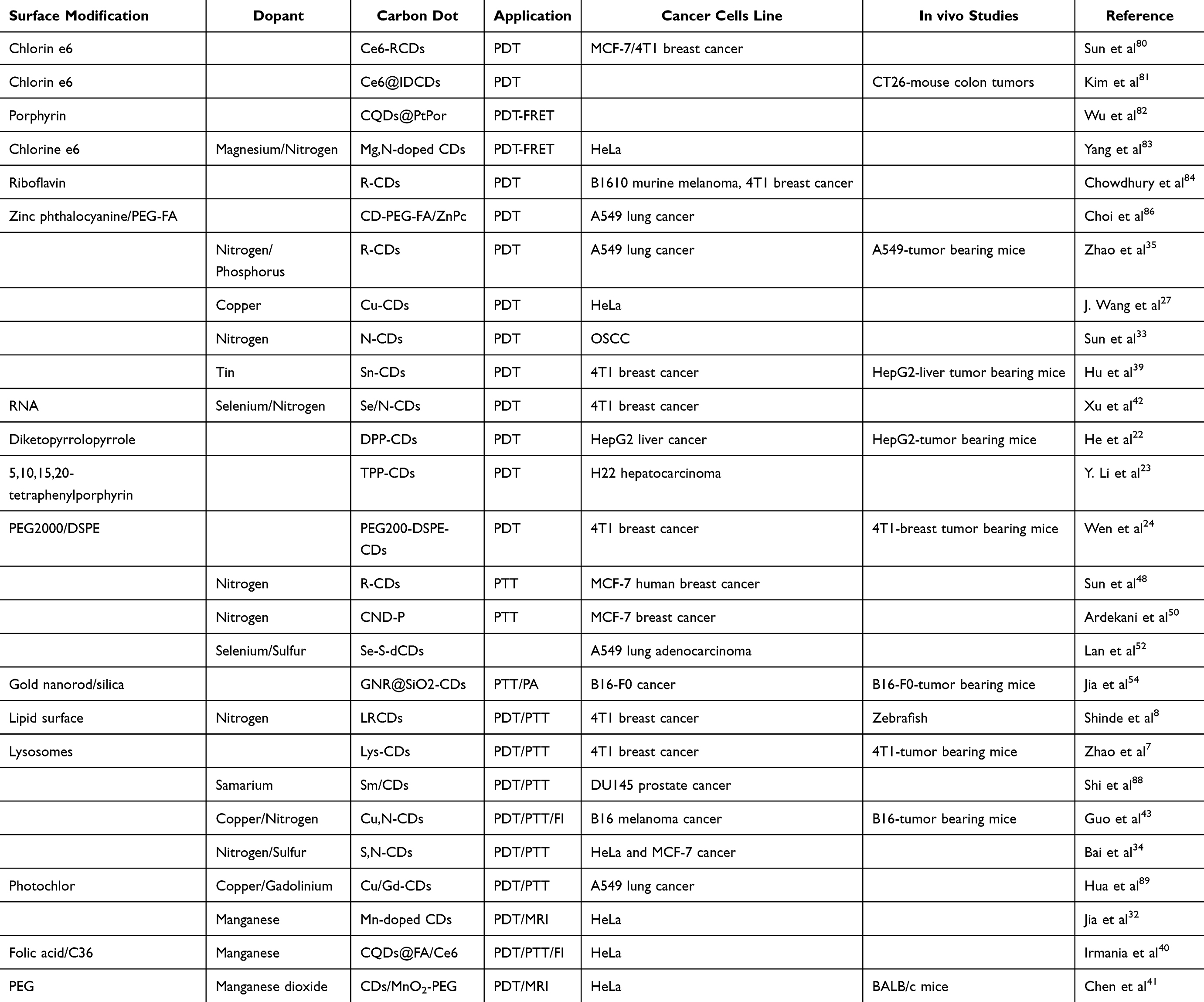 |
Table 3 Photodynamic and Photothermal Therapy Applications of Surface-Modified Carbon Dots |
Metal-doped CDs can generate ROS depending on the dose, metal speciation, and exposure route. For instance, J. Wang et al27 developed copper-doped carbon dots (Cu-CDs) using acrylic acid and copper nitrate as precursors. The Cu-CDs exhibited a 1O2 QY of 36% under LED light irradiation (400–700 nm). In vitro studies revealed that Cu-CDs effectively induced apoptosis in HeLa cancer cells through ROS generation. In vivo experiments demonstrated substantial tumor growth inhibition in mice treated with Cu-CDs, indicating their potential as effective photosensitizers for PDT. In a similar study, Sun et al33 investigated nitrogen-doped carbon dots for their application in oral squamous cell carcinoma (OSCC). The CDs demonstrated significant ROS production under 660 nm laser irradiation, leading to effective apoptosis through mitochondrial pathways. Both in vitro and in vivo studies confirmed that these nitrogen-doped CDs promoted DNA damage and inhibited tumor growth. Hu et al39 also synthesized tin-doped carbon dots (Sn@CDs) that exhibited a high 1O2 quantum yield of 58.3% with low cytotoxicity toward healthy cells. In vitro studies confirmed that Sn@CDs effectively reduced the viability of 4T1 cancer cells upon irradiation with LED light (490 nm, 40 mW/cm2). The results from both in vitro and in vivo experiments indicated significant tumor reduction of HepG2 liver cancer, demonstrating the promise of Sn@CDs as effective photosensitizers for PDT applications.
Heteroatoms doped CDs, such as N-CDs, Se-N-CDs, and S, N-CDs, have been developed as PS for PDT in cancer therapy.40,42 Li et al90 investigated nitrogen-doped CDs (NCDs) synthesized via solvothermal transformation of coal resources, achieving a 1O2 QY of 19%. Their findings suggest that NCDs possess significant potential as photosensitizers for PDT due to their ability to generate ROS effectively. However, the combination of sulfur and nitrogen-doped CDs (S, N-CDs) can enhance the generation of ROS in hypoxic tumor environments. Bai et al34 developed sulfur and nitrogen co-doped carbon dots (S, N-CDs) using a hydrothermal method, which showed improved therapeutic efficiency compared to single doping methods. These multifunctional heteroatom-doped CDs produced 1O2 with a QY of 27% and demonstrated effective photothermal conversion capabilities. In a similar study, Xu et al42 designed selenium- and nitrogen-doped carbon dots (Se/N-CDs) as PSs and used RNA as a carrier to enhance cellular uptake near the nucleus during laser treatment. However, this study reported lower 1O2 production (10.6%) compared to other PSs, indicating challenges in their efficiency relative to established agents.
These findings underscore the potential of doped carbon dots as versatile photosensitizers capable of generating ROS independently under light irradiation, offering promising avenues for improving photodynamic therapy outcomes across various cancer types. Many doped CDs, such as Sn@CDs and Cu-CDs, exhibit low cytotoxicity towards healthy cells while maintaining strong ROS-generating capabilities against cancer cells. This selective cytotoxicity is crucial for minimizing side effects in clinical applications.
Integrating organic PSs as precursors in synthesizing CDs has garnered significant attention in PDT. While PSs such as diketopyrrolopyrrole (DPP), 5,10,15,20-tetraphenylporphyrin (TPP), and phthalocyanine exhibit excellent photostability and high 1O2 QY, their poor water solubility often leads to aggregation in aqueous environments, which diminishes their efficacy in generating 1O2. PSs, as a carbon source, can develop multifunctional CDs with improved ROS generation, quantum yields, and water solubility. He et al22 synthesized blue-emitting diketopyrrolopyrrole-based CDs (DPP-CDs) using DPP and chitosan as precursors. This combination improved the water solubility and biocompatibility of the resulting CDs and retained their ability to produce 1O2 with a QY of 27.6%. In vitro experiments demonstrated that DPP-CDs effectively killed HepG2 cancer cells under 540-nm laser irradiation (15 mW/cm2 for 10 min). Additionally, in vivo studies on tumor-bearing mice showed significant inhibition of tumor growth, confirming the therapeutic potential of DPP-CDs as effective PSs. In similar research, Y. Li et al23 developed porphyrin-based carbon dots (TPP-CDs) through a solvothermal method using TPP and chitosan as precursors. The TPP-CDs exhibited strong blue fluorescence and improved water solubility due to the abundant hydroxyl groups on their surface. TPP-CDs demonstrated superior 1O2 generation to pure TPP, indicating that the CDs enhanced the PS’s performance in PDT applications. These findings suggest that TPP-CDs can be effective therapeutic agents against H22 hepatocarcinoma cells under 625 nm light irradiation (60 mW/cm2 for 1 h).
The CDs synthesized with PSs as a precursor could exhibit hydrophobic qualities. To improve the solubility of PSs, Wen et al24 synthesized red-emitting CDs using pheophytin as a carbon source through a microwave-assisted hydrothermal method. These CDs were then assembled with DSPE-PEG2000 to enhance hydrophilicity through hydrophobic-hydrophobic interactions. The resulting CDs demonstrated a high 1O2 QY of 62% under 671-nm laser irradiation (0.1 W/cm2 for 10 min), indicating strong ROS generation capacity. The CDs effectively killed 4T1 breast cancer cells, highlighting the advantages of NIR-induced PDT. In vivo studies on tumor-bearing mice showed efficient accumulation of these CDs at tumor sites, enabling deep tissue penetration and effective PDT. Tumor growth was significantly inhibited, further confirming the efficacy of PS-derived CDs. The combination maintained efficient 1O2 generation while exhibiting strong NIR fluorescence properties, making them suitable for deep tissue PDT applications.
Hydrophobic photosensitizers, such as porphyrins and phthalocyanines, frequently experience low water solubility, which restricts their usage in biological environments. The utilization of these molecules as precursors for synthesizing CDs enhances solubility and expands their applicability in aqueous biological systems. Although PS-derived CDs demonstrate high singlet oxygen quantum yields, the precise quantum yield may differ depending on the synthesis process and the type of the photosensitizer employed.
Surface Modification-CDs in PTT
PTT is a promising cancer treatment modality that leverages the ability of PTAs to convert light energy into heat, effectively destroying cancer cells. CDs have gained attention for their dual role as nanocarriers and PTAs in PTT, particularly when modified to enhance their optical properties and therapeutic efficacy. Choi et al45 developed pH/redox-responsive CDs loaded with indocyanine green IR825 through π-π stacking and hydrophobic interactions. In this complex, the fluorescence of IR825 was quenched by the CDs. Upon reaching the acidic tumor microenvironment, the imine and disulfide bonds were cleaved, leading to the release of IR825 and activation of fluorescence. Under 808-nm laser irradiation, the released IR825 demonstrated effective photothermal effects, indicating the potential for controlled release in mice bearing MDA-MB-231 breast cancer tumors and enhanced therapeutic outcomes. In a similar study, polydopamine-folate carbon dots (PFCDs) were developed as theranostic nanocarriers for image-guided PTT targeting prostate cancer cells expressing prostate-specific membrane antigen (PSMA). The PFCDs exhibited excellent photothermal effects upon exposure to an 808 nm laser, resulting in complete tumor elimination in treated cells while showing high biocompatibility and low toxicity.46 Similarly, Shen et al47 developed supra-carbon dots (SCDs) with strong near-infrared absorption by self-assembling smaller carbon dots under acidic conditions and modifying them with targeting peptides for cancer cells and mitochondria. The SCDs demonstrated high photothermal efficiency and specificity in targeting liver cancer cells (HepG2). In vivo studies confirmed significant tumor reduction due to mitochondrial damage caused by hyperthermia induced by NIR irradiation.
Metals-doped CDs have been developed as versatile agents in PTT, paving the way for innovative strategies that enhance treatment efficacy while minimizing side effects associated with traditional therapies. Sun et al48 prepared red-emitting nitrogen-doped CDs (R-CDs) using citric acid and formamide as a precursor through a microwave-assisted solvothermal method. The R-CDs exhibited a fluorescence QY of 22.9% and a high photothermal conversion efficiency (η = 43.9%) under 671 nm (2.5 W/cm2 for 10 min) laser irradiation. In vitro studies showed that nearly 90% and 100% of the MCF-7 human breast cells are killed with 100 and 200 μg/mL of the R-CDs. In a similar study, Ardekani et al50 utilized nitrogen-doped and surface-passivated carbon nanohybrid dots (CND-P) to enhance quantum yield. These CND-P were developed for two-photon excitation-triggered chemo-photothermal therapy against breast cancer cells (MCF-7). The passivation of CDs enhanced the QY by 53% and enabled on-demand drug delivery through near-infrared two-photon excitation. This resulted in an improved therapeutic effect compared to conventional treatments.
Adding various metal ions on the surface of CDs allows for improved NIR absorption and photothermal conversion efficiency. Geng et al49 synthesized biocompatible nitrogen-oxygen co-doped carbon dots (N-O-CDs) and demonstrated a photothermal conversion efficiency of 38.3% under low-power density NIR laser (808 nm, 0.8 W/cm²). In similar research, Bao et al51 fabricated copper-sulfur-doped carbon dot-crosslinked nanosheets (CuCD-NSs). The CuCD-NSs exhibited excellent near-infrared absorption and a photothermal conversion efficiency of 41.3%. However, heteroatom-doped selenium-sulfur co-doped CDs showed a remarkable photothermal efficiency of 58.2% under light irradiation of 635 nm for 5 min. In vitro and in vivo studies in A549 lung adenocarcinoma cells showed low cytotoxicity and killed cancer cells.52 These studies noted that the large π-π conjugated structure resulted in a low energy gap that prevented 1O2 generation, allowing absorbed light energy to be primarily converted into heat. This characteristic is particularly advantageous to enhance PTT in cancer therapy with high biocompatibility that offers CDs (Table 3).
Surface Modification of CDs and Nanocomposite for Synergistic Phototherapy
Integrating surface modifications in CDs has significantly advanced their application in phototherapy. The hypoxic microenvironment characteristic of tumors can hinder the efficacy of PDT, while heat-shock proteins can influence the outcomes of PTT. However, combining PDT and PTT is an excellent strategy that can mitigate these challenges, as the heat generated by CDs may enhance oxygen concentration. In contrast, ROS produced by CDs can disrupt heat-shock proteins, enhancing the antitumor immune response and preventing tumor metastases.
Consequently, developing multifunctional nanocomposite CDs that exhibit both PDT and PTT capabilities is crucial for improving therapeutic efficacy against malignant tumors. Jia et al54 synthesized multifunctional gold nanorod@silica-carbon dots (GNR@SiO2-CDs). The positively charged CDs were electrostatically loaded onto negatively charged gold nanorods (GNRs), which were used for PTT and photoacoustic (PA) imaging. The silica layer enhanced the chemical stability of both GNRs and CDs while preventing fluorescence quenching. Under 635 nm laser irradiation for PDT and 808 nm laser irradiation for PTT, GNR@SiO2-CDs demonstrated effective therapeutic outcomes in vitro against B16-F0 cells and in vivo in tumor-bearing nude mice. The combination treatment led to a mortality rate of approximately 70% in the PDT group and about 50% cell death in the PTT group, showcasing the potential of this multifunctional system for enhanced cancer therapy. Shinde et al8 developed lipid-coated red fluorescent carbon dots (LRCDs) to address the hydrophobicity associated with red fluorescent carbon dots (RCDs). The lipid coating improved bioavailability and therapeutic effectiveness. LRCDs generated heat when exposed to near-infrared (690 nm) laser irradiation and showed temperature elevation above 47°C at 5 min, demonstrating more potent photothermal transduction and higher ROS generation. In vitro studies proved that 4 T1 cells showed significant cell death (50%) under 690 nm light irradiations for 5 min. Biocompatibility was studied utilizing Zebrafish (Danio rerio) embryos, demonstrating lower toxicity, bioimaging, and distribution of LRCDs with bright fluorescence at 6 h and enhanced with increased time points. This study highlights the importance of surface modification in enhancing the therapeutic potential of carbon dots, biocompatibility and exceptional optical characteristics as an affective phototherapy agent. These results are confirmed by the study by S. Zhao et al,7 who developed lysosome-targeted CDs capable of generating 1O2, hydroxyl radicals –OH), and heat upon 635 nm laser irradiation. With an 1O2 quantum yield of 5.7% and a photothermal conversion efficiency of 73.5%, these CDs exhibited excellent potential as multifunctional phototheranostics agents for synergistic PDT and PTT. In vivo studies on 4T1 tumor-bearing mice demonstrated significant tumor growth inhibition, confirming their effectiveness as a targeted therapy. These studies support the idea that CDs can be integrated with other nanomaterials (eg, gold nanorods, lipids, lysosomes) to enhance their dual-functionality. These composites are designed to generate ROS for PDT and heat production for PTT.
Doping CDs with metals or elements like nitrogen, phosphorus, and sulfur can enable simultaneous light-triggered heat generation and ROS production, enhancing the therapeutic impact of both PDT and PTT. Shi et al88 demonstrated the high efficiency of samarium-doped CDs (Sm/CDs) with a photothermal conversion rate of 34.12% and a remarkable 93% tumor reduction in DU145 prostate cancer models, showcasing strong ROS production and heat generation under 808 nm laser irradiation at 2.0 W/cm2 for 10 min. Similarly, Guo et al43 synthesized copper and nitrogen co-doped CDs that enable both ROS generation and heat production at 808 nm (1.0 W/cm2), and the temperature rose from 25 to 54 °C in 10 min, effectively inhibiting melanoma (B16) growth in mice through combined PDT and PTT. Lv et al91 took a different approach by integrating N-doped CDs with Co3S4, forming a nano heterostructure that doubled ROS production compared to pure Co3S4, highlighting the potential for enhanced tumor reduction in liver cancer models. Similarly, Bai et al34 proved that the S,N-CDs showed higher therapeutic efficiency than N-doped CDs. The efficiency of producing 1O2 was 27%, and the photothermal conversion efficiency reached 34.4% under 660 nm laser irradiation, where the temperature of the S,N-CD solution increased rapidly to 46.7°C within 3 min and reached 51.7°C at 5 min. In vitro studies proved the cytotoxicity levels in HeLa cells and MCF-7 cells with higher cancer cell inhibition, and in vivo studies through the subcutaneous injection model of tumor-bearing nude mice enhanced antitumor efficacy. In a more multifunctional approach, Hua et al89 developed Cu/Gd-CDs conjugated with the photosensitizer HPPH (Photochlor), demonstrating successful tumor ablation in lung cancer models (A549) through a combination of photothermal and photodynamic effects aided by fluorescence, photoacoustic, and magnetic resonance imaging. These studies underscore metal-doped CDs’ ability to amplify therapeutic effects through simultaneous heat and ROS generation, paving the way for synergistic cancer therapies that utilize PDT and PTT.
Doped-CDs for Bioimaging in PDT and PTT
Doped CDs can be functionalized to enhance imaging modalities such as fluorescence, MRI, or CT while simultaneously being used as photosensitizers in PDT and PTT.92 The development of image-guided doped CDs as photosensitizers represents a significant advancement in PDT and PTT, facilitating precise targeting cancer cells and improving therapeutic outcomes. L. Zhang et al25 synthesized carbon dots (CDs) using hyaluronic acid (HA) as a precursor through a hydrothermal method, resulting in HA-modified CDs (CDs-HA) that retained the structural integrity of HA. This modification facilitated the active targeting of cancer cells that overexpress CD44, a cell surface adhesion receptor implicated in regulating metastasis in various cancers. In vitro studies revealed that the fluorescence intensity of HA-CDs was significantly enhanced when taken up by CD44-overexpressing cancer cells, primarily through receptor-mediated endocytosis. PDT studies demonstrated that these CDs-HA could generate superoxide free radicals, effectively inhibiting the growth of 4T1 breast cancer cells under 650 nm irradiation at an intensity of 1.0 W/cm² for 10 minutes. However, the hypoxic microenvironment within tumors poses challenges for the O2-dependent processes necessary for generating singlet oxygen.
Some studies mention using heteroatoms doped CDs for imaging guidance in PDT and PTT. Combining these ions and metal atoms can enhance the photoluminescent properties and ROS generation efficiency. Bao et al51 developed PEG-modified copper carbon dots (CuCDs) and demonstrated their effectiveness in targeting and accumulating at tumor sites in nude mice bearing subcutaneous C6 cancer. Over time, there was a significant increase in these modified CuCDs at the tumor site, with notable accumulation in the liver and kidneys observed after 12 hours. The tumor contrast-enhanced rapidly within 3 hours post-injection, attributed to the substantial accumulation of carbon dots in the tumor tissue. In vivo PTT experiments were conducted using a 10-minute laser irradiation at 1 W/cm² on the tumor sites. The results indicated a rapid temperature increase from 37.0°C to 49.0°C, which resulted in severe damage to the tumor. However, combining copper and nitrogen-doped CDs can enhance bioimaging efficiently. Guo et al43 utilized Cu,N-CDs as a fluorescence cell-imaging agent and infrared thermal imaging agent to visualize the treatment process both in vitro and in vivo. The Cu,N-CDs were injected into melanoma tumors in mice, resulting in a rapid temperature increase of 24°C in 10 min post-irradiation. The metal ion in CDs is utilized to enhance NIR absorption of CDs and further promote the effect of NIR mediated synergistic PTT/PDT of cancer.
Moreover, the imaging properties of Mn-doped CDs have been proved. Jia et al32 utilized Mn(II) phthalocyanine to prepare Mn-doped carbon dots through thermal solvent. These Mn-CDs exhibited the ability to produce 1O2 even under low oxygen concentrations by catalyzing hydrogen peroxide to generate molecular oxygen. The study demonstrated that these Mn-CDs could serve as smart contrast agents for near-infrared fluorescence and T1-weighted magnetic resonance imaging, enhancing their utility as dual-function agents for imaging-guided PDT. Irmania et al40 developed manganese-doped carbon quantum dots (Mn-CQDs) conjugated with FA and Ce6, forming the Mn-CQDs@FA/Ce6 complex. This dual-modal imaging agent exhibited red photoluminescence and produced 1O2 generation via folate-mediated endocytosis in HeLa cancer cells.
In a similar study, Chen et al41 developed CDs/manganese dioxide (MnO2) nanocomposites functionalized with polyethylene glycol (PEG) to improve oxygenation in cancer cells within hypoxic microregions. The nanocomposites exhibited strong fluorescence emission and effective 1O2 generation under NIR treatment, leading to enhanced apoptosis in HeLa cells during in vitro studies. In vivo experiments on BALB/c mice showed that these nanocomposites effectively alleviated tumor hypoxia by converting hydrogen peroxide into molecular oxygen, creating a more favorable environment for PDT. These studies highlighted the importance of ROS generation in achieving effective PDT, demonstrating enhanced photodynamic cytotoxicity under varying pH conditions and Mn-CDs’ ability to be utilized as MRI alongside PDT, which offers a significant advantage in monitoring treatment efficacy.
The studies reviewed highlight the advancements made with heteroatom and metal ion-doped carbon dots as effective photosensitizers for image-guided photodynamic therapy. Each research effort demonstrates unique strategies for enhancing ROS generation while addressing challenges posed by hypoxic tumor environments (Table 3).
Chemodynamic Therapy Applications
Chemodynamic therapy (CDT) is an emerging cancer treatment strategy that utilizes metal-catalyzed reactions to generate ROS within the tumor microenvironment (TME). CDT exploits the overproduction of hydrogen peroxide (H2O2) and the mildly acidic pH in tumors trigger Fenton or Fenton-like reactions, producing highly reactive hydroxyl radicals (•OH) that induce cancer cell apoptosis.57
Doped CDs have attracted significant attention in CDT due to their ability to enhance ROS generation, improve tumor targeting and enable multimodal imaging. Wu et al58 decorated CDs with Fe3+ to integrated light-harvesting chromophores with Fenton-catalytic properties, optimizing CDT for cancer therapy. The Fe³⁺ ions were spatially distributed on the CD shell, serving as catalytic sites for H2O2 conversion into •OH radicals. Meanwhile, the deep-red photoluminescent chromophores in the CD core harvested visible light energy, enhancing charge separation and promoting Fenton activity, thereby boosting ROS production. This mechanism significantly increased catalytic efficiency and cancer cell cytotoxicity. In vitro studies on HepG2 cancer cells showed only 20% cell viability after treatment. In a related study, CDs functionalized with Metal Organic Framework (MIL-100) that containing Fe3+ ions, demonstrated a tumor-responsive CDT approach. In the presence of glutathione (GSH), a biomarker highly expressed in tumors, Fe3+ was reduced to Fe2+, triggering the collapse of the RCDs@MIL-100 structure and releasing fluorescent RCDs and Fe²⁺ ions into the tumor microenvironment. The released Fe²⁺ ions catalyzed H2O2 decomposition via the Fenton reaction, producing cytotoxic •OH radicals. Simultaneously, the fluorescence of RCDs was activated, enabling real-time tumor imaging. This dual fluorescence imaging and CDT-PTT therapeutic approach allowed precise tumor visualization and efficient treatment.93 Additionally, ferrocene-carbon dots (Fc-CDs) were synthesized to enhance the CDT specificity and efficiency while incorporating PTT for synergistic effects. Ferrocene (Fc) catalyzed the Fenton-like reactions, converting tumor-abundant of H2O2 into •OH radicals, inducing oxidative stress and cancer cell apoptosis. Upon 660 nm laser irradiation, the photothermal effect generated localized heat, accelerating ROS production and amplifying CDT. In vitro and in vivo studies on breast cancer cells (MCF-7/4T1) and tumor bearing mice showed a 90% inhibition of tumor growth after 10 minutes of irradiation (see Figure 4).94
 |
Figure 4 Fc-CDs: (A) Schematic representation of Fc-CD application for enhanced tumor accumulation, imaging-guided, PTT and CDT. (B) In vitro studies in 4T1 cells line (highlighted by red square) and tumor growth inhibition after PTT/CDT at different time points (highlighted by black square). (C) Photothermal studies: hemolysis rate, PA images, and FL images of major organs after injection of Fc-CDs. (D) PA images and FL signal of tumor bearing mice after injection of Fc‐CD NPs at different time points. The strongest PA signal was observed 4 hours post-injection of Fc‐CD NPs (indicated by Orange highlighting), (E) FL images of major organs: H (heart), Li (liver), S (spleen), Lu (lung), K (kidneys) and T (tumors) gathered from mice at different time points post-injection. Tumor areas are marked by red circles. The strongest FL signal was detected 4 hours after injection (indicated by orange highlighting). Reproduced from Sun S, Chen Q, Li Y, et al. Tumor-specific and photothermal-augmented chemodynamic therapy by ferrocene-carbon dot-crosslinked nanoparticles. SmartMat. 2022;3:311–322. © 2022 The Authors. SmartMat published by Tianjin University and John Wiley & Sons Australia, Ltd. Creative Commons CC BY license.94 |
Bao et al95 synthesize CuSO4-based carbon dots and functionated with FA and gallic acid to create FG@Cu-CDs for TME-responsive CDT, PTT and immunotherapy. Cu2+ in FG@Cu-CDs reacted with tumor-overexpressed GSH, reducing it to Cu1+., which depleted GSH and prevented ROS scavenging, allowing •OH radicals to accumulate. FG@Cu-CDs also exhibited strong NIR absorption, enabling PTT upon 808 nm laser irradiation. The resulting heat further promoted CDT and enhanced therapy effectiveness. In vitro studies demonstrated high biocompatibility (90% cell viability in fibroblasts), while in vivo studies on a mouse colon cancer model (CT26) showed 89% apoptosis in treated tumors.
These studies highlight the promising role of doped CDs in CDT and open new possibilities for multi-functional nanoplatforms in precision medicine (Table 4). Future work will focus on optimizing catalytic efficiency, enhancing tumor selectivity, and integrating personalized immunotherapy strategies.
 |
Table 4 Overview of CDT, SDT, and Gas Therapy Applications of Surface-Modified Carbon Dots |
Sonodynamic Therapy Applications
SDT is a non-invasive cancer treatment that utilizes ultrasound (US) to active sonosensitizers, triggering the generation of ROS that induce cancer cell apoptosis. This approach benefits deep tissue penetration, minimal invasiveness, selective tumor targeting and low systemic toxicity. However, conventional sonosensitizers (eg TiO2-based materials) suffer from low ROS generation efficiency, poor stability, and high bandgaps (~3.2 eV), requiring higher-energy activation, which limits their clinical potential.
Doped CDs serve as sonosensitizers by absorbing ultrasound energy and converting it into excitation energy, generating electron–hole (e−/h+) pairs. These pairs which react with molecular oxygen and water to form ROS, including singlet oxygen (1O2), •OH, and H2O2. The incorporation of elements such as Ti, N, Cu or SO3 in the surface of CDs enhances charge separation and significantly boosts ROS yield.
In a study by Geng et al,96 N-doped CDs were coupled with Ti3C2 nanosheets (CD@Ti3C2Tx) to amplify ROS production and deplete tumor-specific GSH. The combination of SDT and PTT led to complete tumor ablation, as PTT alleviated hypoxia, further improving SDT efficiency. CD@Ti3C2Tx formed a heterojunction, reducing the bandgap from 3.2 eV to 0.94 eV, enhancing its sonodynamic properties. In a related study, the same authors97 developed Pt/N-CD@TiO2-x by functionalizing nitrogen-doped CDs with TiO2 and crosslinking them with platinum (Pt). In this system, pyridine N-doped CDs acted as a p-type semiconductors, while oxygen-deficient TiO2-x nanosheets served as n-type semiconductors. Ultrasound excitation promoted rapid electron–hole (e−/h+) pairs separation, boosting ROS generation. The GSH-cleavable Pt crosslinks enabled GSH detection and depletion, preventing ROS neutralization and amplifying oxidative stress. In vitro studies in colon cancer cells demonstrated higher cytotoxicity compared to conventional TiO2-based SDT agents.
Additionally, phosphorescent NIR-emitting sonosensitizers were developed using p-n-CDs, which were synthesized with electron-donating pyrrolic-N and decorated with SO3− functional groups. These modifications enabled spatial charge separation, reducing the bandgap to 1.62 eV and extending the excited-triple state lifetime (11.4 μs). This enhancement significantly boosted ROS generation, improved tumor targeting, and facilitated the complete eradication of solid osteosarcoma (143B) tumors following a single injection and ultrasound irradiation. These results are shown in Figures 5A–C.59
Transition metal-doped CDs have emerged as promising sonosensitizers for enhancing SDT due to their ability to generate ROS and disrupt cellular redox homeostasis. Metals such as Cu, Mn, Ni and Fe play crucial roles in modulating the electronic and catalytic properties of CDs (Table 4). Cheng et al98 developed Cu-doped CDs (Cu-CDs), to enhance SDT-induced oxidative stress and glioblastoma cell apoptosis. Due to their small size and surface charge, Cu-CDs effectively penetrated glioblastoma cells. Upon ultrasound activation, Cu2+ ions were released, amplifying ROS production and inducing mitochondrial damage. The generated ROS disrupted Fe-S cluster proteins (key components of the electron transport chain) causing their misfolding and aggregation- This led to mitochondrial stress, metabolic collapse, and significant tumor growth reduction in vivo. Mice treated with Cu-CDs showed prolonged survival compared to control groups (see Figure 5D–F). Ma et al99 further developed Mn-doped CDs (MnCDs) integrated into oxygen releasing microbubbles (MnCDs@MBs). MnCDs catalyze the decomposition of H2O2 into oxygen, alleviating tumor hypoxia, while ultrasound irradiation amplified ROS generation. In vivo studies on breast cancer-bearing mice showed increased tumor apoptosis.
 |
Figure 5 P-n-CDs: (A) Mechanism of p-n-CDs for sonodynamic therapy Reproduced from Geng B, Hu J, Li Y, et al. Near-infrared phosphorescent carbon dots for sonodynamic precision tumor therapy. Nat Commun. 2022;13.Copyright © 2022, The Author(s). Creative Commons CC BY license. 59(B) In vitro and In vivo studies on p-n-CDs: cell viability of 143B cells and tumor growth curves of mice after treatment, (C) Ex vivo NIR imaging of tumors with p-n-CDs. Cu-CDs: (D) In vivo antiglioma effects of Cu-CDs: bioluminescent imaging of the brain in orthotopic U87-luc xenograft nude mice, (E) Histological analysis of mouse gliomas after Cu-CDs application, (F) Quantification of bioluminescence intensity within the brains of nude mice after SDT. (D,E,F) Reproduced from Cheng M, Liu Y, You Q, et al. Metal-doping strategy for carbon-based sonosensitizer in sonodynamic therapy of glioblastoma. Adv. Sci. © 2024 The Author(s). Advanced Science published by Wiley‐VCH GmbH. Creative Commons CC BY license.98 C-dots MBs: (G) In vitro studies of C-dots MBs: Cytotoxicity effect in TRAMP cells. (H) In vivo studies: lifetime of C-dots MBs detected by US imaging and distribution of C-dots MBs in tumor tissue detected by US imaging; (I) In vivo studies of C-dots MBs in mice.94 |
Moreover, Fe-Ni-CDs were explored as dual-function sonosensitizer, absorbing ultrasound energy to generate electron–hole pairs and ROS. These CDs exhibited dual nanozyme activity, enhancing ROS production through peroxidase-like activity, which converted H2O2 into •OH, and catalase-like activity, which facilitated H2O2 decomposition into O2. This cascade amplification of ROS intensifies tumor cell damage. In vitro experiments on breast cancer cells confirmed increased ROS production, apoptosis and mitochondrial disruption.60 These advancements highlight the potential of metal-doped CDs as next-generation sonosensitizers for more effective and targeted SDT applications.
Ultrasound therapy utilizes high-frequency sound waves to induce cavitation, a process where microbubbles oscillate and collapse, generating localized mechanical forces and energy release. This phenomenon enhances the therapeutic effects of SDT by improving sonosensitizer activation and ROS generation. In a study by Fan et al,106 carbon dot-integrated microbubbles (C-dot MBs) were developed to optimize ultrasound energy absorption and amplify cavitation-mediated ROS production. Upon ultrasound exposure, the gas-core microbubbles ruptured, releasing energy that significantly increased ROS generation. Additionally, the mechanical forces generated by cavitation facilitated deeper penetration of sonosensitizers into tumor tissue, improving therapeutic efficacy. This approach led to a notable increase in lipid peroxidation and apoptosis, with cell death rates reaching 55%, highlighting the potential of C-dot MBs in enhancing SDT outcomes (see Figure 5G–I).
Gas Therapy Applications
Gas therapy utilizes gasotransmitters such as nitric oxide (NO), carbon monoxide (CO), and hydrogen sulfide (H2S) to regulate cellular functions and selectively destroy cancer cells.107 These gases influence tumor growth, apoptosis, and oxidative stress responses through distinct mechanisms. NO plays a dual role in cancer, promoting cell proliferation at low concentrations while inducing nitrosative stress, DNA damage, and apoptosis at higher levels. It also modulates angiogenesis and immune responses.108,109 CO regulates mitochondrial respiration and ROS production. At controlled levels, it induces apoptosis in tumor cells and enhances CDT.110 H2S affects cell metabolism, angiogenesis, and redox balance. While low concentrations support tumor growth, higher levels cause mitochondrial dysfunction and apoptosis.109 These gasotransmitters can be delivered using nanocarriers such as carbon dots (CDs) to achieve controlled release within the tumor microenvironment, enhancing their therapeutic potential while minimizing systemic toxicity.61
Doped or surface-modified CDs serve as effective carriers and activators in gas therapy, enabling controlled release of NO, CO, and release or detection of H2S under specific conditions, such as acid pH, elevated GSH, or H2O2 presence. Liu et al100 developed L-arginine-derived CDs (Arg-dots) to enhance NO-based cancer therapy. These Arg-dots released high levels of NO in response to H2O2, leading to effective cancer cell destruction. In vitro studies confirmed their efficacy against MCF-7, BGC-823, A549, and K562 cancer cells. The fluorescence properties of these ultra-small (2.5 nm) CDs also facilitate imaging applications. In another study, nitrated CDs (CDs-NO) were synthesized from methylene blue via hydrothermal process. These CDs-NO exhibited strong NIR-II absorption, releasing NO that reacts with ROS to generate peroxynitrite (ONOO−), a highly cytotoxic radical. In vivo experiments confirmed significant tumor inhibition and immune response activation, highlighting their potential for metastasis control.
Similarly, lysosome-targeted red-emissive CDs were synthesized from metformin and methylene blue demonstrated high NO release efficiency under LED irradiation. This led to lysosomal membrane disruption, apoptosis and oxidative stress amplification through peroxynitrite formation. In vivo studies on hepatoma HepG2-bearing mice showed significant tumor reduction, underscoring their precision in lysosome-targeted therapy.101
Recent studies have explored innovative strategies for controlled CO and H2S release using CDs, aiming to enhance therapeutic efficiency while minimizing side effects. The high luminescence quantum yield of CDs has been leveraged for CO delivery. In one study, fac-[MN(CO)3bpyBr] complexes were functionalized in the surface of CDs. Upon irradiation at 510 nm, these CDs-sensitized Mn(I) complexes accelerated CO release. While this strategy enhances CO-releasing molecules (CORMs) for therapy, challenges remain due to UV-vis phototoxicity and limited tissue penetration depth.102 Similarly, Wu et al103 investigated CdTe-doped CQDs, revealing that exposure to these nanomaterials decreased L929 cell viability and increased apoptosis via oxidative stress and activation of Akt, p38 and JNK proteins, that induces apoptosis, and autophagy. The introduction of H2S mitigated apoptosis by inhibiting these pathways, suggesting potential strategies to reduce CQD toxicity in biomedical applications.
H2O is an endogenous gasotransmitter involved in various physiological and pathological processes, including cancer progression. Tumor cells overexpress enzymes like cystathionine β-synthase (CBS) and cystathionine γ-lyase (CSE), which produce H2S, promoting redox balance, metabolism and resistance to oxidative stress. CDs provide a highly sensitive and selective approach for detecting H2S in tumor environments through fluorescence-based sensing.107 Li et al104 developed sulfur and nitrogen-doped CDs (S,N-CQDs) for H2S detection in colon cancer cells. Upon interaction with H2S, these S,N-CQDs were etched, leading to fluorescence recovery, enabling highly sensitive H2S for potential cancer diagnostics. In a related study developed by Thota et al105 synthesized Cl-doped CQDs functionalized with Ag nanoparticles (Ag@Cl-CQDs), where Ag enhanced affinity for H2S molecules. Under UV-light irradiation (254nm), these nano sensors exhibited heightened H2S sensitivity due to efficient electron transfer. This mechanism could be adapted for biomedical applications requiring selective gas detection and controlled release.111,112
Gas therapy, particularly NO, CO and H2S-based approaches, has emerged as a promising strategy for cancer treatment.113 The development of carbon dots with controlled gas release and multifunctional capabilities enables targeted, effective, and synergistic therapeutic interventions (Table 4). By integrating phototherapy, chemotherapy, and gasotransmitter-based mechanisms, these nanomaterials offer new avenues for precision medicine in oncology.
Conclusion and Future Perspective
Surface-modified CDs have demonstrated high efficacy in targeted drug delivery by improving solubility, stability, and selective targeting in cancer therapy. The integration of polymers, ligands, and targeting agents has enhanced the drug delivery capabilities of CDs, enabling controlled release of drugs in tumor microenvironments. However, challenges such as complex surface modification processes and limited drug-loading capacity remain. Future studies should focus on enhancing drug-loading efficiency, particularly for large-molecule therapeutics, to optimize their clinical applications.
CDs can be nanocarrier system for genetic material delivery, protecting genes from degradation and improving cellular uptake. Functionalized CDs with hyaluronic acid, polyethyleneimine, and other targeting have shown success in gene therapy, particularly in targeting specific cancer receptors. While in vitro and in vivo results indicate effective gene transfection, further research is needed to assess long-term biocompatibility and safety in human models.
Incorporating heteroatoms and metal doping enhances the imaging potential of CDs for tumor detection. These modifications improve photoluminescence and magnetic properties, facilitating dual-mode imaging for more precise diagnostics. Advances in doping with elements like copper, nitrogen, and gadolinium have expanded the application of CDs in multimodal imaging, increasing their utility for tumor visualization and therapy guidance. Future directions should focus on optimizing biocompatibility and balancing imaging efficacy with therapeutic applications, particularly in MRI and CT imaging for improved tumor detection and therapy monitoring.
Surface-modified and doped CDs have shown remarkable potential in PDT and PTT. Combining both modalities increases therapeutic efficacy, as ROS generated in PDT disrupt cancer cell defenses, making them more susceptible to heat-induced death from PTT. However, addressing the hypoxic tumor environment, which limits PDT efficacy, remains a priority. Strategies to mitigate hypoxia and develop CDs capable of functioning effectively under low oxygen conditions will be critical for enhancing PDT outcomes.
CDs have developed additional applications in CDT, SDT and gas therapy. In CDT, metal-doped CDs serve as Fenton catalysts to produce ROS for the apoptosis of cancer cells, with future strategies focusing on dual-doped or hybrid nanocomposites to enhance catalytic efficiency. SDT has developed a novel non-invasive approach in which ultrasound-activated CDs produce ROS through a cavitation effect. Further developments in SDT should also focus on designing next-generation sonosensitizers with increased ROS quantum yields, developing oxygenation strategies to overcome hypoxia, and studying the use of bimetallic and heterojunction-based CDs for enhanced charge separation and sustained ROS production. Gas therapy has demonstrated potential in modulating cellular pathways and enhancing tumor apoptosis using gas-releasing nanoplatforms. Future research should focus on developing controlled-release gas nanocarriers, integrating gas therapy with CDT and SDT to amplify oxidative stress, and investigating the immunomodulatory effects of gas-releasing CDs to enhance anti-tumor immune responses.
The use of CDs in multimodal imaging can be further advanced by integrating hybrid imaging platforms that combine fluorescence, MRI, and photoacoustic imaging, developing near-infrared emissive CDs for deeper tissue penetration, and designing targeted imaging probes that selectively accumulate in tumor cells for enhanced specificity. In drug delivery applications, efforts should be directed toward creating stimuli-responsive drug release systems that activate in response to pH, temperature, or enzymatic activity, functionalizing CDs with tumor-specific ligands for improved therapeutic precision and developing multifunctional CDs that combine drug delivery with SDT, CDT, or gas therapy for enhanced treatment efficacy.
For clinical translation, future research should prioritize the scalability and reproducibility of synthesis methods to ensure batch-to-batch consistency, conduct long-term biocompatibility and toxicity studies to address safety concerns, integrate precision medicine, personalized therapeutic strategies, and establish regulatory approval pathways to expedite the transition from preclinical research to clinical applications. In conclusion, the future of surface-modified CDs in cancer therapy lies in the convergence of multimodal, stimuli-responsive, and patient-specific approaches. By overcoming current challenges and leveraging strategic advancements, CDs hold immense potential for revolutionizing cancer treatment through improved selectivity, efficacy, and clinical applicability.
Acknowledgement
The authors thank the National Polytechnic School, Vice-Rectorate of Research for their contribution.
Disclosure
The authors report no conflicts of interest in this work.
References
1. Acharya B, Behera A, Behera S, et al. Carbon quantum dots: a systematic overview of recent developments in synthesis, properties, and novel therapeutic applications. Inorg Chem Commun. 2024;165:112492. doi:10.1016/j.inoche.2024.112492
2. Chen M, Ma J, Chen C, et al. Cutting-edge innovations in red carbon dots: synthesis, perfection, and breakthroughs in optoelectronics and electrocatalysis. Chem Eng J. 2024;498:155302. doi:10.1016/j.cej.2024.155302
3. Liu ML, Bin CB, Li CM, et al. Carbon dots: synthesis, formation mechanism, fluorescence origin and sensing applications. Green Chem. 2019;21:449–471. doi:10.1039/C8GC02736F
4. Zaca-Moran O, Díaz-Monge F, Rodríguez-Juárez A, et al. Synthesis and characterization of fluorescent carbon dots obtained from citrus x sinensis by an eco-friendly method. Results Chem. 2024;11:101788. doi:10.1016/j.rechem.2024.101788
5. Yan F, Jiang Y, Sun X, et al. Surface modification and chemical functionalization of carbon dots: a review. Mikrochim Acta. 185. doi:10.1007/s00604-018-2953-9
6. Wang S, Chen L, Wang J, et al. Enhanced-fluorescent imaging and targeted therapy of liver cancer using highly luminescent carbon dots-conjugated foliate. Mater Sci Eng C. 2020;116. doi:10.1016/j.msec.2020.111233
7. Zhao S, Yan L, Cao M, et al. Near-Infrared Light-triggered lysosome-targetable carbon dots for photothermal therapy of cancer. ACS Appl Mater Interfaces. 2021;13:53610–53617. doi:10.1021/acsami.1c15926
8. Shinde VR, Khatun S, Thanekar AM, et al. Lipid-coated red fluorescent carbon dots for imaging and synergistic phototherapy in breast cancer. Photodiagnosis Photodyn Ther. 2023;41. doi:10.1016/j.pdpdt.2023.103314
9. Li S, Amat D, Peng Z, et al. Transferrin conjugated nontoxic carbon dots for doxorubicin delivery to target pediatric brain tumor cells. Nanoscale. 2016;8:16662–16669. doi:10.1039/C6NR05055G
10. Zhang J, Zhang H, Jiang J, et al. Doxorubicin-Loaded carbon dots lipid-coated calcium phosphate nanoparticles for visual targeted delivery and therapy of tumor. Int J Nanomed. 2020;15:433–444. doi:10.2147/IJN.S229154
11. Xu D, Guo D, Zhang J, et al. Innovative tumor interstitial fluid-triggered carbon dot-docetaxel nanoassemblies for targeted drug delivery and imaging of HER2-positive breast cancer. Int J Pharm. 2024;657:124145. doi:10.1016/j.ijpharm.2024.124145
12. Chowdhury M, Kumar Das P. Paclitaxel-loaded biotinylated fe2+-doped carbon dot: combination therapy in cancer treatment. ACS Appl Bio Mater. 2021;4:5132–5144. doi:10.1021/acsabm.1c00348
13. Kailasa SK, Bhamore JR, Jr K, et al. Carbon dots as carriers for the development of controlled drug and gene delivery systems. Biomedical Applications Nanoparticles. 2019;295–317.
14. Luo T, Nie Y, Lu J, et al. Iron doped carbon dots based nanohybrids as a tetramodal imaging agent for gene delivery promotion and photothermal-chemodynamic cancer synergistic theranostics. Mater Des. 208. doi:10.1016/j.matdes.2021.109878
15. Zhang M, Zhao X, Fang Z, et al. Fabrication of HA/PEI-functionalized carbon dots for tumor targeting, intracellular imaging and gene delivery. RSC Adv. 2017;7:3369–3375. doi:10.1039/C6RA26048A
16. Kim S, Choi Y, Park G, et al. Highly efficient gene silencing and bioimaging based on fluorescent carbon dots in vitro and in vivo. Nano Res. 2017;10:503–519. doi:10.1007/s12274-016-1309-1
17. Wang HJ, He X, Luo TY, et al. Amphiphilic carbon dots as versatile vectors for nucleic acid and drug delivery. Nanoscale. 2017;9:5935–5947. doi:10.1039/C7NR01029J
18. Yang X, Wang Y, Shen X, et al. One-step synthesis of photoluminescent carbon dots with excitation-independent emission for selective bioimaging and gene delivery. J Colloid Interface Sci. 2017;492:1–7. doi:10.1016/j.jcis.2016.12.057
19. Zhao H, Duan J, Xiao Y, et al. Microenvironment-driven cascaded responsive hybrid carbon dots as a multifunctional theranostic nanoplatform for imaging-traceable gene precise delivery. Chem Mater. 2018;30:3438–3453. doi:10.1021/acs.chemmater.8b01011
20. Ghosh S, Ghosal K, Mohammad SA, et al. Dendrimer functionalized carbon quantum dot for selective detection of breast cancer and gene therapy. Chem Eng J. 2019;373:468–484. doi:10.1016/j.cej.2019.05.023
21. Zheng M, Ruan S, Liu S, et al. Self-Targeting fluorescent carbon dots for diagnosis of brain cancer cells. ACS Nano. 2015;9:11455–11461. doi:10.1021/acsnano.5b05575
22. He H, Zheng X, Liu S, et al. Diketopyrrolopyrrole-based carbon dots for photodynamic therapy. Nanoscale. 2018;10:10991–10998. doi:10.1039/C8NR02643B
23. Li Y, Zheng X, Zhang X, et al. Porphyrin-based carbon dots for photodynamic therapy of hepatoma. Adv Healthc Mater. 6. doi:10.1002/adhm.201600924
24. Wen Y, Jia Q, Nan F, et al. Pheophytin derived near-infrared-light responsive carbon dot assembly as a new phototheranotic agent for bioimaging and photodynamic therapy. Chem Asian J. 2019;14:2162–2168. doi:10.1002/asia.201900416
25. Zhang L, Lin Z, Yu YX, et al. Multifunctional hyaluronic acid-derived carbon dots for self-targeted imaging-guided photodynamic therapy. J Mater Chem B. 2018;6:6534–6543. doi:10.1039/C8TB01957F
26. Hooiveld-Noeken JS, Eggen AC, Rácz E, et al. Towards less mutilating treatments in patients with advanced non-melanoma skin cancers by earlier use of immune checkpoint inhibitors. Crit Rev Oncol/Hematol. 2022;180. doi:10.1016/j.critrevonc.2022.103855
27. Wang J, Xu M, Wang D, et al. Copper-doped carbon dots for optical bioimaging and photodynamic therapy. Inorg Chem. 2019;58:13394–13402. doi:10.1021/acs.inorgchem.9b02283
28. Su Y, Liu S, Guan Y, et al. Renal clearable Hafnium-doped carbon dots for CT/Fluorescence imaging of orthotopic liver cancer. Biomaterials. 255. doi:10.1016/j.biomaterials.2020.120110
29. Du F, Zhang L, Zhang L, et al. Engineered gadolinium-doped carbon dots for magnetic resonance imaging-guided radiotherapy of tumors. Biomaterials. 2017;121:109–120. doi:10.1016/j.biomaterials.2016.07.008
30. Jiao M, Wang Y, Wang W, et al. Gadolinium doped red-emissive carbon dots as targeted theranostic agents for fluorescence and MR imaging guided cancer phototherapy. Chem Eng J. 2022;440. doi:10.1016/j.cej.2022.135965
31. Ji Z, Ai P, Shao C, et al. Manganese-doped carbon dots for magnetic resonance/optical dual-modal imaging of tiny brain glioma. ACS Biomater Sci Eng. 2018;4:2089–2094. doi:10.1021/acsbiomaterials.7b01008
32. Jia Q, Ge J, Liu W, et al. A magnetofluorescent carbon dot assembly as an acidic H2o2-driven oxygenerator to regulate tumor hypoxia for simultaneous bimodal imaging and enhanced photodynamic therapy. Adv Mater. 2018;30. doi:10.1002/adma.201706090
33. Sun L, Zhao Y, Yan J, et al. Nitrogen-doped carbon dots as a highly efficient photosensitizer for photodynamic therapy to promote apoptosis in oral squamous cell carcinoma. Colloids Surf a Physicochem Eng Asp. 697. doi:10.1016/j.colsurfa.2024.134409
34. Bai Y, Zhao J, Wang S, et al. Carbon Dots with absorption red-shifting for two-photon fluorescence imaging of tumor tissue ph and synergistic phototherapy. ACS Appl Mater Interfaces. 2021;13:35365–35375. doi:10.1021/acsami.1c08076
35. Zhao J, Li F, Zhang S, et al. Preparation of N-doped yellow carbon dots and N, P co-doped red carbon dots for bioimaging and photodynamic therapy of tumors. New J Chem. 2019;43:6332–6342. doi:10.1039/C8NJ06351F
36. Wang H, Revia R, Wang K, et al. Paramagnetic properties of metal-free boron-doped graphene quantum dots and their application for safe magnetic resonance imaging. Adv Mater. 29. doi:10.1002/adma.201605416
37. Espina-Casado J, Fontanil T, Fernández-González A, et al. Carbon dots as multifunctional platform for intracellular pH sensing and bioimaging. In vitro and in vivo studies. Sens Actuators B Chem. 2021;346. doi:10.1016/j.snb.2021.130555
38. Zhou Z, Zhang L, Zhang Z, et al. Advances in photosensitizer-related design for photodynamic therapy. Asian J Pharm Sci. 2021;16:668–686. doi:10.1016/j.ajps.2020.12.003
39. Hu X, Wang S, Luo Q, et al. Synthesis of Sn nanocluster@carbon dots for photodynamic therapy application. Chin Che Lett. 2021;32:2287–2291. doi:10.1016/j.cclet.2021.01.039
40. Irmania N, Dehvari K, Gedda G, et al. Manganese-doped green tea-derived carbon quantum dots as a targeted dual imaging and photodynamic therapy platform. J Biomed Mater Res B Appl Biomater. 2020;108:1616–1625. doi:10.1002/jbm.b.34508
41. Chen S, Jia Q, Zheng X, et al. PEGylated carbon dot/MnO2 nanohybrid: a new pH/H2O2-driven, turn-on cancer nanotheranostics. Sci China Mater. 2018;61:1325–1338. doi:10.1007/s40843-018-9261-x
42. Xu N, Du J, Yao Q, et al. Precise photodynamic therapy: penetrating the nuclear envelope with photosensitive carbon dots. Carbon N Y. 2020;159:74–82. doi:10.1016/j.carbon.2019.12.002
43. Guo XL, Ding ZY, Deng SM, et al. A novel strategy of transition-metal doping to engineer absorption of carbon dots for near-infrared photothermal/photodynamic therapies. Carbon N Y. 2018;134:519–530. doi:10.1016/j.carbon.2018.04.001
44. Zhao S, Wu S, Jia Q, et al. Lysosome-targetable carbon dots for highly efficient photothermal/photodynamic synergistic cancer therapy and photoacoustic/two-photon excited fluorescence imaging. Chem Eng J. 388. doi:10.1016/j.cej.2020.124212
45. Choi CA, Lee JE, Mazrad ZAI, et al. Dual-responsive carbon dot for ph/redox-triggered fluorescence imaging with controllable photothermal ablation therapy of cancer. ChemMedChem. 2018;13:1459–1468. doi:10.1002/cmdc.201800202
46. Phan LMT, Gul AR, Le TN, et al. One-pot synthesis of carbon dots with intrinsic folic acid for synergistic imaging-guided photothermal therapy of prostate cancer cells. Biomater Sci. 2019;7:5187–5196. doi:10.1039/C9BM01228A
47. Shen Y, Zhang X, Liang L, et al. Mitochondria-targeting supra-carbon dots: enhanced photothermal therapy selective to cancer cells and their hyperthermia molecular actions. Carbon N Y. 2020;156:558–567. doi:10.1016/j.carbon.2019.09.079
48. Sun S, Zhang L, Jiang K, et al. Toward high-efficient red emissive carbon dots: facile preparation, unique properties, and applications as multifunctional theranostic agents. Chem Mater. 2016;28:8659–8668. doi:10.1021/acs.chemmater.6b03695
49. Geng B, Yang D, Pan D, et al. NIR-responsive carbon dots for efficient photothermal cancer therapy at low power densities. Carbon N Y. 2018;134:153–162. doi:10.1016/j.carbon.2018.03.084
50. Ardekani SM, Dehghani A, Hassan M, et al. Two-photon excitation triggers combined chemo-photothermal therapy via doped carbon nanohybrid dots for effective breast cancer treatment. Chem Eng J. 2017;330:651–662. doi:10.1016/j.cej.2017.07.165
51. Bao YW, Hua XW, Li YH, et al. Hyperthemia-promoted cytosolic and nuclear delivery of copper/carbon quantum dot-crosslinked nanosheets: multimodal imaging-guided photothermal cancer therapy. ACS Appl Mater Interfaces. 2018;10:1544–1555. doi:10.1021/acsami.7b15332
52. Lan M, Zhao S, Zhang Z, et al. Two-photon-excited near-infrared emissive carbon dots as multifunctional agents for fluorescence imaging and photothermal therapy. Nano Res. 2017;10:3113–3123. doi:10.1007/s12274-017-1528-0
53. Yang K, Dong Y, Li X, et al. Dual-targeted delivery of paclitaxel and indocyanine green with aptamer-modified ferritin for synergetic chemo-phototherapy. Colloids Surf B Biointerfaces. 2023;229. doi:10.1016/j.colsurfb.2023.113437
54. Jia Q, Ge J, Liu W, et al. Gold nanorod@silica-carbon dots as multifunctional phototheranostics for fluorescence and photoacoustic imaging-guided synergistic photodynamic/photothermal therapy. Nanoscale. 2016;8:13067–13077. doi:10.1039/C6NR03459D
55. Nave M, Costa FJP, Alves CG, et al. Simple preparation of POxylated nanomaterials for cancer chemo-PDT/PTT. Eur J Pharm Biopharm. 2023;184:7–15. doi:10.1016/j.ejpb.2023.01.009
56. Li B, Zhao S, Huang L, et al. Recent advances and prospects of carbon dots in phototherapy. Chem Eng J. 408. doi:10.1016/j.cej.2020.127245
57. Wang X, Zhong X, Liu Z, et al. Recent progress of chemodynamic therapy-induced combination cancer therapy. Nano Today. 2020;35:100946. doi:10.1016/j.nantod.2020.100946
58. Wu X, Yu F, Han Y, et al. Enhanced chemodynamic and photoluminescence efficiencies of Fe–O4 coordinated carbon dots via the core–shell synergistic effect. Nanoscale. 2023;15:376–386. doi:10.1039/D2NR05281D
59. Geng B, Hu J, Li Y, et al. Near-infrared phosphorescent carbon dots for sonodynamic precision tumor therapy. Nat Commun. 2022;13. doi:10.1038/s41467-022-33474-8
60. Huang Y, Jia L, Zhang S, et al. Bimetallic doped carbon dot nanozymes for enhanced sonodynamic and nanocatalytic therapy. J Mater Chem B. 2025;13:588–598. doi:10.1039/D4TB01916D
61. Chen L, Zhou S-F, Su L, et al. Gas-mediated cancer bioimaging and therapy. ACS Nano. 2019;13:10887–10917. doi:10.1021/acsnano.9b04954
62. Kong T, Hao L, Wei Y, et al. Doxorubicin conjugated carbon dots as a drug delivery system for human breast cancer therapy. Cell Prolif. 2018;51. doi:10.1111/cpr.12488
63. Li J, Li M, Tian L, et al. Facile strategy by hyaluronic acid functional carbon dot-doxorubicin nanoparticles for CD44 targeted drug delivery and enhanced breast cancer therapy. Int J Pharm. 2020;578. doi:10.1016/j.ijpharm.2020.119122
64. Duan Q, Ma L, Zhang B, et al. Construction and application of targeted drug delivery system based on hyaluronic acid and heparin functionalised carbon dots. Colloids Surf B Biointerfaces. 2020;188. doi:10.1016/j.colsurfb.2019.110768
65. Zeng Q, Shao D, He X, et al. Carbon dots as a trackable drug delivery carrier for localized cancer therapy: in vivo. J Mater Chem B. 2016;4:5119–5126. doi:10.1039/C6TB01259K
66. Hettiarachchi SD, Graham RM, Mintz KJ, et al. Triple conjugated carbon dots as a nano-drug delivery model for glioblastoma brain tumors. Nanoscale. 2019;11:6192–6205. doi:10.1039/C8NR08970A
67. Thakur NS, Patel G, Kushwah V, et al. Facile development of biodegradable polymer-based nanotheranostics: hydrophobic photosensitizers delivery, fluorescence imaging and photodynamic therapy. J Photochem Photobiol B. 2019;193:39–50. doi:10.1016/j.jphotobiol.2019.02.007
68. Yuan Y, Guo B, Hao L, et al. Doxorubicin-loaded environmentally friendly carbon dots as a novel drug delivery system for nucleus targeted cancer therapy. Colloids Surf B Biointerfaces. 2017;159:349–359. doi:10.1016/j.colsurfb.2017.07.030
69. Qiao L, Sun T, Zheng X, et al. Exploring the optimal ratio of D-glucose/L-aspartic acid for targeting carbon dots toward brain tumor cells. Mater Sci Eng C. 2018;85:1–6. doi:10.1016/j.msec.2017.12.011
70. Kumari S, Rajit PS, Mandal D, et al. Carbon dot-DNA-protoporphyrin hybrid hydrogel for sustained photoinduced antimicrobial activity. J Colloid Interface Sci. 2019;553:228–238. doi:10.1016/j.jcis.2019.06.034
71. Shen C, Wang J, Cao Y, et al. Facile access to B-doped solid-state fluorescent carbon dots toward light emitting devices and cell imaging agents. J Mater Chem C Mater. 2015;3:6668–6675. doi:10.1039/C5TC01156F
72. Tiron A, Stan CS, Luta G, et al. Manganese-doped n-hydroxyphthalimide-derived carbon dots—theranostics applications in experimental breast cancer models. Pharmaceutics. 2021;13. doi:10.3390/pharmaceutics13111982
73. Su H, Liao Y, Wu F, et al. Cetuximab-conjugated iodine doped carbon dots as a dual fluorescent/CT probe for targeted imaging of lung cancer cells. Colloids Surf B Biointerfaces. 2018;170:194–200. doi:10.1016/j.colsurfb.2018.06.014
74. Schäferling M. The art of fluorescence imaging with chemical sensors. Angewandte Chemie - Int Ed. 2012;51:3532–3554. doi:10.1002/anie.201105459
75. Yan F, Sun Z, Zhang H, et al. The fluorescence mechanism of carbon dots, and methods for tuning their emission color: a review. Mikrochim Acta. 186. doi:10.1007/s00604-019-3688-y
76. Frangioni JV. In vivo near-infrared fluorescence imaging. Curr Opin Chem Biol. 2003;7:626–634. doi:10.1016/j.cbpa.2003.08.007
77. Balafar MA, Ramli AR, Saripan MI, et al. Review of brain MRI image segmentation methods. Artif Intell Rev. 2010;33:261–274. doi:10.1007/s10462-010-9155-0
78. Manwar R, Zafar M, Xu Q. Signal and image processing in biomedical photoacoustic imaging: a review. Optics. 2021;2:1–24. doi:10.3390/opt2010001
79. Zeitouni NC, Oseroff AR, Shieh S. Photodynamic therapy for nonmelanoma skin cancers: current review and update. Mol Immunol. 2003;39:1133–1136. doi:10.1016/S0161-5890(03)00083-X
80. Sun S, Chen J, Jiang K, et al. Ce6-modified carbon dots for multimodal-imaging-guided and single-nir-laser-triggered photothermal/photodynamic synergistic cancer therapy by reduced irradiation power. ACS Appl Mater Interfaces. 2019;11:5791–5803. doi:10.1021/acsami.8b19042
81. Kim DH, Seo J, Na K. PH-sensitive carbon dots for enhancing photomediated antitumor immunity. Mol Pharm. 2020;17:2532–2545. doi:10.1021/acs.molpharmaceut.0c00227
82. Wu F, Yue L, Su H, et al. Carbon Dots @ platinum porphyrin composite as theranostic nanoagent for efficient photodynamic cancer therapy. Nanoscale Res Lett. 13. doi:10.1186/s11671-018-2761-5
83. Yang K, Li F, Che W, et al. Increment of the FRET efficiency between carbon dots and photosensitizers for enhanced photodynamic therapy. RSC Adv. 2016;6:101447–101451. doi:10.1039/C6RA20412K
84. Chowdhury M, Sarkar S, Das PK. Photosensitizer tailored surface functionalized carbon dots for visible light induced targeted cancer therapy. ACS Appl Bio Mater. 2019;2:4953–4965. doi:10.1021/acsabm.9b00689
85. Yue J, Li L, Jiang C, et al. Riboflavin-based carbon dots with high singlet oxygen generation for photodynamic therapy. J Mater Chem B. 2021;9:7972–7978. doi:10.1039/D1TB01291F
86. Choi Y, Kim S, Choi MH, et al. Highly biocompatible carbon nanodots for simultaneous bioimaging and targeted photodynamic therapy in vitro and in vivo. Adv Funct Mater. 2014;24:5781–5789. doi:10.1002/adfm.201400961
87. Jia Q, Ge J, Liu W, et al. Self-assembled carbon dot nanosphere: a robust, near-infrared light-responsive, and vein injectable photosensitizer. Adv Healthc Mater. 6. doi:10.1002/adhm.201601419
88. Shi H, Yin Y, Xu H, et al. Samarium doped carbon dots for near-infrared photo-therapy. Chem Eng J. 488. doi:10.1016/j.cej.2024.150661
89. Hua XW, Bao YW, Zeng J, et al. Ultrasmall all-in-one nanodots formed via carbon dot-mediated and albumin-based synthesis: multimodal imaging-guided and mild laser-enhanced cancer therapy. ACS Appl Mater Interfaces. 2018;10:42077–42087. doi:10.1021/acsami.8b16065
90. Li M, Yu C, Hu C, et al. Solvothermal conversion of coal into nitrogen-doped carbon dots with singlet oxygen generation and high quantum yield. Chem Eng J. 2017;320:570–575. doi:10.1016/j.cej.2017.03.090
91. Lv K, Lin H, Qu F. Biodegradable hollow Co3S4@N-doped carbon as enhanced PTT/PDT agent for multimodal MR/thermal imaging and synergistic antitumor therapy. Chem Eng J. 392. doi:10.1016/j.cej.2020.124555
92. Lu F, Li Z, Kang Y, et al. Black phosphorus quantum dots encapsulated in anionic waterborne polyurethane nanoparticles for enhancing stability and reactive oxygen species generation for cancer PDT/PTT therapy. J Mater Chem B. 2020;8:10650–10661. doi:10.1039/D0TB02101F
93. Bai Y, Zhao J, Zhang L, et al. A smart near-infrared carbon dot-metal organic framework assemblies for tumor microenvironment-activated cancer imaging and chemodynamic-photothermal combined therapy. Adv Healthc Mater. 2022;11:2102759. doi:10.1002/adhm.202102759
94. Sun S, Chen Q, Li Y, et al. Tumor-specific and photothermal-augmented chemodynamic therapy by ferrocene-carbon dot-crosslinked nanoparticles. SmartMat. 2022;3:311–322. doi:10.1002/smm2.1119
95. Bao Y, Zhang H, Wu X, et al. Dual-responsive cu(i) and cu(ii) co-doped carbon dots for synergistic chemodynamic-photothermal antitumor therapy. ACS Appl Nano Mater. 2023;6:14410–14420. doi:10.1021/acsanm.3c02418
96. Geng B, Xu S, Shen L, et al. Multifunctional carbon dot/MXene heterojunctions for alleviation of tumor hypoxia and enhanced sonodynamic therapy. Carbon N Y. 2021;179:493–504. doi:10.1016/j.carbon.2021.04.070
97. Geng B, Xu S, Li P, et al. Platinum crosslinked carbon Dot@TiO2− p-n junctions for relapse-free sonodynamic tumor eradication via high-yield ros and GSH depletion. Small. 2022;18:2103528. doi:10.1002/smll.202103528
98. Cheng M, Liu Y, You Q, et al. Metal-doping strategy for carbon-based sonosensitizer in sonodynamic therapy of glioblastoma. Adv. Sci. doi:10.1002/advs.202404230
99. Ma G, Cheng K, Wang X, et al. Dual oxygen supply system of carbon dot-loaded microbubbles with acoustic cavitation for enhanced sonodynamic therapy in diabetic wound healing. Biomaterials. 2025;318:123145. doi:10.1016/j.biomaterials.2025.123145
100. Liu X, Liu Y, Thakor AS, et al. Endogenous NO-releasing carbon nanodots for tumor-specific gas therapy. Acta Biomater. 2021;136:485–494. doi:10.1016/j.actbio.2021.09.051
101. Cai H, Wu X, Jiang L, et al. Lysosome-targeted carbon dots with a light-controlled nitric oxide releasing property for enhanced photodynamic therapy. Chin Che Lett. 2024;35:108946. doi:10.1016/j.cclet.2023.108946
102. Ruggi A, Zobi F. Quantum-CORMs: quantum dot sensitized CO releasing molecules. Dalton Trans. 2015;44:10928–10931. doi:10.1039/C5DT01681A
103. Wu T, Zhan Q, Zhang T, et al. The protective effects of resveratrol, H2S and thermotherapy on the cell apoptosis induced by CdTe quantum dots. Toxicol in Vitro. 2017;41:106–113. doi:10.1016/j.tiv.2017.02.013
104. Li Y, Yuan Y, Liang X, et al. Temperature- and pH-sensitive nitrogen and sulfur codoped carbon quantum dots for sequential detection of Fe 3+ and H 2 S. ACS Appl Nano Mater. 2022;5:14507–14519. doi:10.1021/acsanm.2c02858
105. Thota C, Modigunta JKR, Reddeppa M, et al. Light stimulated room-temperature H2S gas sensing ability of Cl-doped carbon quantum dots supported Ag nanoparticles. Carbon N Y. 2022;196:337–346. doi:10.1016/j.carbon.2022.05.008
106. Fan CH, Wu N, Yeh CK. Enhanced sonodynamic therapy by carbon dots-shelled microbubbles with focused ultrasound. Ultrason Sonochem. 2023;94. doi:10.1016/j.ultsonch.2023.106342.
107. Gao F, Liu J, Gong P, et al. Carbon dots as potential antioxidants for the scavenging of multi-reactive oxygen and nitrogen species. Chem Eng J. 2023;462:142338. doi:10.1016/j.cej.2023.142338
108. Wang L, Kang K, Ma Y, et al. In-situ NO release and conversion for highly efficient synergistic gas therapy and phototherapy. Chem Eng J. 2022;444:136512. doi:10.1016/j.cej.2022.136512
109. Dos Reis RA, Sarkar I, Rodrigues MG, et al. NO- and H2S- releasing nanomaterials: a crosstalk signaling pathway in cancer. Nitric Oxide. 2024;151:17–30. doi:10.1016/j.niox.2024.08.002
110. Zhou Y, Yu W, Cao J, et al. Harnessing carbon monoxide-releasing platforms for cancer therapy. Biomaterials. 2020;255:120193. doi:10.1016/j.biomaterials.2020.120193
111. Wu Y, Wang Q, Wu T, et al. Detection and imaging of hydrogen sulfide in lysosomes of living cells with activatable fluorescent quantum dots. ACS Appl Mater Interfaces. 2018;10:43472–43481. doi:10.1021/acsami.8b16971
112. Wang C, Niu M, Wang W, et al. In situ activatable ratiometric nir-ii fluorescence nanoprobe for quantitative detection of H2S in colon cancer. Anal Chem. 2021;93:9356–9363. doi:10.1021/acs.analchem.1c00427
113. Amilan Jose D, Sharma N, Sakla R, et al. Fluorescent nanoprobes for the sensing of gasotransmitters hydrogen sulfide (H2S), nitric oxide (NO) and carbon monoxide (CO). Methods. 2019;168:62–75. doi:10.1016/j.ymeth.2019.06.003
 © 2025 The Author(s). This work is published and licensed by Dove Medical Press Limited. The
full terms of this license are available at https://www.dovepress.com/terms.php
and incorporate the Creative Commons Attribution
- Non Commercial (unported, 4.0) License.
By accessing the work you hereby accept the Terms. Non-commercial uses of the work are permitted
without any further permission from Dove Medical Press Limited, provided the work is properly
attributed. For permission for commercial use of this work, please see paragraphs 4.2 and 5 of our Terms.
© 2025 The Author(s). This work is published and licensed by Dove Medical Press Limited. The
full terms of this license are available at https://www.dovepress.com/terms.php
and incorporate the Creative Commons Attribution
- Non Commercial (unported, 4.0) License.
By accessing the work you hereby accept the Terms. Non-commercial uses of the work are permitted
without any further permission from Dove Medical Press Limited, provided the work is properly
attributed. For permission for commercial use of this work, please see paragraphs 4.2 and 5 of our Terms.
Recommended articles
Near-Infrared Light-Activated Oxygen Generator a Multidynamic Photo-Nanoplatform for Effective Anti-Cutaneous Squamous Cell Carcinoma Treatment
Zhang X, Bu X, Jia W, Ying Y, Lv S, Jiang G
International Journal of Nanomedicine 2022, 17:5761-5777
Published Date: 28 November 2022
Combined Photodynamic and Photothermal Therapy and Immunotherapy for Cancer Treatment: A Review
Kong C, Chen X
International Journal of Nanomedicine 2022, 17:6427-6446
Published Date: 16 December 2022
A Multifunctional MIL-101-NH2(Fe) Nanoplatform for Synergistic Melanoma Therapy

Shang J, Chen Y, Wang F, Yang J, Li Y, Yang L, Liu X, Zhong Z, Yue C, Zhou M
International Journal of Nanomedicine 2025, 20:969-988
Published Date: 22 January 2025
A Strategy to Design Non-Symmetric Compound by Modifying the End-Group Functional Atoms for Photothermal and Photodynamic Therapy of Tumor
Xie X, Zeng J, Xu MY, Han XH, Fan B, Li X, Wei DD, Han XJ, Huang S
International Journal of Nanomedicine 2025, 20:3877-3890
Published Date: 30 March 2025
Antibacterial and Antitumor Application of Carbon Dots Based on Natural Products for Photodynamic/Photothermal Effects
Romero MP, Lagos KJ, Cuadrado CF, Garzón-Romero CC, Salazar MA, Solorzano G, Gardener JA, González MA, Rivera M
International Journal of Nanomedicine 2025, 20:7893-7914
Published Date: 19 June 2025