")
Back to Journals » Research and Reports in Urology » Volume 16
Transperineal Drainage of Seminal Vesicle Abscess Under Local Anaesthetic: A Case Report
Authors Hooshyari A , Vasconcelos Ordones F, Vermeulen LP
Received 9 October 2024
Accepted for publication 27 November 2024
Published 3 December 2024 Volume 2024:16 Pages 337—341
DOI https://doi.org/10.2147/RRU.S499990
Checked for plagiarism Yes
Review by Single anonymous peer review
Peer reviewer comments 2
Editor who approved publication: Dr Panagiotis J Vlachostergios
Ali Hooshyari,1 Flavio Vasconcelos Ordones,1,2 Lodewikus Petrus Vermeulen1,2
1Urology Department, Tauranga Hospital, Tauranga, New Zealand; 2Honorary Clinical Lecturer, University of Auckland, Auckland, New Zealand
Correspondence: Ali Hooshyari, Email [email protected]
Abstract: A 44-year-old healthy Caucasian gentleman presented with fevers and right lower quadrant pain. He had mildly elevated inflammatory markers and computed tomography demonstrated cystic enlargement of the right seminal vesicle concerning for seminal vesicle abscess (SVA). SVA is a rare diagnosis and generally requires drainage for adequate source control. The patient was commenced on intravenous antibiotics and underwent uncomplicated transperineal drainage of seminal vesicle abscess under local anaesthetic. Urine culture confirmed infection with Citrobacter koseri and the patient was discharged on the first post-operative day with a 14-day course of oral co-trimoxazole. Six-week follow-up with multiparametric magnetic resonance imaging of the prostate shows no evidence of any prostatic lesions, prostatitis or recurrence of abscess.
Keywords: seminal vesicle abscess, transperineal drainage, local anaesthetic
Introduction
Seminal vesicle abscesses (SVA) are a rare but important cause of urological infection.1 A clinical diagnosis of SVA is prone to being missed due to the non-specific symptomatology and this could explain the paucity of cases in the literature with less than 40 reported since the first case in 1978.2 Due to their deep and non-palpable anatomical location, SVA are usually diagnosed by cross-sectional imaging looking for more common pathologies in males presenting with fevers and vague abdominal or perineal pain.3 Though rare, clinching the diagnosis is important as these abscesses require both antibiotics and definitive drainage which as we highlight can be done in a minimally invasive, low risk and time-efficient manner under local anaesthetic. We utilized the transperineal approach due to its minimally invasive nature compared with transurethral or transrectal drainage which compromise urothelial and rectal mucosal integrity and can lead to superimposition of other infective organisms.
Case Presentation
A 44-year-old fit and healthy Caucasian male presented to the emergency department with sudden onset right lower quadrant abdominal pain radiating to the groin and testicle. He complained of subjective fevers and painful ejaculation. He denied nausea, vomiting, change in bowel habit, haematuria, dysuria, recurrent UTI or any risk of sexually transmitted infection.
On examination, the patient was well perfused and not in distress. The patient was febrile at 38.3°C but there was no evidence of sepsis. The abdomen was tender in the right lower quadrant, but there was no peritonism. There were no hernias, and scrotal examination was entirely normal.
Blood work up showed a normal white cell count, a raised C-Reactive Protein at 63 mg/L and normal renal function. Prostate-specific antigen (PSA) was mildly elevated at 3.71μg/L from a baseline of 0.90μg/L.
Urinalysis showed 180x106/L white cells and later went on to grow Citrobacter koseri susceptible to co-trimoxazole. Post-void bladder scanning shows a mild residual volume of 60mL urine.
Computed tomography (CT) of the abdomen and pelvis was performed to investigate for appendicitis. The only positive finding was of cystic dilation of the right seminal vesicle measuring 38×13 mm with concern for seminal vesicle abscess (Figure 1).
 |
Figure 1 Computed tomography – axial view of pelvis with red arrow demonstrating right-sided seminal vesicle cystic enlargement. |
The patient was consented for transperineal drainage of right seminal vesicle abscess under local anaesthetic. Ultrasound (USS) was used with a biplanar transrectal probe to identify the seminal vesicle and surrounding anatomy in axial and sagittal views (Figure 2). Eight milliliters of blood-stained purulent fluid was aspirated from the cystic structure using a 20g spinal needle (Figure 3) with a procedural time of nine minutes.
 |
Figure 2 (A) and (B). Biplanar transrectal ultrasound – Seminal vesicle abscess pre-drainage (A) and post-drainage (B) with red arrows demonstrating position of abscess. |
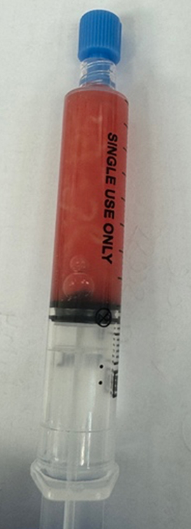 |
Figure 3 Syringe with 8mL of blood-stained purulent fluid. |
The following morning, once afebrile for 24 hours, the patient was discharged home with 14-days of oral co-trimoxazole 960 mg twice daily. Multiparametric magnetic resonance imaging (MRI) of the prostate confirms a normal appearing 36cc prostate with no suspicious lesions, no evidence of prostatitis and no recurrence of the seminal vesicle abscess six-weeks post drainage and the patient reports no symptoms.
Discussion
The seminal vesicles (SV) are a pair of accessory male sex glands which sit in the pelvis. Anatomically they are found anterior to the rectum, inferior to the fundus of the bladder and posterior to the prostate. SVs are usually 1cm in diameter and are made up of a single coiled blind ending tube. The main function of the SVs is to produce fluid which makes up approximately 70% of semen and has important properties to facilitate the function and survival of sperm. Despite a range of benign and malignant pathologies from stones, to infections to carcinomas, diseases of the seminal vesicles are both rarely occurring and rarely reported.4
The pathophysiology of SVA is not certain however it is likely that acute or chronic infections of the male lower urinary tract lead to seminal vesiculitis which predisposes the structure to infection and abscess formation. Given the proximity and ductal connections, the prostate may also be involved with prostatitis or abscess.1
As mentioned, the clinical presentation of SVA is non-specific and the symptoms are often vague and overlapping with more common pathologies. Symptoms may include fever, abdominal or perineal pain, dysuria, other lower urinary tract symptoms, haematospermia or dysorgasmia. As with most infections of the lower urinary tract, the most common risk factors for SVA include immune compromise and urethral instrumentation. Additionally, in endemic areas, Mycobacterium Tuberculosis and Schistosomiasis must be considered as causes of SVA.5,6 It must be noted that the gram negative facultative anaerobe, Citrobacter koseri, is a rare cause of urinary tract infection and as part of the Enterobacteriaceae family, is usually found as part of the normal flora of the human digestive tract.7
As a result of the vague symptomatology and equivocal examination findings, the diagnosis of SVA usually comes as a surprise to the clinician who requested imaging in the form of CT, MRI or USS looking for a more common responsible pathological process. No universal guidelines exist; however, the aforementioned imaging modalities are all reliable options to investigate specifically for SVA. USS via the transrectal route (TRUS) provides the unique ability to gather diagnostic images as well as facilitating treatment with drainage as in our case.8
Treatment of SVA should involve empiric antibiotics with both broad urogenital organism coverage until cultures and sensitivities of urine and pus samples are available. While it is possible to manage SVA conservatively,9 the old surgical adage “never let the sun set on an undrained abscess” should remain a cornerstone in the management of SVA to avoid complications worsening abscess, pelvic collection, peritonitis, bacteraemia and septic shock as well as impaired fertility.10 Such drainage can be achieved via a number of routes including transvesical, transurethral, transrectal, transgluteal, transperineal as well as open surgical excision.
TRUS has long been a skill in the armamentarium of urologists. Within the last few years, there has been a rise in transperineal prostate biopsy around the world for the diagnosis of prostate cancer. This is due to the significant reduction in post-procedure sepsis and a greater ability to sample the anterior and apical regions of the prostate.11 As such, many urologists are now skilled in the transperineal approach to the prostate, which also allows access to the seminal vesicles and can be done in a minimally invasive, low risk and time-efficient manner under local anaesthetic using the principles described by Ordones et al.12 We would recommend the transperineal approach for SVA drainage for those who have covered the learning curve for the transperineal approach to prostate biopsy; however, it is important that source control is achieved efficiently, particularly in an unwell patient. As such, the treatment provider should utilize a technique with which they are comfortable.
A literature search was performed using PubMed®. Search terms “transperineal drainage of seminal vesicle abscess”, “TRUS drainage of seminal vesicle abscess” and “perineal drainage of seminal vesicle abscess” were used. The search revealed only two articles reported in 2001 and 2017 in which a total of just five patients have been reported to have undergone transperineal drainage of SVA.8,13
Conclusion
We have presented a case with an extremely common emergency presentation being fever and right lower quadrant pain, however the diagnosis in this case is one of significant rarity. Even the most astute of clinicians would require a significantly high index of suspicion to clinically diagnose an SVA based on the history of fever, abdominal pain and dysorgasmia. As alluded to, in our case the diagnosis was discovered as a surprise finding on CT and this was crucial in leading to successful treatment and a positive patient outcome at six-week follow-up. The gap in the literature arising from the rarity of this condition as well as the near-novel technique of transperineal drainage highlights the importance of clinical case reporting and adding our case to the literature to aid future clinicians in both diagnostic and therapeutic decision-making.
Ethics
No patient information or ethical approval required.
Acknowledgments
We would like to thank our patient for their informed consent to publish their clinical case.
Author Contributions
All authors made a significant contribution to the work reported, whether that is in the conception, study design, execution, acquisition of data, analysis and interpretation, or in all these areas; took part in drafting, revising or critically reviewing the article; gave final approval of the version to be published; have agreed on the journal to which the article has been submitted; and agree to be accountable for all aspects of the work.
Funding
There is no funding to report.
Disclosure
The authors report no conflicts of interest in this work.
References
1. Rajiwate FL, Bolbandi D. Primary seminal vesicle abscess: a diagnostic dilemma—a review of current literature. J Med Sci. 2019;5(3):67–69.
2. Rajfer J, Eggleston JC, Sanders RC, Walsh PC. Fever and prostatic mass in a young man. J Urol. 1978;119(4):555–558. doi:10.1016/S0022-5347(17)57546-1
3. Sağlam M, Uğurel Ş, Kilciler M, Taşar M, Somuncu I, Üçöz T. Transrectal ultrasound-guided transperineal and transrectal management of seminal vesicle abscesses. Eur J Radiol. 2004;52(3):329–334. doi:10.1016/j.ejrad.2003.11.006
4. McKay AC, Sharma S. Anatomy, Seminal Vesicle. Treasure Island (FL): Statpearls; 2018.
5. Eastham JA, Spires KS, Abreo F, Johnson JB, Venable DD. Seminal vesicle abscess due to tuberculosis: role of tissue culture in making the diagnosis. Southern Med J. 1999;92(3):328–329. doi:10.1097/00007611-199903000-00015
6. Shebel HM, Elsayes KM, Abou El Atta HM, Elguindy YM, El-Diasty TA. Genitourinary schistosomiasis: life cycle and radiologic-pathologic findings. Radiographics. 2012;32(4):1031–1046. doi:10.1148/rg.324115162
7. Deveci A, Coban AY. Optimum management of Citrobacter koseri infection. Exp Rev Anti-Infective Ther. 2014;12(9):1137–1142. doi:10.1586/14787210.2014.944505
8. Monzo JI, García EL, Cabello Benavente R, Moralejo Garate M, Diez Cordero JM, Hernández Fernández C. Primary seminal vesicle abscess: diagnosis and treatment by transrectal ultrasound. Actas Urologicas Espanolas. 2005;29(5):523–525. doi:10.1016/S0210-4806(05)73286-1
9. Kennelly MJ, Oesterling JE. Conservative management of a seminal vesicle abscess. J Urol. 1989;141(6):1432–1433. doi:10.1016/S0022-5347(17)41337-1
10. Li K, Liu NB, Liu JX, Chen QN, Shi BM. Acute diffuse peritonitis secondary to a seminal vesicle abscess: a case report. World J Clin Cases. 2023;11(3):645. doi:10.12998/wjcc.v11.i3.645
11. Wei JT, Barocas D, Carlsson S, et al. Early detection of prostate cancer: AUA/SUO guideline part i: prostate cancer screening. J Urol. 2023;210(1):46–53. doi:10.1097/JU.0000000000003491
12. Ordones FV, Vermeulen L, Bressington M, et al. How I do it: transperineal prostate biopsy using local anesthetic in an outpatient setting. Can J Urol. 2023;30(1):11453–11456.
13. Kuribayashi S, Tanigawa G, Okuda Y, et al. Seminal vesicle abscess associated with Zinner syndrome. Hinyokika Kiyo. Acta Urologica Japonica. 2017;63(10):439–443. doi:10.14989/ActaUrolJap_63_10_439
 © 2024 The Author(s). This work is published and licensed by Dove Medical Press Limited. The
full terms of this license are available at https://www.dovepress.com/terms.php
and incorporate the Creative Commons Attribution
- Non Commercial (unported, 3.0) License.
By accessing the work you hereby accept the Terms. Non-commercial uses of the work are permitted
without any further permission from Dove Medical Press Limited, provided the work is properly
attributed. For permission for commercial use of this work, please see paragraphs 4.2 and 5 of our Terms.
© 2024 The Author(s). This work is published and licensed by Dove Medical Press Limited. The
full terms of this license are available at https://www.dovepress.com/terms.php
and incorporate the Creative Commons Attribution
- Non Commercial (unported, 3.0) License.
By accessing the work you hereby accept the Terms. Non-commercial uses of the work are permitted
without any further permission from Dove Medical Press Limited, provided the work is properly
attributed. For permission for commercial use of this work, please see paragraphs 4.2 and 5 of our Terms.